Review
. 2024 Jul 13;15(7):914.
doi: 10.3390/genes15070914.

Sulfotransferase 4A1 Coding Sequence and Protein Structure Are Highly Conserved in Vertebrates

Affiliations
Review

Sulfotransferase 4A1 Coding Sequence and Protein Structure Are Highly Conserved in Vertebrates

Robert C A M van Waardenburg et al. Genes (Basel). .

Abstract

Cytosolic sulfotransferases (SULTs) are Phase 2 drug-metabolizing enzymes that catalyze the conjugation of sulfonate to endogenous and xenobiotic compounds, increasing their hydrophilicity and excretion from cells. To date, 13 human SULTs have been identified and classified into five families. SULT4A1 mRNA encodes two variants: (1) the wild type, encoding a 284 amino acid, ~33 kDa protein, and (2) an alternative spliced variant resulting from a 126 bp insert between exon 6 and 7, which introduces a premature stop codon that enhances nonsense-mediated decay. SULT4A1 is classified as an SULT based on sequence and structural similarities, including PAPS-domains, active-site His, and the dimerization domain; however, the catalytic pocket lid 'Loop 3' size is not conserved. SULT4A1 is uniquely expressed in the brain and localized in the cytosol and mitochondria. SULT4A1 is highly conserved, with rare intronic polymorphisms that have no outward manifestations. However, the SULT4A1 haplotype is correlated with Phelan-McDermid syndrome and schizophrenia. SULT4A1 knockdown revealed potential SULT4A1 functions in photoreceptor signaling and knockout mice display hampered neuronal development and behavior. Mouse and yeast models revealed that SULT4A1 protects the mitochondria from endogenously and exogenously induced oxidative stress and stimulates cell division, promoting dendritic spines' formation and synaptic transmission. To date, no physiological enzymatic activity has been associated with SULT4A1.

Keywords: gene structure; polymorphism; protein structure; tissue/cell distribution.


Conflict of interest statement

The authors declare no conflicts of interest.

Figures

Figure 1
Figure 1
Schematic reaction mechanism for general cytosolic sulfotransferase action. (A) 3′-Phosphoadenosine 5′-phosphosulfate (PAPS) synthesis is a two-step reaction that in vertebrates is catalyzed by one enzyme, PAPS synthetase (PAPSS). Human cells have two isoforms: PAPSS1 and PAPSS2. Generation of PAPS involves two ATP and one sulfate molecule. (B) General mechanism of cytosolic SULT-catalyzed sulfonation of a molecule (R-SO4) involves transfer of sulfonate (donated by co-factor PAPS) to a hydroxyl-group of the substrate molecule (R-OH), e.g., phenol, cholesterol, or steroid.
Figure 2
Figure 2
SULT4A1 gene and mRNA structure. Gene structure of human SULT4A1 exhibits 7 exons and 6 introns, with intron 6 housing an alternative exon 6p that is inserted by alternative splicing and results in a premature stop codon that primes the SULT4A1 variant 1 mRNA for nonsense-mediated mRNA decay (NMD). This alternative splicing mechanism regulates expression of the wild-type SULT4A1 protein of 284 amino acids in all tissues. The variant 1 mRNA, if translated, would express a 260 amino acid protein that is identical to the wild-type protein until Arg248, in loss of the dimerization domain in its alternative C-terminal domain.
Figure 3
Figure 3
Alignment of the SULT4A1 from lamprey with the human SULT4A1 amino acid sequence. Lamprey, an early primitive vertebrate, contains the oldest known SULT4A1 protein with an amino acid sequence that is highly conserved through its evolution to human SULT4A1 (69% identical). PAPS binding pocket residues (orange), active site His domain (Cyan), and the dimerization domain (green). The residues of the Loop 3 catalytic pocket lid (Blue) with the additional residues (Red) that are in Loop 3 of Lamprey SULT4A1 but were seemingly lost in the subsequent evolutionary step to sharks. *: identical amino acids, .: similar amino acid.
Figure 4
Figure 4
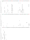
Alignment of vertebrate SULT4A1 amino acid sequences. (1) Human, (2) Pongo, (3) Red Fox, (4) Mouse, (5) Sperm whale, (6) Short-tailed opossum, (7) Great Tit, (8) Zebrafinch, (9) European Starling, (10) Anole lizard, (11) Brown Pit Viper, (12) Eastern Brown Snake, (13) High Himalayan Frog, (14) Xenopus, (15) Catfish, (16) Northern Pike, (17) Zebrafish, and (18) Whale shark. Blue is conserved, Red is multiple changes, and Green is a single change in one of the 18 species. SULT4A1 aa sequence identity conservation shown in Table 2.
Figure 4
Figure 4
Alignment of vertebrate SULT4A1 amino acid sequences. (1) Human, (2) Pongo, (3) Red Fox, (4) Mouse, (5) Sperm whale, (6) Short-tailed opossum, (7) Great Tit, (8) Zebrafinch, (9) European Starling, (10) Anole lizard, (11) Brown Pit Viper, (12) Eastern Brown Snake, (13) High Himalayan Frog, (14) Xenopus, (15) Catfish, (16) Northern Pike, (17) Zebrafish, and (18) Whale shark. Blue is conserved, Red is multiple changes, and Green is a single change in one of the 18 species. SULT4A1 aa sequence identity conservation shown in Table 2.
Figure 5
Figure 5
Structural comparison of Loop 3 between SULT4A1 and the conserved SULTs 1A1 and 2A1. Shown is a cartoon representation of the resolved crystal protein structures of SULT1A1 (PDB 4GRA), SULT2A1 (PDB 3F3Y), and SULT4A1 (PDB 1ZD1) with the prediction model of the 4A1 N-terminal domain attached (unpublished data Tibbs and Falany). The overlay of all three SULTs (not shown is the protein cartoon of SULT2A1 for clarity) to highlight the difference between the highly conserved SULT Loop 3 and unique SULT4A1 loop 3, or catalytic pocket lid. Loop 3 for 4A1 (Orange), 1A1 (Light blue) and 2A1 (Red). Conserved domains are highlighted in blue (PAPS binding pocket), yellow (active site His domain), and wheat (dimerization domain). All structure cartoons were generated using PyMol 2.5.2 (Molecular Graphics System, Schrödinger, LLC, New York, NY, USA).
Figure 6
Figure 6
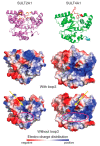
Surface electro-charge distribution comparison of SULTs 2A1 and 4A1. Resolved crystal protein structures of SULT2A1 (PDB 2QP4) (without PAP(S) but with DHEA) and SULT4A1 (PDB 1ZD1) displayed as cartoon (with Loop 3 in orange for 2A1 and in red for 4A1) and electro-charge surface with or without (bottom) Loop 3 [3,42]. All structure cartoons and electro-charge displays were generated using PyMol 2.5.2 (Molecular Graphics System, Schrödinger, LLC). Yellow arrow points to the electro-charge distribution of the PAPS-binding site in ‘opened’ catalytic pockets. Pink circle shows ‘substrate-binding site’.

Similar articles

References

    1. Falany C.N. Enzymology of human cytosolic sulfotransferases. FASEB J. 1997;11:206–216. doi: 10.1096/fasebj.11.4.9068609. - DOI - PubMed
    1. Pedersen L.C., Yi M., Pedersen L.G., Kaminski A.M. From Steroid and Drug Metabolism to Glycobiology, Using Sulfotransferase Structures to Understand and Tailor Function. Drug Metab. Dispos. 2022;50:1027–1041. doi: 10.1124/dmd.121.000478. - DOI - PMC - PubMed
    1. Allali-Hassani A., Pan P.W., Dombrovski L., Najmanovich R., Tempel W., Dong A., Loppnau P., Martin F., Thornton J., Edwards A.M., et al. Structural and chemical profiling of the human cytosolic sulfotransferases. PLoS Biol. 2007;5:e97. doi: 10.1371/journal.pbio.0050097. - DOI - PMC - PubMed
    1. Corbeil D., Thiele C., Huttner W.B. Tyrosine O-sulfation. Curr. Protoc. Protein Sci. 2005;39:14.7.1–14.7.14. doi: 10.1002/0471140864.ps1407s39. - DOI - PubMed
    1. Honke K., Taniguchi N. Sulfotransferases and sulfated oligosaccharides. Med. Res. Rev. 2002;22:637–654. doi: 10.1002/med.10020. - DOI - PubMed

LinkOut - more resources