Review
. 2024 Jul 10;25(14):7563.
doi: 10.3390/ijms25147563.

Bioluminescent Systems for Theranostic Applications

Affiliations
Review

Bioluminescent Systems for Theranostic Applications

Hyemin Kim et al. Int J Mol Sci. .

Abstract

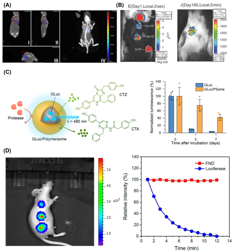
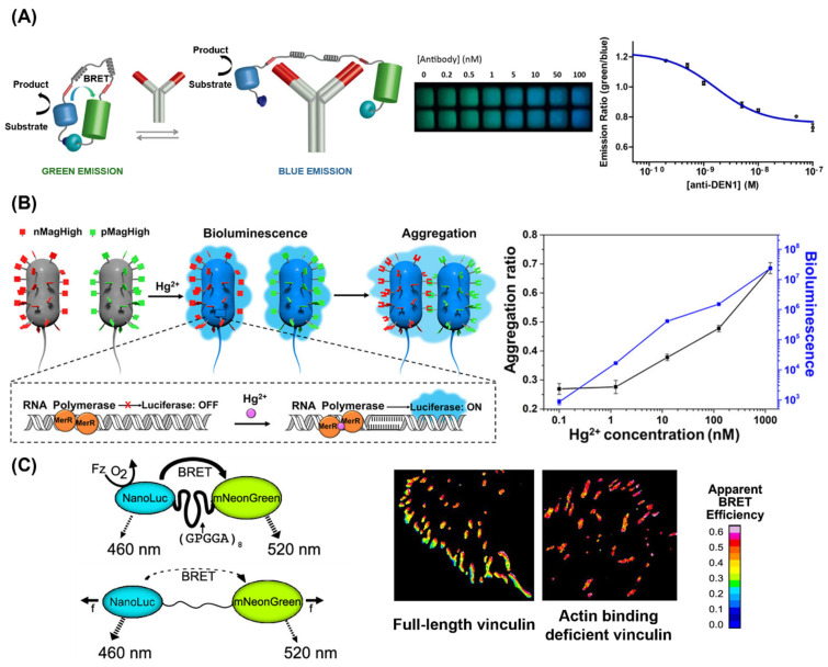
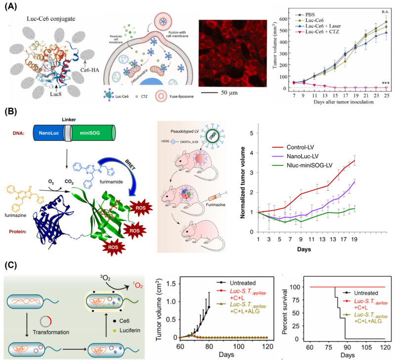
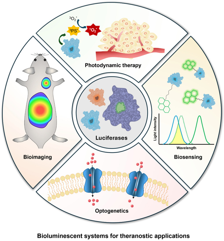
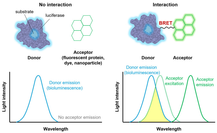
Bioluminescence, the light produced by biochemical reactions involving luciferases in living organisms, has been extensively investigated for various applications. It has attracted particular interest as an internal light source for theranostic applications due to its safe and efficient characteristics that overcome the limited penetration of conventional external light sources. Recent advancements in protein engineering technologies and protein delivery platforms have expanded the application of bioluminescence to a wide range of theranostic areas, including bioimaging, biosensing, photodynamic therapy, and optogenetics. This comprehensive review presents the fundamental concepts of bioluminescence and explores its recent applications across diverse fields. Moreover, it discusses future research directions based on the current status of bioluminescent systems for further expansion of their potential.

Keywords: bioimaging; bioluminescence; biosensing; luciferase; optogenetics; photodynamic therapy; theranostics.


Conflict of interest statement

The authors declare no conflicts of interest.

Figures

Figure 2
Figure 2
(A) Orthotopic breast cancer model established using luciferase-expressing cancer cells visualized by micro-CT and 3-dimensional bioluminescence imaging [42]. Copyright 2017, V. P. Baklaushev et al. (B) Bioluminescence images on day 1 (left) and day 180 (right) after the subcutaneous injection of a FLuc AAV vector [49]. Copyright 2005 John Wiley & Sons, Ltd. (C) Schematic illustration of GLuc-loaded polymersome (GLuc/PSome) and its long-term luminescence after uptake into cells [56]. Copyright 2022 H. Kim et al., Wiley-VCH. (D) Bioluminescence imaging of a mouse subcutaneously injected with stem cells labeled with FLuc-coated fluorescent nanodiamonds (FNDs). Decays of the fluorescence (FND) and bioluminescence (luciferase) intensities are also shown over time [57]. Copyright 2019 American Chemical Society. Adapted with permission from [42,49,56,57].
Figure 5
Figure 5
(A) Schematic representation of the BRET sensor for detecting antibodies (left) and the results obtained using a smartphone for the titration of the antibody against dengue virus type I (anti-DEN1) in a buffer (right) [97]. Copyright 2016 American Chemical Society. (B) Schematic representation of the bioluminescent bacterial sensor for detecting Hg2+ (left) and the dose-dependent results of bacterial aggregation ratio and bioluminescence (right) [109]. Copyright 2020 American Chemical Society. (C) Schematic representation of the BRET molecular tension sensor (left) and results with full-length vinculin and actin-binding-deficient vinculin (right) [111]. Copyright 2019 American Chemical Society. Adapted with permission from [97,109,111].
Figure 6
Figure 6
(A) Schematic representation of the BRET-based PDT system. Luciferase-chlorin e6 conjugates (Luc-Ce6) in membrane-fusion liposomes (left). The BRET system (red) was efficiently delivered into cytosol of 4T1 cells (center) and showed therapeutic effect on early-stage 4T1 tumor (right) (*** p < 0.001 and n.s.: not significant (two-way ANOVA)) [115]. Copyright 2023 Elsevier Ltd. (B) Schematic representations of the gene encoding NanoLuc luciferase fused to miniSOG phototoxic protein and its lentiviral delivery to HER-2 positive tumor (left). The lentiviral delivery of NanoLuc-miniSOG inhibited the tumor growth (right) [126]. Copyright 2022, E. I. Shramova et al. (C) Schematic representation of bioluminescent bacteria (LucS.T.ppGpp) (left) and its PDT effect on inhibition of tumor metastasis and recurrence (right). Tumor growth curves and survival rate of rechallenged tumor 60 days post-treatment are represented (C: Ce6, L: luciferin, and ALG: alginate hydrogel) [127]. Copyright 2021 Elsevier Ltd. Adapted with permission from [115,126,127].
Figure 1
Figure 1
Overview of bioluminescent systems for theranostic applications. Unpaired electrons are represented by an asterisk (*).
Figure 3
Figure 3
Schematic illustration of bioluminescence resonance energy transfer (BRET).
Figure 4
Figure 4
(A) Chemical structure of AkaLumine-HCl (Aka-HCl) and D-luciferin (D-luci) (left). Aka-HCl was used for ex vivo and in vivo bioluminescence imaging of deep tissue tumors (center). Quantitative analysis of bioluminescence shows stronger signal of Aka-HCl than D-luci (right) (* p < 0.05 (t-test)) [82]. Copyright 2016, T. Kuchimaru et al. (B) Schematic illustration of FLuc-luciferin-regenerating enzyme (LRE)-introduced bacteria (EcN) for stable bioluminescence imaging (left). EcN-FLuc-LRE was used for in vivo bioluminescence imaging of the mouse intestinal tract (center). Quantitative analysis of bioluminescence shows high signal of EcN-FLuc-LRE (right) [85]. Copyright 2021, T. Jiang et al. (C) Bioluminescence imaging of hepatic copper deficiency using copper-caged luciferin-1 (CCL-1) in a diet-induced murine model of nonalcoholic fatty liver disease. Bioluminescence signal was obtained after CCL-1 treatment as shown in the quantitative analysis (left) and representative images (center), and it was compared with hepatic copper levels measured by ICP-MS (right) (* p < 0.05, ** p < 0.01, *** p < 0.001 and **** p < 0.0001 (two-tailed Student’s t-test)) [87]. Copyright 2016, M. C. Heffern et al. Adapted with permission from [82,85,87].
Figure 7
Figure 7
(A) Virus-mediated expression of bioluminescent optogenetic systems (LMO3 or iLMO) modulates neuronal firing rates in vivo, inducing increases (left) or decreases (right) upon the addition of coelenterazine (CTZ) (** p < 0.01 and *** p < 0.001 (two-way ANOVA followed by post hoc test)) [131]. Copyright 2016, K. Berglund et al. (B) Schematic representation of in situ bioluminescence-driven optogenetic therapy of retinoblastoma based on camouflage nanoparticles (top). The delivery of optogenetic plasmids results in the expression of FAS-CIB1-EGFP-CAAX in the plasma membrane and the expression of CRY2-mCherry-FADD in the cytoplasm. Blue bioluminescence induces the binding of the blue light receptor proteins CIB1 and CRY2, and the subsequent binding of FADD to FAS activates the caspase-3 apoptotic signaling pathway. In vivo evaluation of camouflage nanoparticle-based optogenetic system demonstrated more significant tumor cell apoptosis upon addition of Nano-Glo (furimazine) than in other groups [141]. Red arrows indicate regions of tumor cell apoptosis. Scale bars, 100 μm. Copyright 2023 American Chemical Society. Adapted with permission from [131,141].

Similar articles

References

    1. Yun S.H., Kwok S.J.J. Light in diagnosis, therapy and surgery. Nat. Biomed. Eng. 2017;1:0008. doi: 10.1038/s41551-016-0008. - DOI - PMC - PubMed
    1. Lee G.-H., Moon H., Kim H., Lee G.H., Kwon W., Yoo S., Myung D., Yun S.H., Bao Z., Hahn S.K. Multifunctional materials for implantable and wearable photonic healthcare devices. Nat. Rev. Mater. 2020;5:149–165. doi: 10.1038/s41578-019-0167-3. - DOI - PMC - PubMed
    1. Wolfbeis O.S. An overview of nanoparticles commonly used in fluorescent bioimaging. Chem. Soc. Rev. 2015;44:4743–4768. doi: 10.1039/C4CS00392F. - DOI - PubMed
    1. Wang S., Li B., Zhang F. Molecular fluorophores for deep-tissue bioimaging. ACS Central. Sci. 2020;6:1302–1316. doi: 10.1021/acscentsci.0c00544. - DOI - PMC - PubMed
    1. Liu S., Pan X., Liu H. Two-dimensional nanomaterials for photothermal therapy. Angew. Chem. Int. Ed. 2020;132:5943–5953. doi: 10.1002/ange.201911477. - DOI - PubMed

LinkOut - more resources