. 2024 Jul 10;25(14):7562.
doi: 10.3390/ijms25147562.

Mechanism of Histone Arginine Methylation Dynamic Change in Cellular Stress

Affiliations

Mechanism of Histone Arginine Methylation Dynamic Change in Cellular Stress

Xiao-Guang Ren et al. Int J Mol Sci. .

Abstract

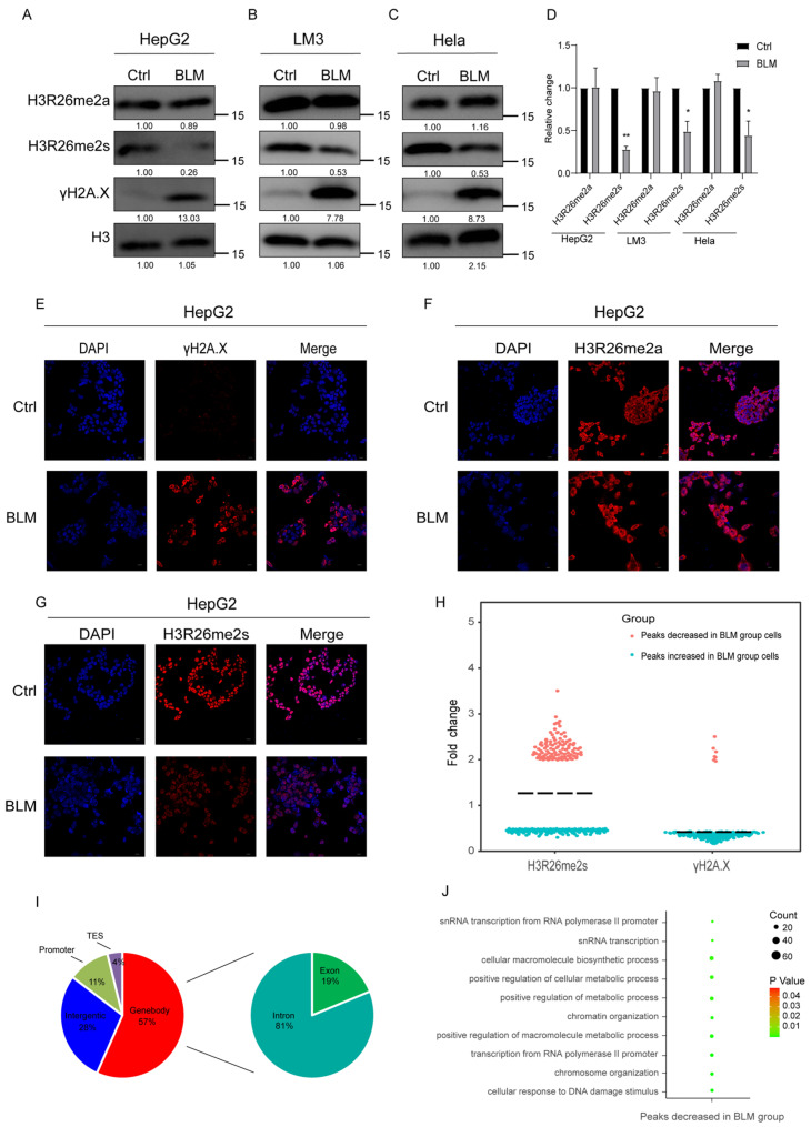
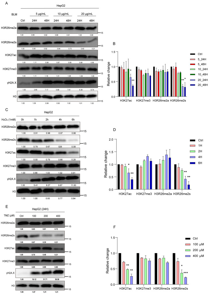
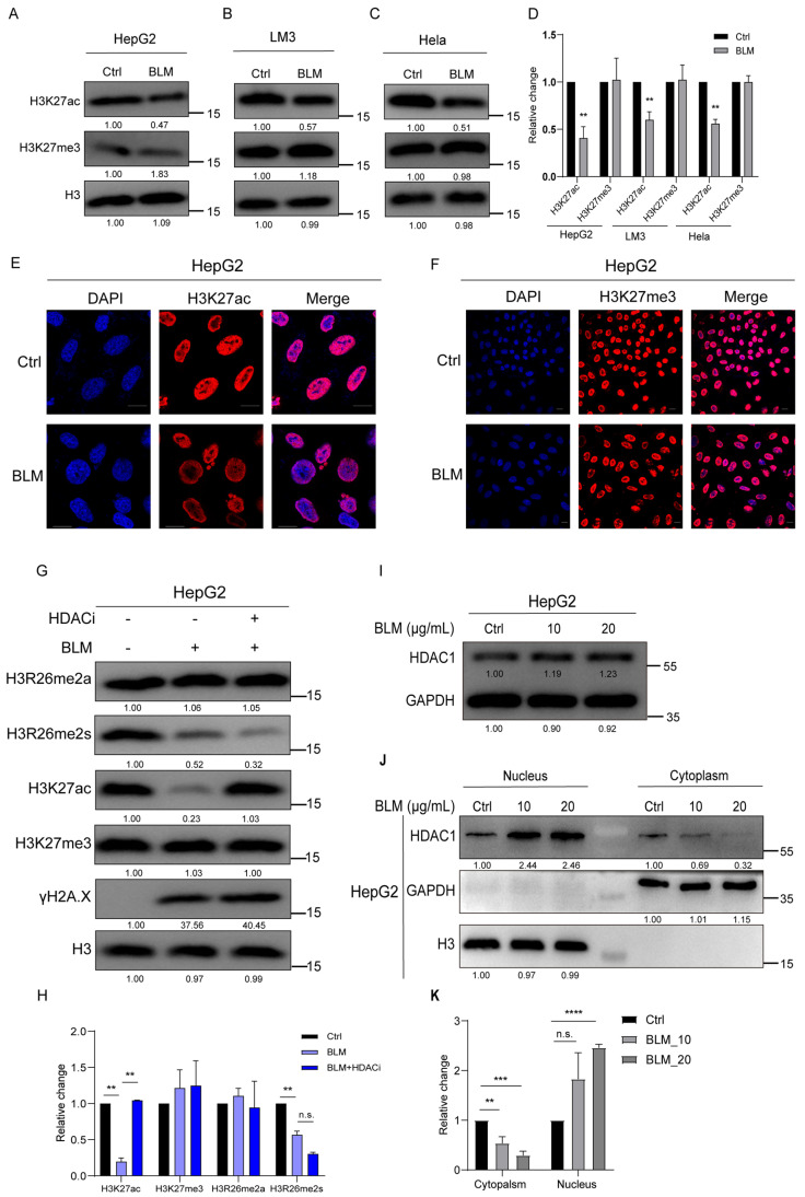
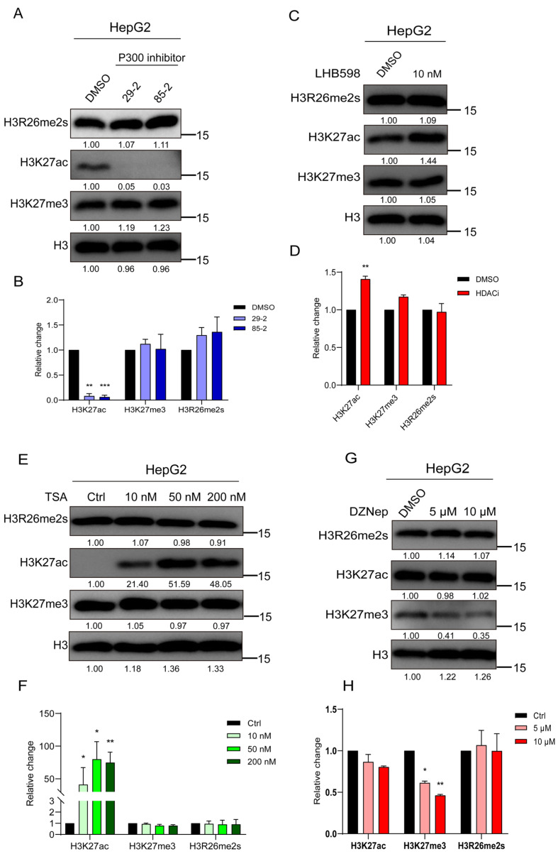
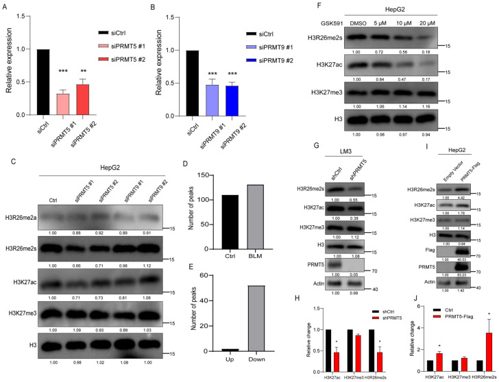
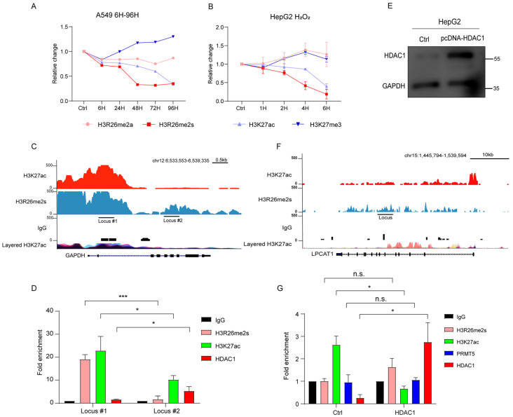
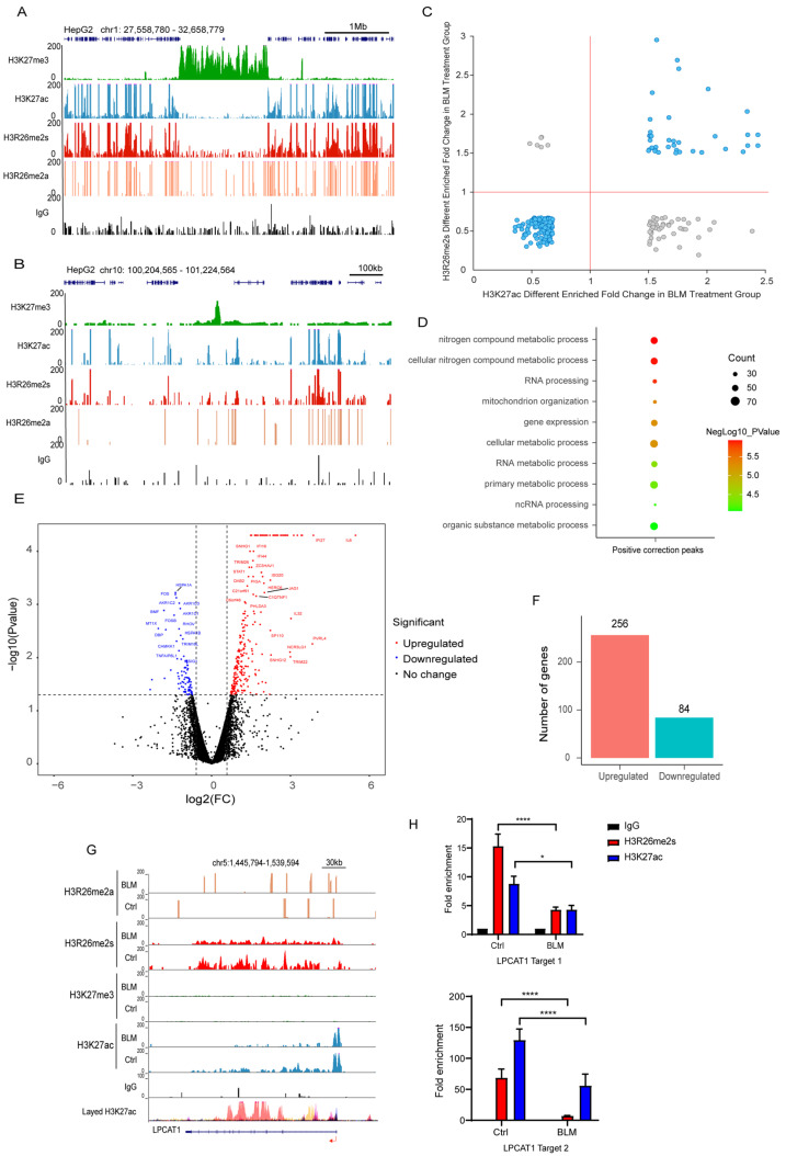
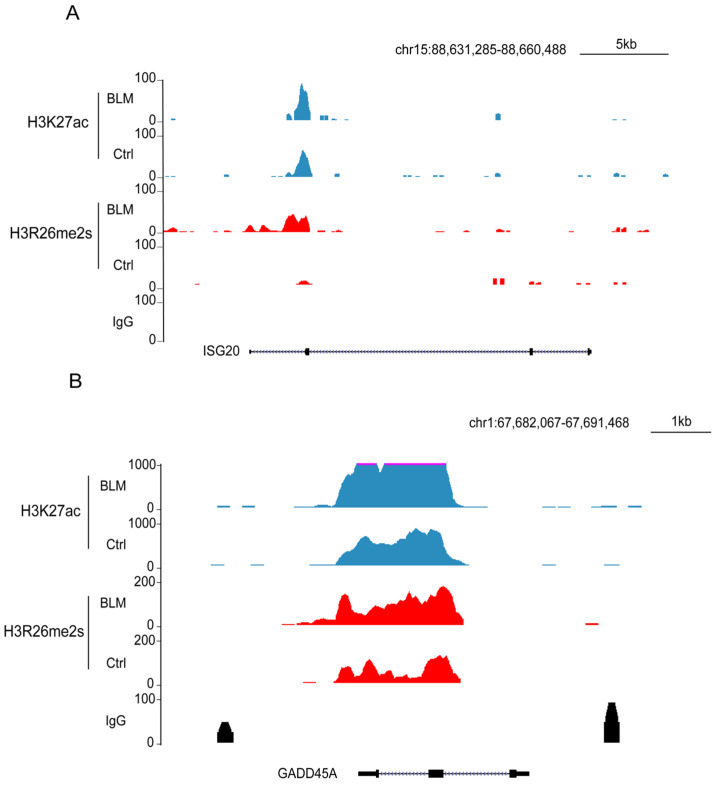
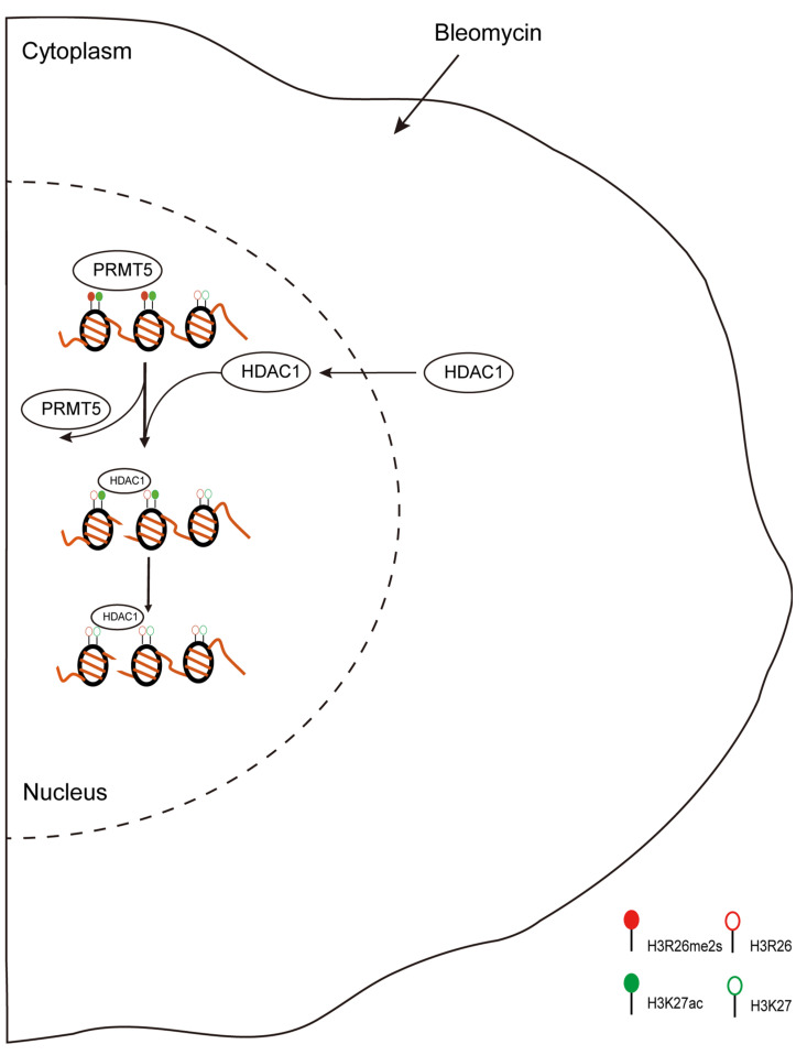
Histone arginine residue methylation is crucial for individual development and gene regulation. However, the dynamics of histone arginine methylation in response to cellular stress remains largely unexplored. In addition, the interplay and regulatory mechanisms between this and other histone modifications are important scientific questions that require further investigation. This study aimed to investigate the changes in histone arginine methylation in response to DNA damage. We report a global decrease in histone H3R26 symmetric dimethylation (H3R26me2s) and hypoacetylation at the H3K27 site in response to DNA damage. Notably, H3R26me2s exhibits a distribution pattern similar to that of H3K27ac across the genome, both of which are antagonistic to H3K27me3. Additionally, histone deacetylase 1 (HDAC1) may be recruited to the H3R26me2s demethylation region to mediate H3K27 deacetylation. These findings suggest crosstalk between H3R26me2s and H3K27ac in regulating gene expression.

Keywords: H3K27ac; H3R26me2s; crosstalk; stress.


Conflict of interest statement

The authors declare that they have no conflicts of interest.

Figures

Figure 1
Figure 1
Histone arginine methylation decreased in DNA damaged cells. (A) H3R26me2s methylation level was decreased in bleomycin-treated HepG2 cells analyzed using immunoblot assay, while for H3R26me2a there was no change. (B) H3R26me2s methylation level was decreased in bleomycin-treated LM3 cells analyzed using immunoblot assay, while for H3R26me2a there was no change. (C) H3R26me2s methylation level was decreased in bleomycin-treated Hela cells analyzed using immunoblot assay, while for H3R26me2a there was no change. (D) Relative changes in H3R26me2a and H3R26me2s in bleomycin-treated HepG2 (A), LM3 (B), and Hela (C) cells. (E) γH2A.X modification was increased in bleomycin-treated HepG2 cells measured using immunofluorescence. (F) Immunofluorescence of H3R26me2a in control and bleomycin-treated cells. (G) Immunofluorescence of H3R26me2s in control and bleomycin-treated cells. (H) Nearly half of the H3R26me2s modification peaks were downregulated in bleomycin-treated cells, as a positive control, 93% percent γH2A.X of peaks were upregulated in bleomycin-treated cells. (I) Percentage of H3R26me2s’s different peaks’ distribution features across the genome. Left Pie Chart: Characteristics of H3R26me2s’ different peaks in genome features of Genebody, Intergenic, Promoter, and TES regions. Right Pie Chart: Percentage of H3R26me2s different peaks distribution in Exon and Intron regions. (J) Analysis of H3R26me2s different peaks associated with gene functions using Gene Oncology. Data information: Scale bars in micrographs = 10 μm. Data are shown as the mean ± SEM of three biological replicates and were analyzed with a two-tailed unpaired Student’s t-test. *: p < 0.05, **: p < 0.01.
Figure 2
Figure 2
Histone modifications respond to the stress of chemical reagents in HepG2 cells. (A) HepG2 cells were treated with 5 μg/mL, 10 μg/mL, and 20 μg/mL bleomycin; for each BLM concentration group, cells were collected at 24 h and 48 h. Histone modifications were detected using immune blot with the indicated antibodies. (B) Relative changes in H3R26me2a, H3R26me2s, H3K27ac, and H3K27me3 in bleomycin-treated HepG2 cells (A). (C) HepG2 cells were treated with 1 mM H2O2 for 1, 2, 4, and 6 h, cell lysate was collected, and histone modifications were detected with the indicated antibodies. (D) Relative changes in H3R26me2a, H3R26me2s, H3K27ac, and H3K27me3 in H2O2-treated HepG2 cells (C). (E) HepG2 cells were treated with 100 μM, 200 μM, and 400 μM TMZ for 24 h, cell lysate was collected, and histone modifications were detected with the indicated antibodies. (F) Relative changes in H3R26me2a, H3R26me2s, H3K27ac, and H3K27me3 in TMZ-treated HepG2 cells (E). Data are shown as the mean ± SEM of two biological replicates and were analyzed with a two-tailed unpaired Student’s t-test. *: p < 0.05, **: p < 0.01, ***: p < 0.001.
Figure 3
Figure 3
H3K27ac undergoes hypoacetylation in response to DNA damage. (A) H3k27ac level was decreased in bleomycin-treated HepG2 cells analyzed using immunoblot assay, and the H3K27me3 level showed no change. (B) H3k27ac level was decreased in bleomycin-treated LM3 cells analyzed using immunoblot assay; H3K27me3 level showed no change. (C) H3k27ac level was decreased in bleomycin Hela cells analyzed using immunoblot assay; H3K27me3 level showed no change. (D) Relative changes in H3K27ac and H3K27me3 in bleomycin-treated HepG2 (A), LM3 (B), and Hela (C) cells. (E) Immunofluorescence of H3K27ac in control and bleomycin-treated cells; H3K27ac was decreased in bleomycin-treated cells. (F) Immunofluorescence of H3K27me3 in control and bleomycin-treated cells; H3K27me3 showed no change in bleomycin-treated cells. (G) H3K27ac level was reversed in treatment with bleomycin and HDAC inhibitor LBH598 cells. (H) Relative changes in H3K27ac, H3K27me3, H3R26me2a, and H3R26me2s in BLM- or BLM+HDACi-treated HepG2 cells (G). (I) Immune blot analysis of HDAC1 expression in bleomycin-treated HepG2 cells. (J) HDAC1 was detected in the cellular nucleus and cytoplasmic content using immunoblot; H3 and GAPDH were used as a nucleus or cytoplasm marker. (K) Relative levels of HDAC1 in cytoplasmic or nucleus in bleomycin-treated HepG2 (J) cells. Data information: Scale bars in micrographs = 10 μm. Data were analyzed with a two-tailed unpaired Student’s t-test. **: p < 0.01, ***: p < 0.001, ****: p < 0.0001, n.s.: no significance.
Figure 4
Figure 4
H3K27 acetylation or trimethylation does not impair H3R26me2s. (A) Cells were treated with P300 inhibitors for 48 h to inhibit histone lysine acetylation, and the H3R26 methylation state detected using immunoblot assay was not impaired. (B) Relative changes in H3R26me2s, H3K27ac, and H3K27me3 in P300 inhibitor-treated HepG2 cells (A). (C) Cells were treated with 10 nM HDAC inhibitor LHB598 for 24 h to evaluate histone lysine acetylation, the H3R26 symmetric dimethylation state detected via immunoblot assay was not impaired. (D) Relative changes in H3R26me2s, H3K27ac, and H3K27me3 in HDAC inhibitor LHB598-treated HepG2 cells (C). (E) Cells were treated with 10 nM, 50 nM, and 200 nM HDAC inhibitor TSA for 24 h to evaluate histone lysine acetylation; the H3R26 symmetric dimethylation state detected via immunoblot assay was not impaired. (F) Relative changes in H3R26me2s, H3K27ac, and H3K27me3 in HDAC inhibitor TSA-treated HepG2 cells (E). (G) Cells were treated with 5 μM and 10 μM EZH2 inhibitor DZNep for 72 h to inhibit histone lysine methylation, the H3R26 methylation state detected via immunoblot assay was not impaired. (H) Relative changes in H3R26me2s, H3K27ac, and H3K27me3 in EZH2 inhibitor DZNep-treated HepG2 cells (G). Data information: Data were analyzed with a two-tailed unpaired Student’s t-test. *: p < 0.05, **: p < 0.01, ***: p < 0.001.
Figure 5
Figure 5
Histone H3R26me2s modulates H3K27 acetylation state. (A) qPCR analysis of PRMT5 knockdown efficiency by siRNA in HepG2 cells. (B) qPCR analysis of PRMT9 knockdown efficiency by siRNA in HepG2 cells. (C) PRMT5 and PRMT9 were knocked down in HepG2 cells by siRNA, and histone modifications were detected with the indicated antibodies. (D) Number of PRMT5 peaks in BLM and control group HepG2 cells; (E) Different PRMT5 peaks in BLM group versus control group HepG2 cells. (F) Cells were treated with 5 μM, 10 μM, and 20 μM PRMT5 inhibitor GSK591 for 48 h to inhibit histone arginine methylation, H3K27ac modification level detected via immunoblot assay was decreased in a dose-dependent manner. (G) Histone H3R26me2s and H3K27ac levels were decreased in the PRMT5 knockdown HepG2 cells. (H) Relative changes in H3R26me2s, H3K27ac, and H3K27me3 in PRMT5 knockdown HepG2 cells (G). (I) Validation of H3R26me2s and H3K27ac levels change in the PRMT5 overexpressed cells through immunoblot. (J) Relative changes in H3R26me2s, H3K27ac, and H3K27me3 in PRMT5 overexpressed HepG2 cells (I). Data information: Data were analyzed with a two-tailed unpaired Student’s t-test. *: p < 0.05, **: p < 0.01, ***: p < 0.001.
Figure 6
Figure 6
H3K27ac deacetylation mediated by HDAC1. (A) Normalized H3K27ac, H3K27me3, H3R26me2a, and H3R26me2s levels in bleomycin-treated A549 cells; A549 cells were treated with a single BLM concentration in a time-course manner, immune blot results are provided in Figure S1B. (B) Normalized H3K27ac, H3K27me3, H3R26me2a, and H3R26me2s levels in H2O2-treated HepG2 cells; HepG2 cells were treated with a single H2O2 concentration in a time-course manner, immune blot results are provided in Figure 2C. (C) Respective of the H3R26me2s and H3K27ac modifications landscape of the GAPDH genomic locus. (D) H3R26me2s, H3K27ac, and HDAC1 enrichment in GAPDH genomic locus. (E) Validation of HDAC1 overexpression in HepG2 cells. (F) Respective of the H3R26me2s and H3K27ac modifications landscape of the LPCAT1 genomic locus. (G) H3R26me2s, H3K27ac, PRMT5, and HDAC1 enrichment in LPCAT1 genomic locus with control or HDAC1 overexpression cells. Data information: Data are shown as the mean ± SEM of three biological replicates and were analyzed with two-tailed unpaired Student’s t-test. *: p < 0.05, ***: p < 0.001, n.s.: no significance. Layered H3K27ac indicates H3K27ac ChIP-seq peaks, each color represents one cell line, and data were downloaded from the UCSC Genome Browser.
Figure 7
Figure 7
H3R26me2s shares a similar pattern with H3K27ac. (A) Respective of the H3R26me2a, H3R26me2s, H3K27ac, and H3K27me3 modifications landscape across a 1 Mb segment of the human genome; H3R26me2s shares a similar distribution pattern with H3K27ac and antagonizes H3K27me3. (B) Respective of the H3R26me2a, H3R26me2s, H3K27ac, and H3K27me3 modifications landscape across a 100 Kb segment of the human genome; H3R26me2s shares a similar distribution pattern with H3K27ac and antagonizes H3K27me3. (C) Overlapped H3R26me2s and H3K27ac up or down peaks; the cyan dots represent that H3R26me2s and H3K27ac have the same change pattern, while the grey dots represent that they have an opposite change pattern. (D) Gene Oncology of H3R26me2s and H3K27ac overlapped positive correction peaks shown in Figure 4F associated genes. (E) Volcano Plot of 256 downregulated and 84 upregulated genes in bleomycin-treated cells compared with control cells. (F) Number of genes up and down in bleomycin-treated HepG2 cells; gene fold changes above two were in the account. (G) Respective of the control and bleomycin-treated cells histone H3R26me2a, H3R26me2s, H3K27me3, and H3K27ac modifications in the downregulated gene LPCAT1 locus. (H) H3R26me2s and H3K27ac modifications were decreased in bleomycin-treated cells at the LPCAT1 locus quantitated using CUT&Tag-qPCR. Data information: Data are shown as the mean ± SEM of three biological replicates and were analyzed with two-tailed unpaired Student’s t-test. *: p < 0.05, ****: p < 0.0001.
Figure 8
Figure 8
Feature and function of H3R26me2s and H3K27ac overlapped peaks. (A) H3R26me2s and H3K27ac peaks were increased in the upregulated gene ISG15 locus. (B) H3R26me2s and H3K27ac peaks were increased in the upregulated gene GADD45A locus.
Figure 9
Figure 9
Proposed scheme for DNA damage-induced histone modifications change. During DNA damage, histone modifications change to regulate gene expression or DNA repair. In this study, we found DNA damage-induced histone arginine demethylation and lysine deacetylation. Moreover, the lower H3R26me2s region then recruited HDAC1 to catalyze H3K27 deacetylation. H3R26me2s and H3K27ac have similar occupancy across the genome, indicating the crosstalk between H3R26me2s and H3K27ac in regulating gene expression.

Similar articles

References

    1. Zhang W., Zhang X., Xue Z., Li Y., Ma Q., Ren X., Zhang J., Yang S., Yang L., Wu M., et al. Probing the Function of Metazoan Histones with a Systematic Library of H3 and H4 Mutants. Dev. Cell. 2019;48:406–419.e405. doi: 10.1016/j.devcel.2018.11.047. - DOI - PMC - PubMed
    1. Li Z., Chen C., Yong H., Jiang L., Wang P., Meng S., Chu S., Li Z., Guo Q., Zheng J., et al. PRMT2 promotes RCC tumorigenesis and metastasis via enhancing WNT5A transcriptional expression. Cell Death Dis. 2023;14:322. doi: 10.1038/s41419-023-05837-6. - DOI - PMC - PubMed
    1. Di Lorenzo A., Bedford M.T. Histone arginine methylation. FEBS Lett. 2011;585:2024–2031. doi: 10.1016/j.febslet.2010.11.010. - DOI - PMC - PubMed
    1. Cao L., Wu G., Zhu J., Tan Z., Shi D., Wu X., Tang M., Li Z., Hu Y., Zhang S., et al. Genotoxic stress-triggered β-catenin/JDP2/PRMT5 complex facilitates reestablishing glutathione homeostasis. Nat. Commun. 2019;10:3761. doi: 10.1038/s41467-019-11696-7. - DOI - PMC - PubMed
    1. Cheng Y., Wang X., Huang S., Zhang L., Lan B., Li X., Chen H., Liu Z., Su Y., Xi L., et al. A CRISPR-Cas9 library screening identifies CARM1 as a critical inhibitor of ferroptosis in hepatocellular carcinoma cells. Mol. Ther. Nucleic Acids. 2023;34:102063. doi: 10.1016/j.omtn.2023.102063. - DOI - PMC - PubMed

LinkOut - more resources