. 2024 Jul 13;25(14):7691.
doi: 10.3390/ijms25147691.

Saturation Mutagenesis and Molecular Modeling: The Impact of Methionine 182 Substitutions on the Stability of β-Lactamase TEM-1

Affiliations

Saturation Mutagenesis and Molecular Modeling: The Impact of Methionine 182 Substitutions on the Stability of β-Lactamase TEM-1

Vitaly G Grigorenko et al. Int J Mol Sci. .

Abstract

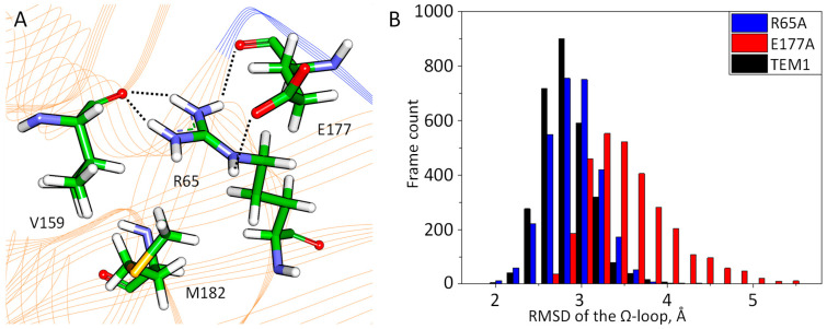
Serine β-lactamase TEM-1 is the first β-lactamase discovered and is still common in Gram-negative pathogens resistant to β-lactam antibiotics. It hydrolyzes penicillins and cephalosporins of early generations. Some of the emerging TEM-1 variants with one or several amino acid substitutions have even broader substrate specificity and resistance to known covalent inhibitors. Key amino acid substitutions affect catalytic properties of the enzyme, and secondary mutations accompany them. The occurrence of the secondary mutation M182T, called a "global suppressor", has almost doubled over the last decade. Therefore, we performed saturating mutagenesis at position 182 of TEM-1 to determine the influence of this single amino acid substitution on the catalytic properties, thermal stability, and ability for thermoreactivation. Steady-state parameters for penicillin, cephalothin, and ceftazidime are similar for all TEM-1 M182X variants, whereas melting temperature and ability to reactivate after incubation at a higher temperature vary significantly. The effects are multidirectional and depend on the particular amino acid at position 182. The M182E variant of β-lactamase TEM-1 demonstrates the highest residual enzymatic activity, which is 1.5 times higher than for the wild-type enzyme. The 3D structure of the side chain of residue 182 is of particular importance as observed from the comparison of the M182I and M182L variants of TEM-1. Both of these amino acid residues have hydrophobic side chains of similar size, but their residual activity differs by three-fold. Molecular dynamic simulations add a mechanistic explanation for this phenomenon. The important structural element is the V159-R65-E177 triad that exists due to both electrostatic and hydrophobic interactions. Amino acid substitutions that disturb this triad lead to a decrease in the ability of the β-lactamase to be reactivated.

Keywords: M182X mutation; antibiotic resistance; molecular modeling; saturating mutagenesis; thermostability; β-lactamase TEM-1.


Conflict of interest statement

The authors declare no conflicts of interest.

Figures

Figure 1
Figure 1
Relative occurrence of TEM-type β-lactamase mutations in 1990–2013 (red) and 2013–2024 (blue) years. The residue numbers responsible for expanded substrate specificity are violet and for resistance to inhibitors are pink; the most common secondary mutations are green.
Figure 2
Figure 2
Time dependence of the relative residual activity of TEM-type β-lactamases with M182 residue substitutions. Enzyme concentration is 0.1 mg/mL, 50 mM sodium phosphate buffer pH 7.0. Incubation temperature is 60 °C.
Figure 3
Figure 3
(A) Interactions of M182 and neighboring residues R65, E177, V159, and M182. Hydrogen bonds are shown in black dashed lines. The protein backbone is shown in cartoon representation and colored orange. The Ω-loop is blue. Here and on next Figures color code is the following: carbon—green, oxygen—red, nitrogen—blue, sulfur—yellow and hydrogen—white. (B) RMSD distributions for the Ω-loop for TEM-1 (black) and its R65A (blue) and E177A (red) variants.
Figure 4
Figure 4
Alignment of MD trajectory frames for the TEM-1 M182E (A), with the mutation M182L (B). Several conformations of the Ω-loop along MD trajectories are shown to demonstrate the loop flexibility. The protein backbone is shown in cartoon representation and colored blue. The 182nd residue is shown in stick representation.
Figure 5
Figure 5
Interactions in the V159-R65-E177 region in the TEM-1 M182X variants. Upper panels correspond to the variants with higher residual activities, M182E (A), M182I (B), and M182T (C). Lower panels correspond to variants with lower residual activities, M182Q (D), M182G (E), and M182L (F). The protein backbone is shown in cartoon representation and colored orange, the Ω-loop is colored blue. Hydrogen bonds are shown by dotted lines. Representative structures and their corresponding occupancies are shown.

References

    1. Eichenberger E.M., Thaden J.T. Epidemiology and Mechanisms of Resistance of Extensively Drug Resistant Gram-Negative Bacteria. Antibiotics. 2019;8:37. doi: 10.3390/antibiotics8020037. - DOI - PMC - PubMed
    1. Bush K. Past and Present Perspectives on β-Lactamases. Antimicrob. Agents Chemother. 2018;62:e01076-18. doi: 10.1128/AAC.01076-18. - DOI - PMC - PubMed
    1. Abraham E.P., Chain E. An Enzyme from Bacteria able to Destroy Penicillin. Nature. 1940;146:837. doi: 10.1038/146837a0. - DOI - PubMed
    1. Bush K. Classification for β-lactamases: Historical perspectives. Expert Rev. Anti. Infect. Ther. 2023;21:513–522. doi: 10.1080/14787210.2023.2194633. - DOI - PubMed
    1. Bonomo R.A. β-Lactamases: A Focus on Current Challenges. Cold Spring Harb. Perspect. Med. 2017;7:a025239. doi: 10.1101/cshperspect.a025239. - DOI - PMC - PubMed

LinkOut - more resources