Review
. 2024 Jul 15;25(14):7737.
doi: 10.3390/ijms25147737.

Autotaxin-Lysophosphatidate Axis: Promoter of Cancer Development and Possible Therapeutic Implications

Affiliations
Review

Autotaxin-Lysophosphatidate Axis: Promoter of Cancer Development and Possible Therapeutic Implications

Carmelo Laface et al. Int J Mol Sci. .

Abstract

Autotaxin (ATX) is a member of the ectonucleotide pyrophosphate/phosphodiesterase (ENPP) family; it is encoded by the ENPP2 gene. ATX is a secreted glycoprotein and catalyzes the hydrolysis of lysophosphatidylcholine to lysophosphatidic acid (LPA). LPA is responsible for the transduction of various signal pathways through the interaction with at least six G protein-coupled receptors, LPA Receptors 1 to 6 (LPAR1-6). The ATX-LPA axis is involved in various physiological and pathological processes, such as angiogenesis, embryonic development, inflammation, fibrosis, and obesity. However, significant research also reported its connection to carcinogenesis, immune escape, metastasis, tumor microenvironment, cancer stem cells, and therapeutic resistance. Moreover, several studies suggested ATX and LPA as relevant biomarkers and/or therapeutic targets. In this review of the literature, we aimed to deepen knowledge about the role of the ATX-LPA axis as a promoter of cancer development, progression and invasion, and therapeutic resistance. Finally, we explored its potential application as a prognostic/predictive biomarker and therapeutic target for tumor treatment.

Keywords: ATX-LPA axis; ENPP2; autotaxin (ATX); cancer; lysophosphatidic acid (LPA); lysophosphatidylcholine (LPC); lysophospholipase D (lysoD); tumor microenvironment.


Conflict of interest statement

The authors declare no conflicts of interest.

Figures

Figure 1
Figure 1
Representation of the ATX–LPA axis. ATX is linked to plasmatic membrane by means of Integrinβ3, near to LPA receptors. ATX converts LPC in LPA, which, in turn, binds to LPAR1–6 very efficiently in consideration of the proximity. Consequently, the LPA-mediated activation of LPAR receptors induces different downstream signaling pathways, including RAS, PI3K, Rho, and PLC through different G proteins. Each LPAR (1–6) activates specific G proteins and it is indicated by the same color lines of the receptor.
Figure 2
Figure 2
Schematic description about LPA effects on tumor cells and tumor microenvironment.
Figure 3
Figure 3
Schematic illustration of ATX–LPA pathway involvement in genesis and progression of breast cancer, ovarian cancer, hepatocellular cancer, pancreatic cancer, and glioblastoma multiforme.
Figure 4
Figure 4
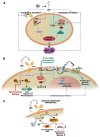
Representation of different drug resistance mechanisms mediated by the ATX–LPA pathway. (A) The ATX–LPA axis inhibits extrinsic and intrinsic apoptosis pathways, downregulating FASL/FAS and BAX/BAD and upregulating BCL2. The lilac-colored arrows refer to pathways activated by LPA (B) LPA favors resistance to Temsirolimus and Sutinib, activating Arf6 GTPase and its downstream pathway; the ATX–LPA axis stimulates transformation of ceramide in SP-1, countering activities of Tamoxifen, Doxorubicin, and Paclitaxel; LPAR1 stimulates Nrf2, the transcription factor of MDR carriers and antioxidant genes activating chemoresistance. The lilac-colored arrows refer to pathways activated by LPA, the green-colored arrows refer to pathways activated by drugs written in green. (C) Downregulation of RGS favors drug resistance in cancer cells by enhancing LPARs activity. Abbreviations: FASL—FAS Ligand; FADD—FAS-associated protein with death domain; BCL2—B-cell lymphoma 2; BAD—BCL2 associated agonist of cell death; BAX—Bcl-2-associated X protein; Apaf—apoptotic protease activating factor; CytC—cytocrome C; LPARs—lysophosphatidic acid receptor; Arf—ADP-ribosylation factor; SK-1—sphingosine kinase-1; SP-1—sphingosine 1-phosphate; Nrf2—nuclear factor erythroid 2-related factor 2; MDR carriers—multi drug resistant carriers; EMT—epithelial-mesenchymal transition; RGS—regulators of G protein signaling.

References

    1. van Meeteren L.A., Moolenaar W.H. Regulation and Biological Activities of the Autotaxin–LPA Axis. Prog. Lipid Res. 2007;46:145–160. doi: 10.1016/j.plipres.2007.02.001. - DOI - PubMed
    1. Zhang X., Li M., Yin N., Zhang J. The Expression Regulation and Biological Function of Autotaxin. Cells. 2021;10:939. doi: 10.3390/cells10040939. - DOI - PMC - PubMed
    1. Choi J.W., Herr D.R., Noguchi K., Yung Y.C., Lee C.W., Mutoh T., Lin M.E., Teo S.T., Park K.E., Mosley A.N., et al. LPA Receptors: Subtypes and Biological Actions. Annu. Rev. Pharmacol. Toxicol. 2010;50:157–186. doi: 10.1146/annurev.pharmtox.010909.105753. - DOI - PubMed
    1. Tomsig J.L., Snyder A.H., Berdyshev E.V., Skobeleva A., Mataya C., Natarajan V., Brindley D.N., Lynch K.R. Lipid Phosphate Phosphohydrolase Type 1 (LPP1) Degrades Extracellular Lysophosphatidic Acid in Vivo. Biochem. J. 2009;419:611–618. doi: 10.1042/BJ20081888. - DOI - PMC - PubMed
    1. Tang X., Benesch M.G.K., Brindley D.N. Lipid Phosphate Phosphatases and Their Roles in Mammalian Physiology and Pathology. J. Lipid Res. 2015;56:2048–2060. doi: 10.1194/jlr.R058362. - DOI - PMC - PubMed

Substances

LinkOut - more resources