. 2024 Jul 16;25(14):7800.
doi: 10.3390/ijms25147800.

Auto/Paracrine C-Type Natriuretic Peptide/Cyclic GMP Signaling Prevents Endothelial Dysfunction

Affiliations

Auto/Paracrine C-Type Natriuretic Peptide/Cyclic GMP Signaling Prevents Endothelial Dysfunction

Franziska Werner et al. Int J Mol Sci. .

Abstract

Endothelial dysfunction is cause and consequence of cardiovascular diseases. The endothelial hormone C-type natriuretic peptide (CNP) regulates vascular tone and the vascular barrier. Its cGMP-synthesizing guanylyl cyclase-B (GC-B) receptor is expressed in endothelial cells themselves. To characterize the role of endothelial CNP/cGMP signaling, we studied mice with endothelial-selective GC-B deletion. Endothelial EC GC-B KO mice had thicker, stiffer aortae and isolated systolic hypertension. This was associated with increased proinflammatory E-selectin and VCAM-1 expression and impaired nitric oxide bioavailability. Atherosclerosis susceptibility was evaluated in such KO and control littermates on Ldlr (low-density lipoprotein receptor)-deficient background fed a Western diet for 10 weeks. Notably, the plaque areas and heights within the aortic roots were markedly increased in the double EC GC-B/Ldlr KO mice. This was accompanied by enhanced macrophage infiltration and greater necrotic cores, indicating unstable plaques. Finally, we found that EC GC-B KO mice had diminished vascular regeneration after critical hind-limb ischemia. Remarkably, all these genotype-dependent changes were only observed in female and not in male mice. Auto/paracrine endothelial CNP/GC-B/cGMP signaling protects from arterial stiffness, systolic hypertension, and atherosclerosis and improves reparative angiogenesis. Interestingly, our data indicate a sex disparity in the connection of diminished CNP/GC-B activity to endothelial dysfunction.

Keywords: C-type natriuretic peptide; angiogenesis; arterial stiffening; atherosclerosis; cyclic GMP; endothelial dysfunction; systolic hypertension.


Conflict of interest statement

The authors declare no conflicts of interest.

Figures

Figure 1
Figure 1
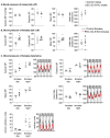
Demonstration of the deletion of the GC-B receptor in aortic endothelial cells. qRT-PCR showed significantly reduced GC-B mRNA expression (normalized to β2-microglobulin) in freshly enriched aortic endothelial cells (Ecs) of EC GC-B KO mice and unaltered GC-B expression in the rest of the aortic wall (n = 4–6, * p < 0.05 versus controls; Mann–Whitney test).
Figure 2
Figure 2
Increased systolic BP in female EC GC-B KO mice. (A,B) Tail cuff plethysmography showed unaltered systolic and diastolic blood pressure (BP) in male EC GC-B KO mice (top panels) but increased systolic and mean BP in female KO mice as compared to respective sex-matched control littermates (lower panels) (n = 5–8). (C) Telemetry confirmed increased systolic BP in female KO mice at daytime. Heart rate and locomotor activity did not differ between genotypes (n = 5). * p < 0.05 versus controls (unpaired t-test).
Figure 3
Figure 3
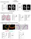
Aortic stiffening and thickening in female EC GC-B KO mice. (A) Non-invasive Doppler measurements showed increased aortic pulse wave velocity (PWV) in female, but not male, EC GC-B KO mice as compared to their control littermates (n = 8–10). (B) Hematoxylin–eosin stainings showed enhanced total, adventitial, and medial thickness of aortae from female EC GC-B KO mice; representative pictures are in right panels. (C) Sirius red and Elastica Van Gieson stainings revealed increased collagen area, unchanged elastin area, and increased numbers of elastin breaks (see arrows) in aortae of female EC GC-B KO mice; representative pictures are on top (n = 5–6). * p < 0.05 versus controls (unpaired t-test).
Figure 4
Figure 4
Enhanced expression of proinflammatory adhesion proteins and decreased eNOS phosphorylation and activity in aortae from female EC GC-B KO mice. (A) Aortic ET-1 mRNA expression levels (qRT-PCR) and ET-1 plasma concentrations (ELISA) were similar in KO and control females (n = 13, unpaired t-test). (B) Unchanged P-selectin but increased E-selectin and VCAM-1 mRNA expression levels in aortae from KO females. Target mRNAs were normalized to S12 and calculated as x-fold vs. controls (n = 16; * p < 0.05 versus controls; unpaired t-test). (C,D) Immunoblots: reduced eNOS-Serine1177 and VASP-Serine239 phosphorylation in aortae (C) and platelets (D) from female KO mice; platelet VASP-Serine157 phosphorylation was unaltered. Target proteins were normalized to GAPDH and calculated as x-fold vs. controls (n = 6–7; * p < 0.05 versus controls; two-way ANOVA).
Figure 5
Figure 5
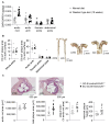
Western diet increased CNP mRNA expression in aortic roots and EC GC-B KO females develop enhanced atherosclerosis in this region. (A) Quantitative qRT-PCR: CNP mRNA expression (normalized to S12) was greater in the aortic root as compared to distal segments and increased after Western diet (n = 12 samples from 6 mice; p < 0.05 vs. + aortic root, or * normal diet; two-way ANOVA). (B) Oil red stainings: plaque areas in the whole aorta and specifically in the aortic arch as well as thoracic and abdominal segments were not different between control and EC GC-B KO females (all with Ldlr−/− and Western diet; n = 9; Mann–Whitney test). Right panels: Representative pictures. (C) Aldehyde-fuchsin stainings showed that in the aortic roots of EC GC-B KO/Ldlr−/− females the total and relative plaque areas as well as plaque heights were increased. Representative pictures are on top (n = 9; * p < 0.05 vs. control/Ldlr−/− females; unpaired t-test).
Figure 6
Figure 6
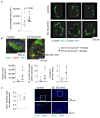
The atherosclerotic plaques in the aortic roots of EC GC-B KO female mice have increased macrophage infiltrations and greater necrotic cores despite unaltered endothelial coverage area. (A) Immunocytochemistry: increased macrophage areas in atherosclerotic aortic roots of EC GC-B KO/Ldlr−/− females; right panels: examples of macrophage (Mac2) stainings (n = 9; p = 0.06 vs. control/Ldlr−/− females; unpaired t-test). (B) Morphometric analyses of aortic root plaques (stained for macrophages (Mac2) and smooth muscle cells (αSMA)) showed increased areas and numbers of necrotic cores (arrows) in such KO females (n = 9, * p < 0.05 vs. controls; unpaired t-test). (C) Stainings of endothelial cells (CD31) did not reveal differences in CD31+ areas within aortic root plaques of the two genotypes; exemplary pictures of green-labeled endothelial cells are shown in the right panels (n = 5; Mann–Whitney test). The two lower panels show the tissue areas labeled with white squares in the upper panels, at higher magnification.
Figure 7
Figure 7
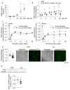
Impaired post-ischemic endothelial regeneration in female EC GC-B KO mice. (A) CNP enhanced cGMP levels in HUVECs (n = 6–8; * p < 0.05 vs. basal (0); one-way ANOVA). (B) BrdU incorporation assays: CNP stimulated HUVEC proliferation and this effect was abolished by pretreatment with the PKGI inhibitor Rp-8-Br-PET-cGMPs (100 µM) (n = 6–9; p < 0.05 vs. * basal (0) or # PBS; two-way ANOVA). (C) Laser Doppler perfusion imaging before/after experimental hind-limb ischemia (HLI): post-ischemic reperfusion was unaltered in male (left panel) but reduced in female EC GC-B KO mice (right panel) in comparison to sex-matched control littermates (n = 6–9; * p < 0.05 vs. controls; two-way ANOVA). (D) Isolectin stainings of gastrocnemius muscles dissected 28 days after HLI revealed reduced capillary/myofiber ratios in female KO mice (n = 18 sections from 9 mice; * p < 0.05 vs. controls; Mann–Whitney test); right panels: exemplary pictures. (E) Immunoblots: Reduced CD31 expression in ischemic gastrocnemius muscles of female KO mice. Levels were normalized to GAPDH and expressed as x-fold vs. controls (n = 9; * p < 0.05 vs. controls; unpaired t-test).

Similar articles

References

    1. Safar M.E. Arterial stiffness as a risk factor for clinical hypertension. Nat. Rev. Cardiol. 2018;15:97–105. doi: 10.1038/nrcardio.2017.155. - DOI - PubMed
    1. Laurent S., Boutouyrie P., Asmar R., Gautier I., Laloux B., Guize L., Ducimetiere P., Benetos A. Aortic stiffness is an independent predictor of all-cause and cardiovascular mortality in hypertensive patients. Hypertension. 2001;37:1236–1241. doi: 10.1161/01.HYP.37.5.1236. - DOI - PubMed
    1. Humphrey J.D., Harrison D.G., Figueroa C.A., Lacolley P., Laurent S. Central Artery Stiffness in Hypertension and Aging: A Problem with Cause and Consequence. Circ. Res. 2016;118:379–381. doi: 10.1161/CIRCRESAHA.115.307722. - DOI - PMC - PubMed
    1. Wu J., Thabet S.R., Kirabo A., Trott D.W., Saleh M.A., Xiao L., Madhur M.S., Chen W., Harrison D.G. Inflammation and mechanical stretch promote aortic stiffening in hypertension through activation of p38 mitogen-activated protein kinase. Circ. Res. 2014;114:616–625. doi: 10.1161/CIRCRESAHA.114.302157. - DOI - PMC - PubMed
    1. Ferreira N.S., Tostes R.C., Paradis P., Schiffrin E.L. Aldosterone, Inflammation, Immune System, and Hypertension. Am. J. Hypertens. 2021;34:15–27. doi: 10.1093/ajh/hpaa137. - DOI - PMC - PubMed

MeSH terms

Substances

LinkOut - more resources