. 2024 Jul 18;25(14):7864.
doi: 10.3390/ijms25147864.

Joint Inflammation Correlates with Joint GPR30 Expression in Males and Hippocampal GPR30 Expression in Females in a Rat Model of Rheumatoid Arthritis

Affiliations

Joint Inflammation Correlates with Joint GPR30 Expression in Males and Hippocampal GPR30 Expression in Females in a Rat Model of Rheumatoid Arthritis

Tanja Grubić Kezele et al. Int J Mol Sci. .

Abstract

It is not entirely clear how the interaction between joint inflammation and the central nervous system (CNS) response in rheumatoid arthritis (RA) works, and what pathophysiology underlies the sex differences in coexisting neuropsychiatric comorbidities. It is known that estrogen hormones reduce inflammation in RA and that this occurs mainly via the stimulation of G protein-coupled receptor-30 (GPR30), also known as G protein-coupled estrogen receptor (GPER) 1. However, changes in GPR30 expression and sex differences induced by local and systemic inflammation in RA are not yet known. Our aim was to reveal sex differences in the expression and association of joint GPR30 with local and systemic inflammation, clinical course and furthermore with hippocampal GPR30 expression during pristane-induced arthritis (PIA) in Dark Agouti (DA) rats, an animal model of RA. Furthermore, we demonstrated sex-specific differences in the association between joint and systemic inflammation and hippocampal microglia during PIA. Our results suggest sex-specific differences not only in the clinical course and serum levels of pro-inflammatory cytokines but also in the expression of GPR30. Female rats show greater synovial inflammation and greater damage to the articular cartilage compared to males during PIA attack. Male rats express higher levels of synovial and cartilaginous GPR30 than females during PIA, which correlates with a less severe clinical course. The correlation between synovial and cartilaginous GPR30 and joint inflammation scores (Krenn and Mankin) in male rats suggests that the more severe the joint inflammation, the higher the GPR30 expression. At the same time, there is no particular upregulation of hippocampal GPR30 in males. On the other hand, female rats express higher levels of neuroprotective GPR30 in the hippocampus than male rats at the basic level and during PIA attack. In addition, females have a higher number of Iba-1+ cells in the hippocampus during PIA attack that strongly correlates with the clinical score, serum levels of IL-17A, and Krenn and Mankin scores. These results suggest that male rats are better protected from inflammation in the joints and female rats are better protected from the inflammation in the hippocampus during a PIA attack, independently of microglia proliferation. However, in the remission phase, synovial GPR30 expression suddenly increases in female rats, as does hippocampal GPR30 expression in males. Further experiments with a longer remission period are needed to investigate the molecular background of these sex differences, as well as microglia phenotype profiling.

Keywords: G protein-coupled estrogen receptor 1; Krenn synovitis score; Mankin osteoarthritis score; cytokines; hippocampus; microglia; neuroinflammation; rheumatoid arthritis.


Conflict of interest statement

Authors declare no conflict of interest.

Figures

Figure 1
Figure 1
Clinical course. The clinical course in the male (n = 45) and female (n = 45) rat groups. Values are presented as mean ± SD using PIA scores of each animal for every day. Mann–Whitney U test revealed no statistical significance (p = 0.847).
Figure 2
Figure 2
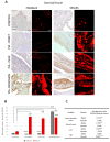
Pristane-induced arthritis upregulates synovial GPR30 expression mostly in male DA rats, which is in strong positive correlation with clinical and Krenn synovitis score. (A) Representative immunofluorescent and immunohistological pictures show staining with anti-GPR30 antibodies in paraffin-embedded sections of metatarsophalangeal joints (synovial tissue) obtained from male and female DA rats: control (treated with saline); PIA onset (between 9th and 12th day after induction); PIA peak (between 16th and 20th day after induction); PIA remission (between 20th and 25th day after induction). Scale bars indicate 50 μm. (B) Quantification of synovial GPR30 expression was performed using Cell F v3.1 software analysis on 12 regions of interest (3 slides/rat × 6 rats/group = 18 slides/group and total 24 rats). Values are expressed as mean value ± SE. One-way ANOVA followed by the post hoc Scheffé test: * difference between male and female rats; X difference between male control and male PIA rats; # difference between female control and female PIA rats; XXX p < 0.001; ### p < 0.001; *** p < 0.001. (C) Pearson’s correlation between expression of synovial GPR30 and Krenn synovitis score, clinical score and serum level of IL-17A and TNF: ** p < 0.01; *** p < 0.001.
Figure 3
Figure 3
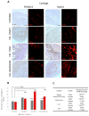
Pristane-induced arthritis upregulates cartilage GPR30 expression mostly in male DA rats, which is in strong positive correlation with clinical and Mankin synovitis score. (A) Representative immunofluorescent and immunohistological pictures show staining with anti-GPR30 antibodies in paraffin-embedded sections of metatarsophalangeal joints (cartilage) obtained from male and female DA rats: control (treated with saline); PIA onset (between 9th and 12th day after induction); PIA peak (between 16th and 20th day after induction); PIA remission (between 20th and 25th day after induction). Insert show staining in slides incubated without primary anti-GPR30 antibodies (negative control). Scale bars indicate 50 μm. (B) Quantification of cartilaginous GPR30 expression was performed using Cell F v3.1 software analysis on 12 regions of interest (3 slides/rat × 6 rats/group = 18 slides/group and total 24 rats). Values are expressed as mean value ± SE. One-way ANOVA followed by the post hoc Scheffé test: * difference between male and female rats; X difference between male control and male PIA rats; # difference between female control and female PIA rats; XXX p < 0.001; ### p < 0.001; *** p < 0.001. (C) Pearson’s correlation between expression of cartilage GPR30 and Mankin synovitis score, clinical score, and serum level of IL-17A and TNF: *** p < 0.001.
Figure 4
Figure 4
Pristane-induced arthritis upregulates hippocampal GPR30 expression in the acute phase but downregulates in the remission phase of female rats, which is in strong negative correlation with the serum level of TNF. (A) Representative immunofluorescent pictures show staining with anti-GPR30 antibodies in paraffin-embedded sections of brain tissue (hippocampus/DG) obtained from male and female DA rats: control (treated with saline); PIA onset (between 9th and 12th day after induction); PIA peak (between 16th and 20th day after induction); PIA remission (between 20th and 25th day after induction). Scale bars indicate 50 μm. (B) Quantification of hippocampal GPR30 expression was performed using Cell F v3.1 software analysis on 12 regions of interest (three slides/rat × six rats/group = 18 slides/group and total 24 rats). Values are expressed as mean value ± SE. One-way ANOVA followed by the post hoc Scheffé test: * difference between male and female rats; X difference between male control and male PIA rats; # difference between female control and female experiment rats; XXX p < 0.001; ### p < 0.001; *** p < 0.001. (C) Pearson’s correlation between hippocampal GPR30 expression, clinical score, serum level of IL-17A and TNF, Krenn synovitis and Mankin osteoarthritis score: ** p < 0.01.
Figure 5
Figure 5
Pristane-induced arthritis upregulates the number of hippocampal Iba-1+ cells mostly in female DA rats, which is in positive correlation with the clinical score, serum level of IL-17A and the Krenn synovitis and Mankin osteoarthritis scores. (A) Representative immunofluorescent pictures show staining with anti-Iba-1 antibody in paraffin-embedded sections of brain tissue (hippocampus/DG) obtained from male and female DA rats: control (treated with saline); PIA onset (between 9th and 12th day after induction); PIA peak (between 16th and 20th day after induction); PIA remission (between 20th and 25th day after induction). Scale bars indicate 50 μm. (B) The number of hippocampal Iba-1+ cells per mm2. Cells were manually counted in regions of interest (12 ROI/4 μm slide x 3 slides/rat × 6 rats/group = 18 slides/group and total 24 rats). Values are expressed as mean value ± SE. One-way ANOVA followed by the post hoc Scheffé test: * difference bewteen male and female rats; X difference between male control and male experiment rats; # difference between female control and female PIA rats; X p < 0.05, ### p < 0.001; and *** p < 0.001. (C) Pearson’s correlation between the number of hippocampal Iba-1+ cells, hippocampal GPR30 expression, clinical score, serum level of IL-17A and TNF, Krenn synovitis and Mankin osteoathritis score: * p < 0.05; ** p < 0.01; *** p < 0.001.
Figure 6
Figure 6
Disease scoring system. The maximum score per limb and rat was 15 and 60, respectively. Scores were not given for deformations if they were not accompanied by erythema or swelling. (A) Normal hind paw without the inflammation. (B) Inflammed and swolen ankle of a hind paw after PIA induction. (C) Illustration of a hind paw from the dorsal side with score points. (D) Illustration of a front paw from the dorsal side with score points. (E) Illustration of a hind paw from the medial side with score points.
Figure 7
Figure 7
Rat hippocampus from the medial side. DG, dentate gyrus, CA, cornu Ammonis. Magnification 40×.

References

    1. Chaurasia N., Singh A., Singh I., Singh T., Tiwari T. Cognitive Dysfunction in Patients of Rheumatoid Arthritis. J. Fam. Med. Prim. Care. 2020;9:2219. doi: 10.4103/jfmpc.jfmpc_307_20. - DOI - PMC - PubMed
    1. Matcham F., Norton S., Scott D.L., Steer S., Hotopf M. Symptoms of Depression and Anxiety Predict Treatment Response and Long-Term Physical Health Outcomes in Rheumatoid Arthritis: Secondary Analysis of a Randomized Controlled Trial. Rheumatology. 2015;55:268–278. doi: 10.1093/rheumatology/kev306. - DOI - PMC - PubMed
    1. Michelsen B., Kristianslund E.K., Sexton J., Hammer H.B., Fagerli K.M., Lie E., Wierød A., Kalstad S., Rødevand E., Krøll F., et al. Do Depression and Anxiety Reduce the Likelihood of Remission in Rheumatoid Arthritis and Psoriatic Arthritis? Data from the Prospective Multicentre NOR-DMARD Study. Ann. Rheum. Dis. 2017;76:1906–1910. doi: 10.1136/annrheumdis-2017-211284. - DOI - PubMed
    1. Giacometti J., Grubić-Kezele T. Olive Leaf Polyphenols Attenuate the Clinical Course of Experimental Autoimmune Encephalomyelitis and Provide Neuroprotection by Reducing Oxidative Stress, Regulating Microglia and SIRT1, and Preserving Myelin Integrity. Oxid. Med. Cell. Longev. 2020;2020:1–20. doi: 10.1155/2020/6125638. - DOI - PMC - PubMed
    1. Süß P., Rothe T., Hoffmann A., Schlachetzki J.C.M., Winkler J. The Joint-Brain Axis: Insights from Rheumatoid Arthritis on the Crosstalk between Chronic Peripheral Inflammation and the Brain. Front. Immunol. 2020;11:612104. doi: 10.3389/fimmu.2020.612104. - DOI - PMC - PubMed

MeSH terms

LinkOut - more resources