Review
. 2024 Jul 18;25(14):7880.
doi: 10.3390/ijms25147880.

Metabolic Derangement of Essential Transition Metals and Potential Antioxidant Therapies

Affiliations
Review

Metabolic Derangement of Essential Transition Metals and Potential Antioxidant Therapies

Adriana Fontes et al. Int J Mol Sci. .

Abstract

Essential transition metals have key roles in oxygen transport, neurotransmitter synthesis, nucleic acid repair, cellular structure maintenance and stability, oxidative phosphorylation, and metabolism. The balance between metal deficiency and excess is typically ensured by several extracellular and intracellular mechanisms involved in uptake, distribution, and excretion. However, provoked by either intrinsic or extrinsic factors, excess iron, zinc, copper, or manganese can lead to cellular damage upon chronic or acute exposure, frequently attributed to oxidative stress. Intracellularly, mitochondria are the organelles that require the tightest control concerning reactive oxygen species production, which inevitably leaves them to be one of the most vulnerable targets of metal toxicity. Current therapies to counteract metal overload are focused on chelators, which often cause secondary effects decreasing patients' quality of life. New therapeutic options based on synthetic or natural antioxidants have proven positive effects against metal intoxication. In this review, we briefly address the cellular metabolism of transition metals, consequences of their overload, and current therapies, followed by their potential role in inducing oxidative stress and remedies thereof.

Keywords: antioxidants; copper; iron; manganese; oxidative stress; transition metals; zinc.


Conflict of interest statement

HZ is scientific consultant for ArborMed Co., Gangnam-gu, Seoul 06237 South Korea.

Figures

Figure 1
Figure 1
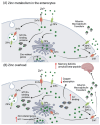
Iron metabolism in the enterocyte and hemochromatosis. (A) Iron is absorbed as Fe2+ through reduction by DcytB/STEAP, and transported via DMT1. In the case iron is bound to heme, it can be transported via heme carrier 1. Another route is via receptor-mediated endocytosis bound to transferrin. Afterwards, iron can be stored in the form of ferritin, used for the biosynthesis of Fe-S clusters, or integrate the cellular labile iron pool. The iron in the labile iron pool can bind to ferritin, and vice-versa. If iron is not stored or used, it will be exported to the circulation via ferroportin, and oxidized to ferric iron by hephaestin (in the intestine) or ceruloplasmin. Iron export via ferroportin is regulated by hepcidin, which is produced by the liver. (B) In hemochromatosis, hepcidin expression is low, thus ferroportin activity is left unregulated, resulting in an increase in iron efflux in the circulation. Abbreviations: DcytB: ferrireductase duodenal cytochrome b; DMT1: divalent metal transporter 1; Fe: iron; STEAP: six-transmembrane epithelial antigen of the prostate. Created in BioRender.com (accessed on 19 May 2024).
Figure 2
Figure 2
Zinc metabolism in the enterocyte and its overload. (A) Zinc is absorbed as Zn2+ via the ZIPs (mainly ZIP4) and can be excreted into the intestinal lumen by the ZnTs. In the enterocytes, Zn2+ can be found as cytoplasmic free zinc (which can bind to low molecular weight ligants), sequestered into vesicles, or bound to Zn-binding proteins, such as MT. Zinc can also be transported to the Golgi apparatus or to the nuclei, where it induces the expression of several zinc-related genes. In circulation, Zn2+ binds to albumin, macroglobulin, or transferrin. (B) In a scenario of zinc overload, there is an increase in the transcription of MTF-1, which leads to an increased MT expression to buffer the cytosolic Zn2+. The overexpression of MT leads to Cu+ binding and decreased absorption. The increase in zinc in circulation can be detrimental for brain function, leading to the development of memory deficits and increased deposition of the amyloid beta peptide, which is linked to different neurodegenerative conditions. Abbreviations: MT: metallothioneins; MTF-1: metal regulatory transcription factor 1; ZIP: Zrt-, Irt-like protein family; Zn: zinc; ZnT: zinc transporter. Created in BioRender.com.
Figure 3
Figure 3
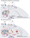
Copper metabolism in the enterocytes and Wilson disease. (A) Copper is reduced to Cu+ by STEAP or DcytB, and transported mainly via CTR1, but also by DMT1/CTR2. Thereupon, copper can be stored in the cell in the form of Cu-MT, bound to ATOX1, or transported to SOD1 in the mitochondria via CCS. In the case copper is not stored or used, it will be exported via ATP7A, oxidized to Cu+ (in the presence of oxygen), and bound to albumin, macroglobulin, or cysteine-rich amino acids such as histidine (His), in circulation. (B) In Wilson disease, in one hand, the ATP7B mutation results in reduced Cu incorporation into ceruloplasmin (Cp), increasing the amount of non-ceruloplasmin bound Cu (NCBC) in circulation. On the other hand, copper sequestration and excretion into the bile by ATP7B is strongly inhibited, leading to its accumulation in the hepatocytes. As consequence, MT and GSH levels increase and Cu accumulates in the mitochondria, leading to mitochondrial dysfunction. Abbreviations: ATOX1: antioxidant copper chaperone 1; ATP7A: ATPase copper-transporting alpha; ATP7B: ATPase copper-transporting beta; CCS: copper chaperone for superoxide dismutase 1; apo-Cp: ceruloplasmin; CTR1/2: high-affinity copper transporter 1/2; Cu: copper; DcytB: ferrireductase duodenal cytochrome b; DMT1: divalent metal transporter 1; GSH: glutathione; His: histidine; MTs: metallothioneins; NCBC: non-ceruloplasmin-bound copper; SOD1: superoxide dismutase 1; STEAP: six transmembrane epithelial antigen of the prostate. Created with BioRender.com.
Figure 4
Figure 4
Manganese metabolism and hypermanganesemia. (A) Manganese can be taken up as Mn2+ via DMT1, ZIP, or Ca2+ channels, or as Mn3+ by binding to transferrin. In mitochondria, Mn2+ works as a cofactor for MnSOD (SOD2). Manganese (Mn2+) can also influence the expression of genes (i.e., antioxidant proteins) by binding to nuclear transcription factors. Basolateral manganese release occurs mainly via ferroportin and zinc transporter 10. (B) In excess, as happens in hypermanganesemia, manganese accumulates in the mitochondria, leading to inhibition of ATP production and disruption of the membrane potential, and in the nucleus, where it dysregulates gene transcription. Both situations lead to an increase in ROS production. Abbreviations: ARE: antioxidant response elements; Ca: calcium; DMT1: divalent metal transporter 1; Nrf2: nuclear factor erythroid 2; ROS: reactive oxygen species; sMaf: small MAF; SOD2: superoxide dismutase 2; ZIP: Zrt-, Irt-like protein family. Created with BioRender.com.
Figure 5
Figure 5
Overview of cellular mechanisms related to toxicity by essential transition metals. (A) Different metabolic pathways in the cell use O2, e.g., oxidative phosphorylation in the mitochondria. The generation of ROS, such as H2O2 and •O2, are mostly kept under control by the internal antioxidant system, e.g., SOD1, catalase, etc. (B) However, in a scenario of decreased antioxidant cellular capacity, an increase in ROS can elicit damage to proteins, lipids, and DNA, leading to cell death, e.g., via apoptosis. (C) Also, through Fenton-based reactions involving transition metals, such as Fe2+, •OH is formed, potentially leading to cellular damage. (D) Zinc, despite being a redox-inert metal, can cause mitochondrial dysfunction by interfering with the oxidative phosphorylation process, biogenesis of Fe-S clusters, and glucose metabolism. In excess, zinc can displace other metals, like Cu+ and Fe2+, from antioxidant enzymes, thus causing cellular metal imbalance. (E) Manganese, with Cu+ and Fe2+, is a redox-active metal that targets mitochondria and causes inhibition of the complexes’ activity leading to a decrease ATP production and the increase in mitochondria permeability transition pore (MPT). Furthermore, a decreased glycolytic activity and GSH content can occur as consequence of metal overload. (F) Mn2+ can also act in cellular signaling by activating Nrf2, which induces the expression of HO-1 through binding to ARE, potentially leading to cell death. ARE: antioxidant response elements, CAT: catalase, Cu: copper, Fe: iron, HO-1: heme oxygenase 1, LIP: labile iron pool, Mn: manganese; MPT: mitochondria permeability transition pore; Nrf2: nuclear factor erythroid 2, ROS: reactive oxygen species, sMaf: small MAF; SOD1: Cu/Zn superoxide dismutase, Zn: zinc. Created with BioRender.com.
Figure 6
Figure 6
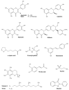
Chemical structures of commonly used antioxidants from (pre-)clinical trials for the treatment of metal toxicity. Structures were illustrated with ChemDraw Professional, Revity Signals Software. Sources: quercetin [184], rutin [185], catechin [186], myricetin [187], silibinin [188], α-lipoic acid [189], N-acetylcysteine [190], resveratrol [191], ferulic acid [192], taurine [193], vitamin E [194], Trolox [195], and ebselen [196].

References

    1. Jomova K., Makova M., Alomar S.Y., Alwasel S.H., Nepovimova E., Kuca K., Rhodes C.J., Valko M. Essential metals in health and disease. Chem. Biol. Interact. 2022;367:110173. doi: 10.1016/j.cbi.2022.110173. - DOI - PubMed
    1. Li L., Yang X. The Essential Element Manganese, Oxidative Stress, and Metabolic Diseases: Links and Interactions. Oxidative Med. Cell. Longev. 2018;2018:7580707. doi: 10.1155/2018/7580707. - DOI - PMC - PubMed
    1. Tchounwou P.B., Yedjou C.G., Patlolla A.K., Sutton D.J. Heavy Metal Toxicity and the Environment. Mol. Clin. Environ. Toxicol. 2012;101:133–164. - PMC - PubMed
    1. Ackerman C.M., Chang C.J. Copper Signaling in the Brain and Beyond. J. Biol. Chem. 2018;293:4628–4635. doi: 10.1074/jbc.R117.000176. - DOI - PMC - PubMed
    1. Bashir K., Rasheed S., Kobayashi T., Seki M., Nishizawa N.K. Regulating Subcellular Metal Homeostasis: The Key to Crop Improvement. Front. Plant Sci. 2016;7:1192. doi: 10.3389/fpls.2016.01192. - DOI - PMC - PubMed

LinkOut - more resources