Review
. 2024 Jul 19;25(14):7914.
doi: 10.3390/ijms25147914.

Profound Properties of Protein-Rich, Platelet-Rich Plasma Matrices as Novel, Multi-Purpose Biological Platforms in Tissue Repair, Regeneration, and Wound Healing

Affiliations
Review

Profound Properties of Protein-Rich, Platelet-Rich Plasma Matrices as Novel, Multi-Purpose Biological Platforms in Tissue Repair, Regeneration, and Wound Healing

Peter A Everts et al. Int J Mol Sci. .

Abstract

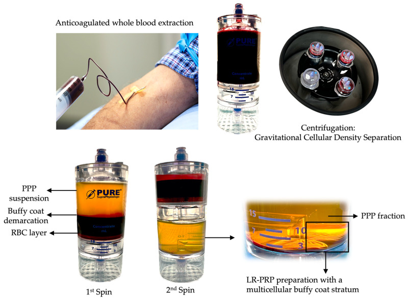
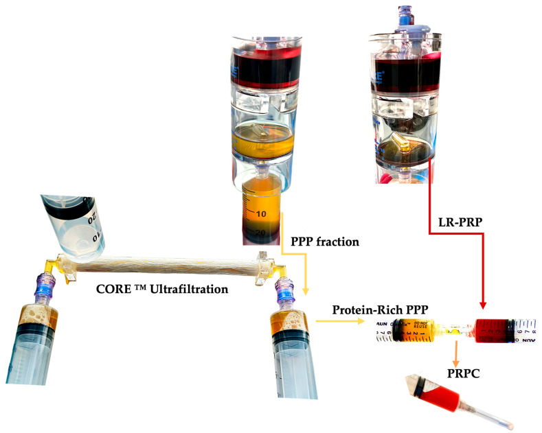
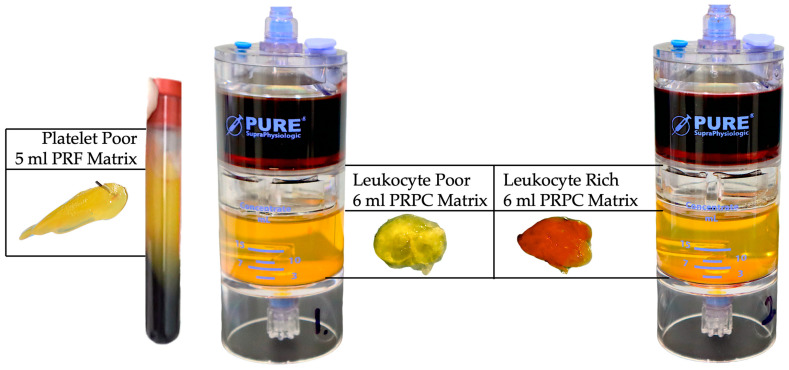
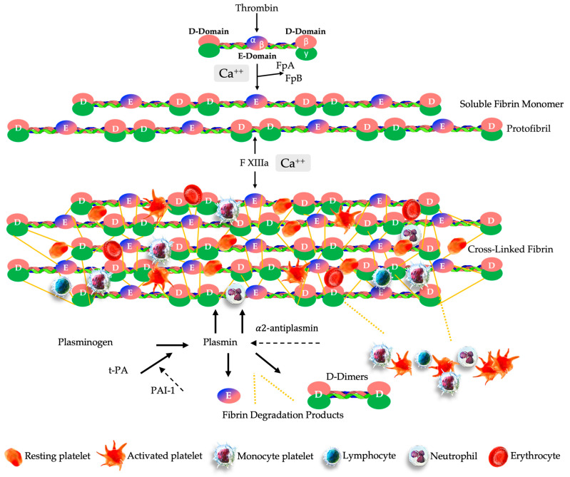
Autologous platelet-rich plasma (PRP) preparations are prepared at the point of care. Centrifugation cellular density separation sequesters a fresh unit of blood into three main fractions: a platelet-poor plasma (PPP) fraction, a stratum rich in platelets (platelet concentrate), and variable leukocyte bioformulation and erythrocyte fractions. The employment of autologous platelet concentrates facilitates the biological potential to accelerate and support numerous cellular activities that can lead to tissue repair, tissue regeneration, wound healing, and, ultimately, functional and structural repair. Normally, after PRP preparation, the PPP fraction is discarded. One of the less well-known but equally important features of PPP is that particular growth factors (GFs) are not abundantly present in PRP, as they reside outside of the platelet alpha granules. Precisely, insulin-like growth factor-1 (IGF-1) and hepatocyte growth factor (HGF) are mainly present in the PPP fraction. In addition to their roles as angiogenesis activators, these plasma-based GFs are also known to inhibit inflammation and fibrosis, and they promote keratinocyte migration and support tissue repair and wound healing. Additionally, PPP is known for the presence of exosomes and other macrovesicles, exerting cell-cell communication and cell signaling. Newly developed ultrafiltration technologies incorporate PPP processing methods by eliminating, in a fast and efficient manner, plasma water, cytokines, molecules, and plasma proteins with a molecular mass (weight) less than the pore size of the fibers. Consequently, a viable and viscous protein concentrate of functional total proteins, like fibrinogen, albumin, and alpha-2-macroglobulin is created. Consolidating a small volume of high platelet concentrate with a small volume of highly concentrated protein-rich PPP creates a protein-rich, platelet-rich plasma (PR-PRP) biological preparation. After the activation of proteins, mainly fibrinogen, the PR-PRP matrix retains and facilitates interactions between invading resident cells, like macrophages, fibroblast, and mesenchymal stem cells (MSCs), as well as the embedded concentrated PRP cells and molecules. The administered PR-PRP biologic will ultimately undergo fibrinolysis, leading to a sustained release of concentrated cells and molecules that have been retained in the PR-PRP matrix until the matrix is dissolved. We will discuss the unique biological and tissue reparative and regenerative properties of the PR-PRP matrix.

Keywords: fibrin(ogen); matrix; platelet-rich plasma; protein-rich platelet concentrate; tissue regeneration; tissue repair; ultrafiltration.


Conflict of interest statement

The authors declare no conflict of interests.

Figures

Figure 1
Figure 1
Portrayal of a 2-spin centrifugation method used to produce autologous PRP. PRP preparation involves the collection of a predetermined volume of peripheral blood in collection syringes containing an anticoagulant, like calcium citrate 3.8%. The predonated whole blood is gently loaded in a PRP device for gravitational cellular density separation using a two-spin centrifugation protocol. After the first spin cycle, the whole blood components are separated into three basic layers: the PPP suspension, the buffy coat demarcation layer, and the RBC layer. During the second centrifugation cycle, the platelet and other cells in the PPP fraction and RBC layer are further separated, resulting in a multicellular buffy coat stratum containing high concentrations of platelets and, eventually, leukocytes. A calculated portion of the PPP fraction is removed, leaving the platelet concentrate within a small volume of plasma for platelet resuspension. Thereafter, the PRP is extracted from the device. In this graphic, LR-PRP has been prepared, and the multicellular fractions consist of a high concentration of platelets, monocytes, lymphocytes, neutrophils, and some red blood cells. Abbreviations: PPP: platelet-poor plasma; RBC: red blood cells; LR-PRP: leukocyte-rich PRP; PRP: platelet-rich plasma.
Figure 2
Figure 2
PR-PRP preparation. After the second spin, a calculated portion of the PPP fraction is removed with a syringe and attached with an empty syringe to the ultrafiltration device, as well as an effluent collection syringe, to collect the eliminated plasma water. The PPP syringes are manually pushed through the device, and plasma water, proteins smaller than 20 kDa, and cytokines are removed through the hollow fiber pores. After a series of passes, the PPP volume is significantly reduced, leading to a small and viscous volume of protein-rich plasma. The remaining PPP volume in the PRP device is used to resuspend the highly concentrated multicellular LR-PRP fraction and capture it from the PRP device. The concentrated protein-rich PPP and LR-PRP are consolidated into one syringe and gently mixed, creating PR-PRP. Abbreviations: LR-PRP: leukocyte-rich PRP; PPP: platelet-poor plasma; PRP: platelet-rich plasma; PR-PRP: protein-rich platelet concentrate. (the ultrafiltration device shown is the CORE™ Ultrafiltration System, developed by EmCyte Corporation®, Fort Myers, FL, USA).
Figure 3
Figure 3
Visualization of PRF, LP-PR-PRP, and LR-PRP PR-PRP matrices. The PRF matrix and leukocyte-poor and leukocyte-rich PR-PRP matrices are exhibited. In a same-patient experiment, 130 mL of anticoagulated whole blood was extracted to prepare 10 mL of PRF, LP-PPR, and LR-PRP, using two 60 mL 2-spin PRP devices (PurePRP®SP, EmCyte Corporation®, Fort Myers, FL, USA). After the first spin, the PRF clot was removed from the test tube. After the 2nd spin of the LP and LR-PRP preparations, the PPP fraction was removed for ultrafiltration to concentrate the plasma to produce protein-rich PPP resuspended with the concentrated PRP fractions, described in detail in Figure 2. Exactly 3 mL of both PRP formulations was consolidated with 3 mL of protein-rich PPP. To create the PR-PRP matrices, normal baseline clotting parameters were restored by adding 0.35 mL of NaCl 10% to the 6 mL PR-PRP volume. Thereafter, 150 IU of bovine thrombin was added to the re-calcified PR-PRP volume, mimicking the effect of TF for matrix formation.
Figure 4
Figure 4
Schematic overview of the multistep physiological process of PR-PRP matrix creation and the sustained release of matrix biological components. The ultrafiltration device concentrates non-activated plasma proteins, including fibrinogen, consisting of two D-domains composed of α, β, γ chains, and plasma growth factors. After consolidating a small volume of high-concentration PRP with the concentrated protein suspension, fibrinogen undergoes a structural change in the presence of thrombin and calcium ions, leading to the cleavage of fibrinogen into FpA and FpB to form stable, complex, soluble fibrin monomers. The soluble monomer polymerizes to form half-staggered protofibrils. Several enzymes, including FXIIIa, and in presence of Ca++ ions, protofibrils are converted to the cross-linked fibrin embedded with highly concentrated PRP cells. Fibrinolysis is initiated when t-PA converts plasminogen into plasmin, whereas PAI-1 inhibits t-PA, preventing the activation of plasminogen and thus fibrinolysis. Hence, FDP, D-dimers, platelets, and other cells are continuously released from the PR-PRP matrix, whereas α2 antiplasmin acts by blocking plasmin activity, reducing the proteolytic fibrinolytic breakdown.
Figure 5
Figure 5
Illustration of the biological features of a PR-PRP matrix. The beneficial effects of an accurately prepared and applied PR-PRP matrix in pathological microenvironments are depicted by several biologically induced processes that are crucial in the repair and regeneration of diseased tissues. The PR-PRP matrix, which is composed of concentrated and activated fibrinogen as its main component, serves as a temporary dense insoluble three-dimensional scaffold with high concentrations of PRP cellular content embedded. Importantly, the matrix enhances the viability and functionality of the PRP cellular content. The PR-PRP matrix is instrumental in providing a molecular link for invading local tissue-resident cells, such as MSCs, macrophages, fibroblasts, and ECs. Inside the matrix microenvironment, a multitude of cells and molecules engage in many cellular interactions. Upon fibrinolytic breakdown of the PR-PRP matrix, an abundance of activated platelets, their PGFs, and other biologically active molecules and cells are released to the local microenvironment. Ultimately, the sustained release of matrix substances leads to an increase in cellular activity and signaling, angiogenetic and immunomodulatory processes, and antimicrobial activities, contributing to the overall tissue repair and regenerative process. Moreover, as a result of the breaking down of fibrin strands, they provide structural support for the development of new tissues and facilitate the adhesion and migration of cells [40,185,191,192,193,194,195].

References

    1. Vallet-Regí M. Evolution of Biomaterials. Front. Mater. 2022;9:864016. doi: 10.3389/fmats.2022.864016. - DOI
    1. Todros S., Todesco M., Bagno A. Biomaterials and Their Biomedical Applications: From Replacement to Regeneration. Processes. 2021;9:1949. doi: 10.3390/pr9111949. - DOI
    1. Vallet-Regí M. Bioceramics: From bone substitutes to nanoparticles for drug delivery. Pure Appl. Chem. 2019;91:687–706. doi: 10.1515/pac-2018-0505. - DOI - PMC - PubMed
    1. Van Belleghem S.M., Mahadik B., Snodderly K.L., Fisher J.P. Biomaterials Science. Elsevier; Amsterdam, The Netherlands: 2020. Overview of tissue engineering concepts and applications; pp. 1289–1316.
    1. Van Blitterswijk C., De Boer J. Tissue Engineering. Academic Press; Cambridge, MA, USA: 2022.

Substances

LinkOut - more resources