

Effect of Acetylsalicylic Acid on Biological Properties of Novel Cement Based on Calcium Phosphate Doped with Ions of Strontium, Copper, and Zinc
- PMID: 39063181
- PMCID: PMC11276672
- DOI: 10.3390/ijms25147940
Effect of Acetylsalicylic Acid on Biological Properties of Novel Cement Based on Calcium Phosphate Doped with Ions of Strontium, Copper, and Zinc
Abstract
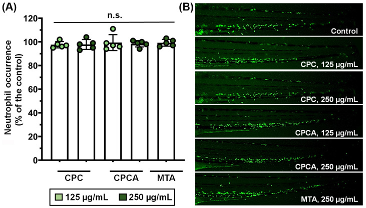
This study aimed to compare the biological properties of newly synthesized cements based on calcium phosphate with a commercially used cement, mineral trioxide aggregate (MTA). Strontium (Sr)-, Copper (Cu)-, and Zinc (Zn)-doped hydroxyapatite (miHAp) powder was obtained through hydrothermal synthesis and characterized by scanning electron microscopy (SEM), X-ray diffraction (XRD), and energy dispersive X-ray spectrometry (EDX). Calcium phosphate cement (CPC) was produced by mixing miHAp powder with a 20 wt.% citric acid solution, followed by the assessment of its compressive strength, setting time, and in vitro bioactivity. Acetylsalicylic acid (ASA) was added to the CPC, resulting in CPCA. Biological tests were conducted on CPC, CPCA, and MTA. The biocompatibility of the cement extracts was evaluated in vitro using human dental pulp stem cells (hDPSCs) and in vivo using a zebrafish model. Antibiofilm and antimicrobial effect (quantified by CFUs/mL) were assessed against Streptococcus mutans and Lactobacillus rhamnosus. None of the tested materials showed toxicity, while CPCA even increased hDPSCs proliferation. CPCA showed a better safety profile than MTA and CPC, and no toxic or immunomodulatory effects on the zebrafish model. CPCA exhibited similar antibiofilm effects against S. mutans and L. rhamnosus to MTA.
Keywords: antibiofilm; calcium phosphate; dental pulp stem cells; hydroxyapatite; zebrafish.
Conflict of interest statement
The authors declare no conflicts of interest.
Figures


References
-
- GBD 2017 Disease and Injury Incidence and Prevalence Collaborators Global, Regional, and National Incidence, Prevalence, and Years Lived with Disability for 354 Diseases and Injuries for 195 Countries and Territories, 1990–2017: A Systematic Analysis for the Global Burden of Disease Study 2017. Lancet. 2018;392:1789–1858. doi: 10.1016/S0140-6736(18)32279-7. - DOI - PMC - PubMed
-
- Schwendicke F., Walsh T., Lamont T., Al-Yaseen W., Bjørndal L., Clarkson J.E., Fontana M., Gomez Rossi J., Göstemeyer G., Levey C., et al. Interventions for Treating Cavitated or Dentine Carious Lesions. Cochrane Database Syst. Rev. 2021;7:CD013039. doi: 10.1002/14651858.CD013039.pub2. - DOI - PMC - PubMed
MeSH terms
Substances
Grants and funding
- 451-03-9/2021-14/200129/Ministry of Science, Technological Development and Innovation, Republic of Serbia
- 451-03-65/2024-03/200135/Ministry of Science, Technological Development and Innovation, Republic of Serbia
- 451-03-66/2024-03/200042/Ministry of Science, Technological Development and Innovation, Republic of Serbia
LinkOut - more resources
Full Text Sources
Molecular Biology Databases
Research Materials
