. 2024 Jul 21;25(14):7965.
doi: 10.3390/ijms25147965.

Plastic Fly: What Drosophila melanogaster Can Tell Us about the Biological Effects and the Carcinogenic Potential of Nanopolystyrene

Affiliations

Plastic Fly: What Drosophila melanogaster Can Tell Us about the Biological Effects and the Carcinogenic Potential of Nanopolystyrene

Massimo Aloisi et al. Int J Mol Sci. .

Abstract

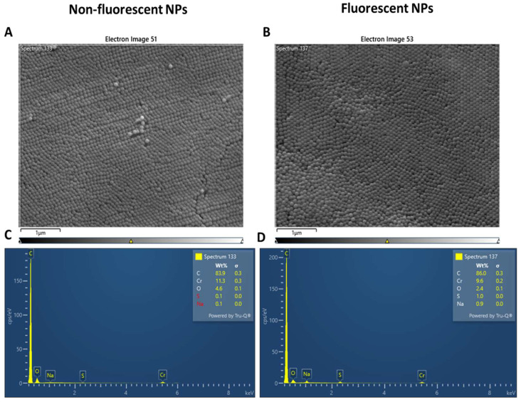
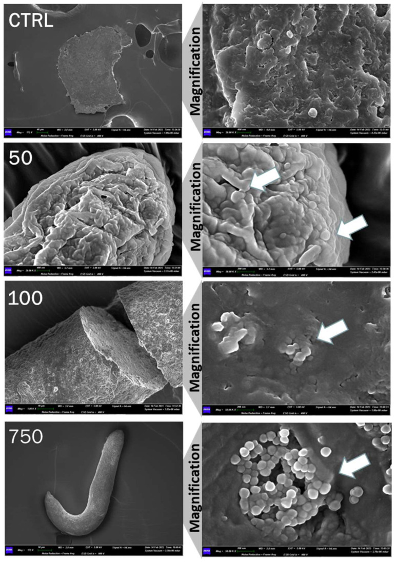
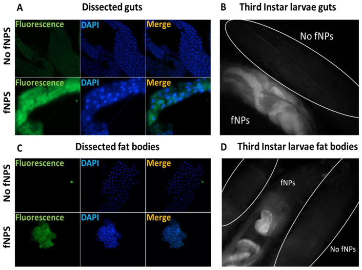
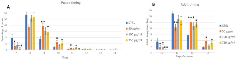
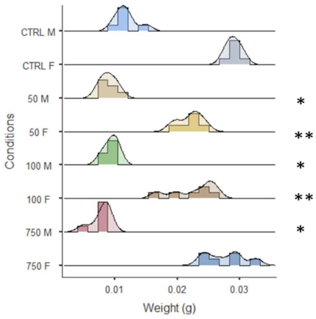
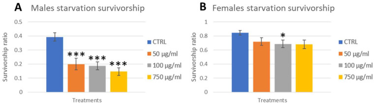
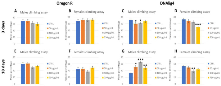
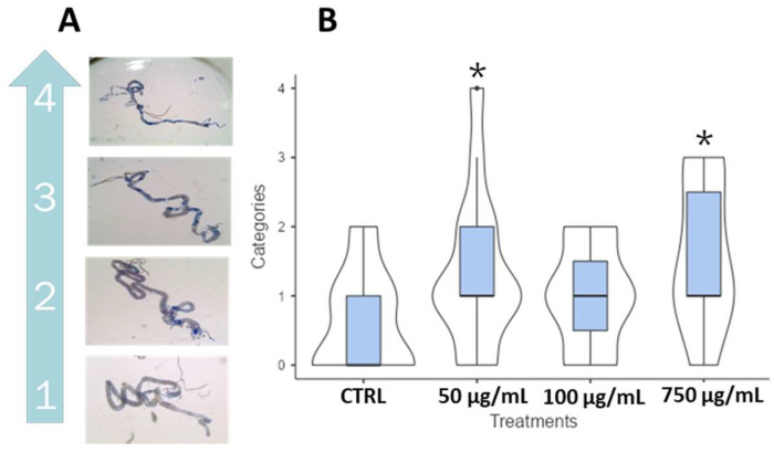
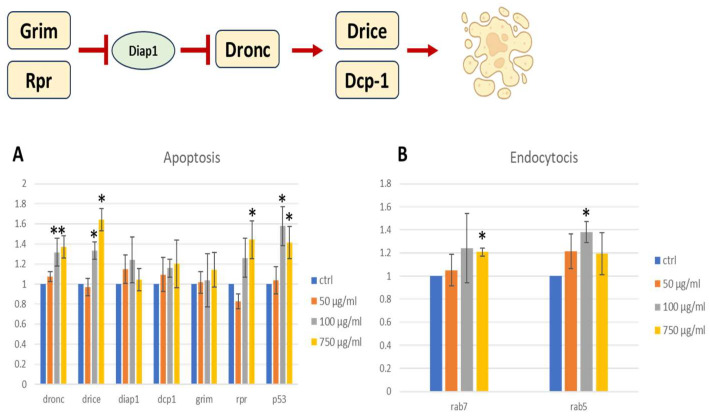
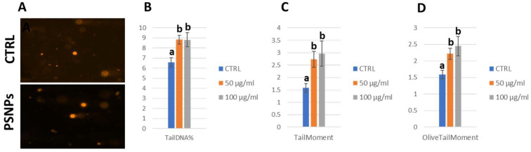
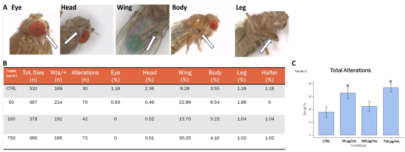
Today, plastic pollution is one of the biggest threats to the environment and public health. In the tissues of exposed species, micro- and nano-fragments accumulate, leading to genotoxicity, altered metabolism, and decreased lifespan. A model to investigate the genotoxic and tumor-promoting potential of nanoplastics (NPs) is Drosophila melanogaster. Here we tested polystyrene, which is commonly used in food packaging, is not well recycled, and makes up at least 30% of landfills. In order to investigate the biological effects and carcinogenic potential of 100 µm polystyrene nanoparticles (PSNPs), we raised Oregon [R] wild-type flies on contaminated food. After prolonged exposure, fluorescent PSNPs accumulated in the gut and fat bodies. Furthermore, PSNP-fed flies showed considerable alterations in weight, developmental time, and lifespan, as well as a compromised ability to recover from starvation. Additionally, we noticed a decrease in motor activity in DNAlig4 mutants fed with PSNPs, which are known to be susceptible to dietary stressors. A qPCR molecular investigation of the larval intestines revealed a markedly elevated expression of the genes drice and p53, suggesting a response to cell damage. Lastly, we used warts-defective mutants to assess the carcinogenic potential of PSNPs and discovered that exposed flies had more aberrant masses than untreated ones. In summary, our findings support the notion that ingested nanopolystyrene triggers metabolic and genetic modifications in the exposed organisms, eventually delaying development and accelerating death and disease.

Keywords: Drosophila melanogaster; carcinogenic nanoplastics; genotoxicity; in vivo models; nanopolystyrene.


Conflict of interest statement

The authors declare no conflicts of interest.

Figures

Figure 1
Figure 1
Morphological analysis (A,B) and energy dispersive X-ray analysis (C,D) of polystyrene nanoparticles, non-fluorescent (A,C) and fluorescent (C,D).
Figure 2
Figure 2
SEM analysis of PSNPs in feces from untreated flies (CTRL) and three different concentrations (50 μg/mL, 100 μg/mL, and 750 μg/mL). Left column has 172 X magnification for CTRL and 750 μg/mL, 20.00 K X for 50 μg/mL, and 1.00 K X for 100 μg/mL; the right column magnification is 50.00 K X for every condition. White arrows point to NPs.
Figure 3
Figure 3
Fluorescence microscopy of fPSNPs in OR-R third instar larvae guts and fat bodies in two conditions: negative control (no fPSNPs, 0 μg/mL) and 750 μg/mL (fPSNPs). (A,C) dissected guts and fat bodies, respectively (400X); (B,D) whole living larvae (100X).
Figure 4
Figure 4
Analysis of development traits of OR-R flies: pupae formation (A) and adult timing (B). Treatments: Control (0 µg/mL); 50 µg/mL, 100 µg/mL, 750 µg/mL. * p < 0.05; ** p < 0.005; *** p < 0.001.
Figure 5
Figure 5
Weight distribution of 3 days old OR-R flies. * p < 0.05; ** p < 0.005.
Figure 6
Figure 6
Starvation assay with OR-R flies. Three days-old adults chronically exposed were starved in empty vials and checked hourly for the first 12 h (Figure S2A,B Supplementary Materials) and after 24 h (A,B). Treatments: CTRL (0 µg/mL); 50 µg/mL; 100 µg/mL; 750 µg/mL. * p < 0.05; *** p < 0.001.
Figure 7
Figure 7
Climbing assay of OR-R (A,B,E,F) and DNAlig4 (C,D,G,H) flies after 3 (AD) and 18 (EH) days from eclosion, divided into males (A,C,E,G) and females (B,D,F,H). * p < 0.05; ** p < 0.005; *** p < 0.001.
Figure 8
Figure 8
Trypan blue assay in wild-type third instar larvae gut fed with PSNPs. (A) dissected guts were incubated with Trypan blue and then scored under a stereoscope based on blue intensity and spreading; (B) violin plot and box plot showing the final scoring of the four conditions used (0, 50, 100, and 750 μg/mL). * p < 0.05.
Figure 9
Figure 9
A schematic drawing of the apoptotic process. Image realized with Biorender.com. Gene expression of apoptosis (A) and endocytosis (B) biomarkers in third instar larvae guts. Student’s t test was employed to compare the values of untreated (ctrl) and treated. * p < 0.05; ** p < 0.005.
Figure 10
Figure 10
Dissected OR-R third instar larvae guts were homogenized to obtain a cell suspension that was subjected to an electrophoretic run and subsequent nuclear stain with ethidium bromide to observe DNA breaks under a fluorescence microscope (A). The percentage of DNA detected in comet tails, the tail moment and the olive tail moment are reported (BD). Non-parametric one-way ANOVA was used for significance analysis. Non-significant conditions were grouped with the same letter; different letters represent significance between conditions. Treatment: Control (0 µg/mL); 50 µg/mL; 100 µg/mL.
Figure 11
Figure 11
warts assay showing carcinogenic potential of PSNPS. (A) representative images of tumors in 3 days old wts defective flies (white arrows); (B) table resuming the percentage of masses in the wts fly population; (C), bar plot showing the percentage of total alterations. Treatment: CTRL (0 µg/mL); 50 µg/mL, 100 µg/mL, 750 µg/mL. * p < 0.05.

Similar articles

Cited by

References

    1. Ostle C., Thompson R.C., Broughton D., Gregory L., Wootton M., Johns D.G. The rise in ocean plastics evidenced from a 60-year time series. Nat. Commun. 2019;10:1622. doi: 10.1038/s41467-019-09506-1. - DOI - PMC - PubMed
    1. De Frond H., Hampton L.T., Kotar S., Gesulga K., Matuch C., Lao W., Weisberg S.B., Wong C.S., Rochman C.M. Monitoring microplastics in drinking water: An interlaboratory study to inform effective methods for quantifying and characterizing microplastics. Chemosphere. 2022;298:134282. doi: 10.1016/j.chemosphere.2022.134282. - DOI - PubMed
    1. Jambeck J.R., Geyer R., Wilcox C., Siegler T.R., Perryman M., Andrady A., Narayan R., Law K.L. Marine pollution. Plastic waste inputs from land into the ocean. Science. 2015;347:768–771. doi: 10.1126/science.1260352. - DOI - PubMed
    1. Wurm F.R., Spierling S., Endres H.J., Barner L. Plastics and the Environment-Current Status and Challenges in Germany and Australia. Macromol. Rapid Commun. 2020;41:e2000351. doi: 10.1002/marc.202000351. - DOI - PubMed
    1. Čerkasova N., Enders K., Lenz R., Oberbeckmann S., Brandt J., Fischer D., Fischer F., Labrenz M., Schernewski G. A Public Database for Microplastics in the Environment. Microplastics. 2023;2:132–146. doi: 10.3390/microplastics2010010. - DOI

LinkOut - more resources