. 2024 Jul 8;14(7):854.
doi: 10.3390/life14070854.

Micromassage Compression Leggings Associated with Physical Exercise: Pilot Study and Example of Evaluation of the Clinical and Instrumental Effectiveness of Conservative Treatment in Lipedema

Affiliations

Micromassage Compression Leggings Associated with Physical Exercise: Pilot Study and Example of Evaluation of the Clinical and Instrumental Effectiveness of Conservative Treatment in Lipedema

Lorenzo Ricolfi et al. Life (Basel). .

Abstract

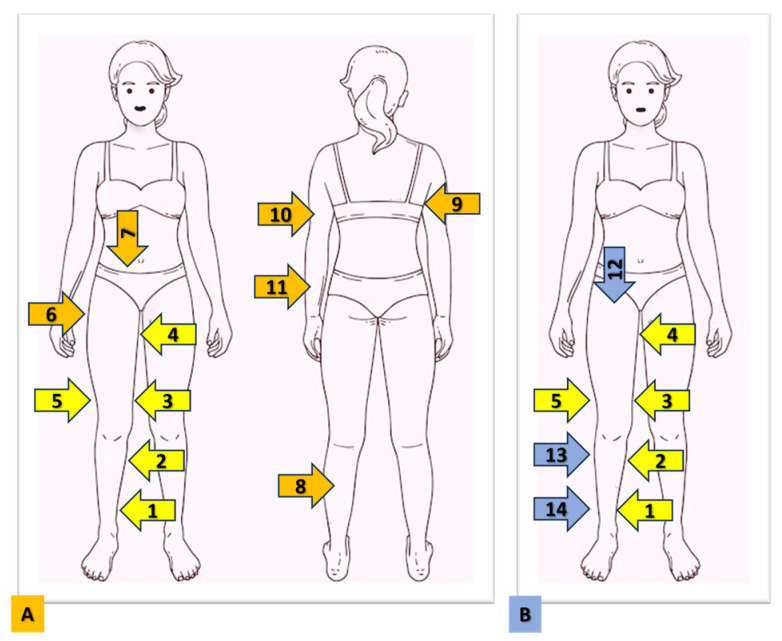
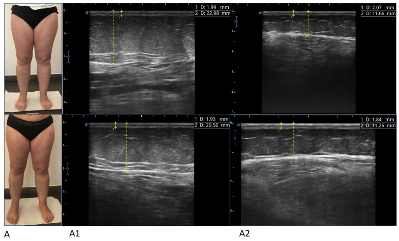
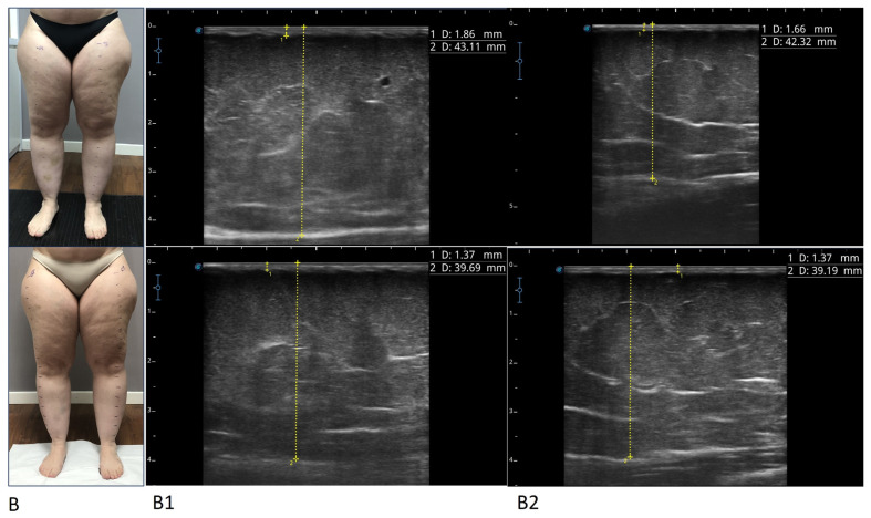
We evaluated the effect of compression leggings with micromassage in association with physical activity on women with lipedema, not previously treated and without recent changes in body weight. The treatment resulted in an improvement in all subjective parameters, in spontaneous and evoked pain, in the volume of the limbs, in the absence of significant changes in body weight, and regardless of the duration of use, age, years of illness, the clinical stage, and body mass index (BMI). Evoked pain did not improve in areas affected by untreated lipedema; foot circumferences did not increase. We found a significant reduction in the thickness of subcutaneous adipose tissue (SAT) and skin evaluated in multiple points of the lower limb. Micromassage compression leggings are proposed as an integral part of conservative treatment. A method is proposed for the clinical evaluation of evoked pain, called the Progressive Pain Check (PPC), which allows for the calculation of a numerical score called the Ricolfi-Patton Score (RPS) and for the ultrasound evaluation of tissues. The method is simple and repeatable and allows for completion of the clinical evaluation of the patient at diagnosis and for an evaluation of the effects of various treatments, even applied to just one side of the body.

Keywords: adipose tissue; compression; exercise; lipedema; micromassaging leggings; pain; treatment; ultrasound.


Conflict of interest statement

The authors declare no conflicts of interest.

Figures

Figure 1
Figure 1
The figure shows the points where pain is detected with the Progressive Pain Check (PPC) method (A) and the points where ultrasound mixing of the thickness of the skin and adipose tissue is carried out (B). The first 5 points (from 1 to 5) are the same. The Lower Body Pain Score is calculated from the sum of point 1 to point 8. The Upper Body Pain Score is calculated from the sum of points 9, 10, and 11. The Total Body Pain Score or Ricolfi–Patton Score (RPS) is calculated with the sum of all points from 1 to 11. 1: medial lower third of the leg; 2: medial upper third of the leg; 3: medial lower third of the thigh; 4: medial upper third of the thigh; 5: later lower third of the thigh; 6: lateral upper third of the thigh 7: lower abdomen; 8: posterior middle third of the leg; 9: lateral edge of the tissue covering the teres major muscle; 10: arm; 11: forearm; 12: anterior upper third of the thigh; 13: lateral upper third of the leg; 14: lateral upper and lower third of the leg.
Figure 2
Figure 2
Photographs and ultrasounds of two patients (A,B), before treatment (images above) and after treatment (images below). The ultrasound measurements are represented by the dashed yellow lines: the thickness of the skin was measured from the skin surface to the lower edge of the epidermis and dermis complex, and the thickness of the suprafascial superficial adipose tissue was measured from the skin surface to the subcutaneous transition structure (fascia). All imaging was performed using a high-frequency linear probe (8–14 MHz), keeping the probe perpendicular to the skin and always without applying pressure. For patient A, ultrasound images relating to the lower medial third of the leg (A1) and the upper lateral third of the leg (A2) before and after the treatment are shown (box above and box below, respectively). For patient B, ultrasound images relating to the lower medial third of the thigh (B1) and the upper medial third of the thigh (B2) before and after the treatment are shown (box above and box below, respectively).
Figure 2
Figure 2
Photographs and ultrasounds of two patients (A,B), before treatment (images above) and after treatment (images below). The ultrasound measurements are represented by the dashed yellow lines: the thickness of the skin was measured from the skin surface to the lower edge of the epidermis and dermis complex, and the thickness of the suprafascial superficial adipose tissue was measured from the skin surface to the subcutaneous transition structure (fascia). All imaging was performed using a high-frequency linear probe (8–14 MHz), keeping the probe perpendicular to the skin and always without applying pressure. For patient A, ultrasound images relating to the lower medial third of the leg (A1) and the upper lateral third of the leg (A2) before and after the treatment are shown (box above and box below, respectively). For patient B, ultrasound images relating to the lower medial third of the thigh (B1) and the upper medial third of the thigh (B2) before and after the treatment are shown (box above and box below, respectively).

Similar articles

Cited by

References

    1. Alcolea J.M., Alonso A.B., Arroyo B.A., Domingo P., Garcia G.A., Graells G.M., Urraca I.C., Nipoti I.E., Castillo M.E., Alvarez M., Jr., et al. Consensus Document on Lipedema 2018. Includes Current Status of Lipedema 2019. LITOGAMA SL; Barcelona, Spain: 2018.
    1. Wounds UK Best. Practice Guidelines: The Management of Lipoedema. 2017. [(accessed on 28 August 2017)]. Available online: https://wounds-uk.com/bestpractice-statements/management-lipoedema/
    1. Reich-Schupke S., Schmeller W., Brauer W.J., Cornely M.E., Faerber G., Ludwig M., Lulay G., Miller A., Rapprich S., Richter D.F., et al. S1 guidelines: Lipedema. J. Dtsch. Dermatol. Ges. 2017;15:758–767. doi: 10.1111/ddg.13036. - DOI - PubMed
    1. Herbst K.L., Kahn L.A., Iker E., Ehrlich C., Wright T., McHutchison L., Schwartz J., Sleigh M., Mc Donahue P., Lisson K.H., et al. Standard of care for lipedema in the United States. Phlebology. 2021;36:779–796. doi: 10.1177/02683555211015887. - DOI - PMC - PubMed
    1. Fonder M.A., Loveless J.W., Lazarus G.S. Lipedema, a frequently unrecognized problem. J. Am. Acad. Dermatol. 2007;57((Suppl. S2)):S1–S3. doi: 10.1016/j.jaad.2006.09.023. - DOI - PubMed

LinkOut - more resources