

Effects of Aerobic Exercise on Brain Age and Health in Middle-Aged and Older Adults: A Single-Arm Pilot Clinical Trial
- PMID: 39063609
- PMCID: PMC11278044
- DOI: 10.3390/life14070855
Effects of Aerobic Exercise on Brain Age and Health in Middle-Aged and Older Adults: A Single-Arm Pilot Clinical Trial
Abstract
Backgrounds: Sleep disturbances are prevalent among elderly individuals. While polysomnography (PSG) serves as the gold standard for sleep monitoring, its extensive setup and data analysis procedures impose significant costs and time constraints, thereby restricting the long-term application within the general public. Our laboratory introduced an innovative biomarker, utilizing artificial intelligence algorithms applied to PSG data to estimate brain age (BA), a metric validated in cohorts with cognitive impairments. Nevertheless, the potential of exercise, which has been a recognized means of enhancing sleep quality in middle-aged and older adults to reduce BA, remains undetermined.
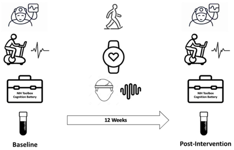
Methods: We conducted an exploratory study to evaluate whether 12 weeks of moderate-intensity exercise can improve cognitive function, sleep quality, and the brain age index (BAI), a biomarker computed from overnight sleep electroencephalogram (EEG), in physically inactive middle-aged and older adults. Home wearable devices were used to monitor heart rate and overnight sleep EEG over this period. The NIH Toolbox Cognition Battery, in-lab overnight polysomnography, cardiopulmonary exercise testing, and a multiplex cytokines assay were employed to compare pre- and post-exercise brain health, exercise capacity, and plasma proteins.
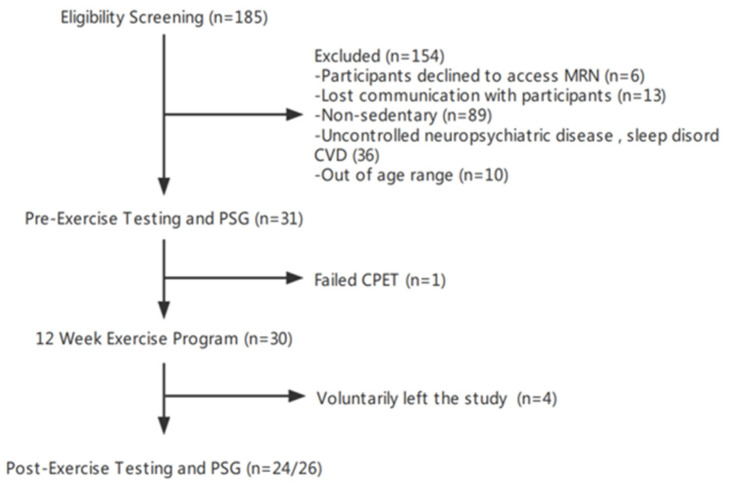
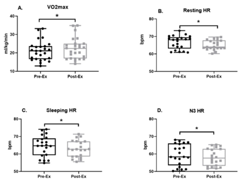
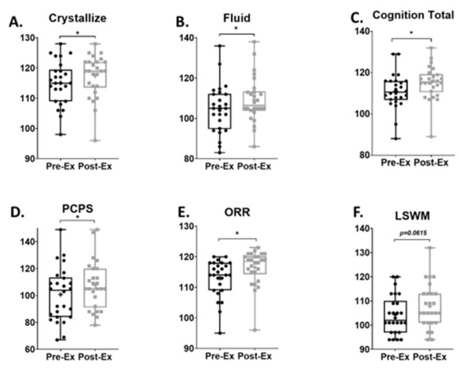
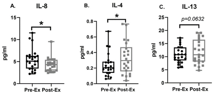
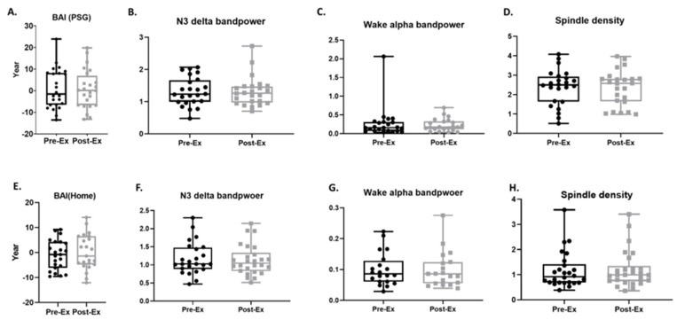
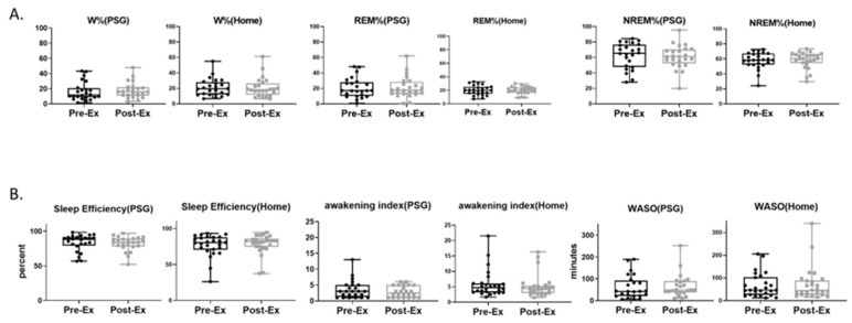
Results: In total, 26 participants completed the initial assessment and exercise program, and 24 completed all procedures. Data are presented as mean [lower 95% CI of mean, upper 95% CI of mean]. Participants significantly increased maximal oxygen consumption (Pre: 21.11 [18.98, 23.23], Post 22.39 [20.09, 24.68], mL/kg/min; effect size: -0.33) and decreased resting heart rate (Pre: 66.66 [63.62, 67.38], Post: 65.13 [64.25, 66.93], bpm; effect size: -0.02) and sleeping heart rate (Pre: 64.55 [61.87, 667.23], Post: 62.93 [60.78, 65.09], bpm; effect size: -0.15). Total cognitive performance (Pre: 111.1 [107.6, 114.6], Post: 115.2 [111.9, 118.5]; effect size: 0.49) was significantly improved. No significant differences were seen in BAI or measures of sleep macro- and micro-architecture. Plasma IL-4 (Pre: 0.24 [0.18, 0.3], Post: 0.33 [0.24, 0.42], pg/mL; effect size: 0.49) was elevated, while IL-8 (Pre: 5.5 [4.45, 6.55], Post: 4.3 [3.66, 5], pg/mL; effect size: -0.57) was reduced.
Conclusions: Cognitive function was improved by a 12-week moderate-intensity exercise program in physically inactive middle-aged and older adults, as were aerobic fitness (VO2max) and plasma cytokine profiles. However, we found no measurable effects on sleep architecture or BAI. It remains to be seen whether a study with a larger sample size and more intensive or more prolonged exercise exposure can demonstrate a beneficial effect on sleep quality and brain age.
Keywords: EEG; brain health; exercise; intervention trial; sleep.
Conflict of interest statement
The authors declare no conflict of interest.
Figures
References
-
- Dinges D.F., Pack F., Williams K., Gillen K.A., Powell J.W., Ott G.E., Aptowicz C., Pack A.I. Cumulative sleepiness, mood disturbance, and psychomotor vigilance performance decrements during a week of sleep restricted to 4–5 hours per night. Sleep. 1997;20:267–277. - PubMed
-
- Motomura Y., Kitamura S., Oba K., Terasawa Y., Enomoto M., Katayose Y., Hida A., Moriguchi Y., Higuchi S., Mishima K. Sleep debt elicits negative emotional reaction through diminished amygdala-anterior cingulate functional connectivity. PLoS ONE. 2013;8:e56578. doi: 10.1371/annotation/5970fff3-0a1c-4056-9396-408d76165c4d. - DOI - PMC - PubMed
Grants and funding
LinkOut - more resources
Full Text Sources
Other Literature Sources
Miscellaneous
