

The Current Role of Contrast-Enhanced Ultrasound (CEUS) in the Diagnosis and Staging of Bladder Cancer: A Review of the Available Literature
- PMID: 39063611
- PMCID: PMC11278273
- DOI: 10.3390/life14070857
The Current Role of Contrast-Enhanced Ultrasound (CEUS) in the Diagnosis and Staging of Bladder Cancer: A Review of the Available Literature
Abstract
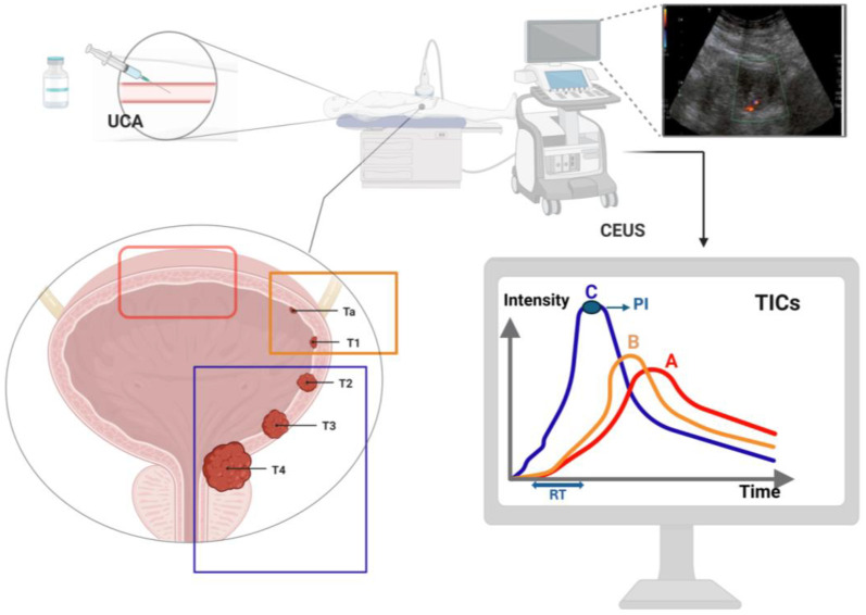
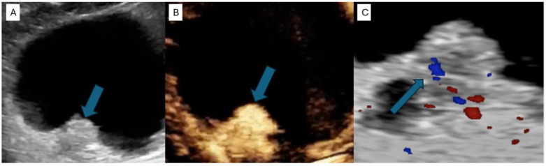
Contrast-enhanced ultrasound (CEUS) is an advanced imaging technique that integrates conventional US with the intravenous injection of specific US contrast agents (UCAs), combining the non-invasiveness of US with the higher accuracy of contrast-enhanced imaging. In contrast with magnetic resonance imaging (MRI), computed tomography (CT) and cystoscopy, CEUS has few contraindications, and UCAs are non-nephrotoxic agents that can be safely used in patients with kidney failure. CEUS is a well-established method for the detection of liver lesions and for echocardiography, and its indications are expanding. The updated 2018 WFUMB-EFSUMB guidelines have added the urinary bladder under non-hepatic applications of CEUS. The technique is able to distinguish between benign tissue, such as clots or hematoma, and malignant lesions by perfusing the mass with contrast agent. Thanks to the different perfusion rates of the various layers of the bladder wall, CEUS is also able to predict tumor invasion depth and stage. Despite that, current urological guidelines do not include CEUS as a plausible imaging technique for bladder urothelial carcinoma. The main reason for this omission might be the presence of scarce randomized evidence and the absence of large validated series. In this review, we describe the rationale behind the use of CEUS in bladder cancer and the added value of this imaging technique in the detection and staging of bladder lesions. In addition, we researched the available literature on the topic and then described the results of randomized clinical trials and a meta-analysis investigating the accuracy of CEUS in bladder cancer diagnosis and staging. The reported studies show that CEUS is a highly accurate diagnostic and staging tool for BC, reaching levels of specificity and sensitivity in differentiating between Ta-T1, or low-grade BC, and T2, or high-grade BC, that are comparable to those shown by the reference standard methods. Nonetheless, several limitations were found and are highlighted in this review. The aim of this study is to further validate and promote the use of CEUS as a quick, economic and effective diagnostic tool for this high-impact disease.
Keywords: CEUS; bladder cancer; diagnosis; review; staging; ultrasound.
Conflict of interest statement
The authors declare no conflicts of interest.
Figures
References
-
- Brierley J.D., Gospodarowicz M.K., Wittekind C. TNM Classification of Malignant Tumours. John Wiley & Sons; Hoboken, NJ, USA: 2017.
Publication types
LinkOut - more resources
Full Text Sources
