Comparison of a Two (32/38 Weeks) versus One (36 Weeks) Ultrasound Protocol for the Detection of Decreased Fetal Growth and Adverse Perinatal Outcome
- PMID: 39063963
- PMCID: PMC11278302
- DOI: 10.3390/jpm14070709
Comparison of a Two (32/38 Weeks) versus One (36 Weeks) Ultrasound Protocol for the Detection of Decreased Fetal Growth and Adverse Perinatal Outcome
Abstract
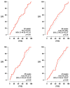
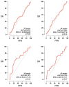
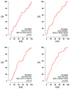
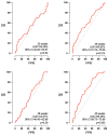
Third-trimester ultrasound has low sensitivity to small for gestational age (SGA) and adverse perinatal outcomes (APOs). The objective of this study was to compare, in terms of cost-effectiveness, two routine third-trimester surveillance protocols for the detection of SGA and evaluate the added value of a Doppler study for the prediction of APO. This was a retrospective observational study of low-risk pregnancies that were followed by a two growth scans protocol (P2) at 32 and 38 weeks or by a single growth scan at 36 weeks (P1). Ultrasound scans included an estimated fetal weight (EFW) in all cases and a Doppler evaluation in most cases. A total of 1011 pregnancies were collected, 528 with the P2 protocol and 483 with the P1 protocol. While the two models presented no differences for the detection of SGA in terms of sensitivity (47.89% vs. 50% p = 0.85) or specificity (94.97 vs. 95.86% p = 0.63), routine performance of two growth scans (P2) led to a 35% cost increase. The accuracy of EFW for the detection of SGA showed a noteworthy improvement when reducing the interval to labor, and the only parameter with predictive capacity of APO was the cerebroplacental ratio at 38 weeks. In low-risk pregnancies, the higher costs of a two-scan growth surveillance protocol at the third trimester are not justified by an increase in diagnostic effectivity.
Keywords: adverse perinatal outcome; low-risk pregnancy; small for gestational age; third-trimester; ultrasound.
Conflict of interest statement
The authors declare no conflicts of interest.
Figures


References
-
- Bricker L., Medley N., Pratt J.J. Routine ultrasound in late pregnancy (after 24 weeks’ gestation) [(accessed on 1 June 2024)];Cochrane Database Syst. Rev. 2015 2015:CD001451. doi: 10.1002/14651858.CD001451.pub4. Available online: https://doi.wiley.com/10.1002/14651858.CD001451.pub4. - DOI - PMC - PubMed
-
- Sokol Karadjole V., Agarwal U., Berberovic E., Poljak B., Alfirevic Z. Does serial 3rd trimester ultrasound improve detection of small for gestational age babies: Comparison of screening policies in 2 European maternity units. Eur. J. Obstet. Gynecol. Reprod. Biol. 2017;215:45–49. doi: 10.1016/j.ejogrb.2017.05.031. - DOI - PubMed
LinkOut - more resources
Full Text Sources

