

Perfusion in Pedicled Skin Flaps: Initial Insights from Smartphone-Based Thermal Imaging Protocol
- PMID: 39063984
- PMCID: PMC11278002
- DOI: 10.3390/jpm14070730
Perfusion in Pedicled Skin Flaps: Initial Insights from Smartphone-Based Thermal Imaging Protocol
Abstract
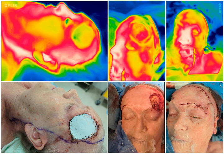
Objective: Successful outcomes in head and neck surgery rely on maintaining perfusion in pedicled skin flaps. Thermal imaging offers a noninvasive means to assess tissue perfusion, potentially aiding in predicting flap viability. This pilot study explores the utility of SBTI (smartphone-based thermal imaging) for predicting flap vitality and monitoring during surgery.
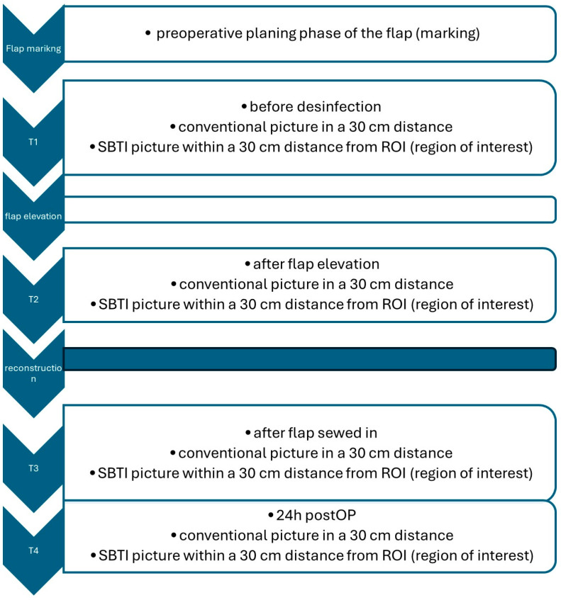
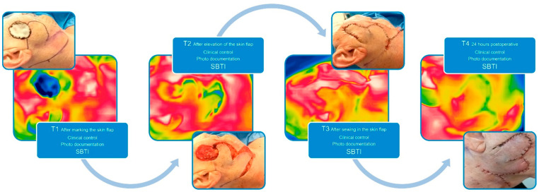
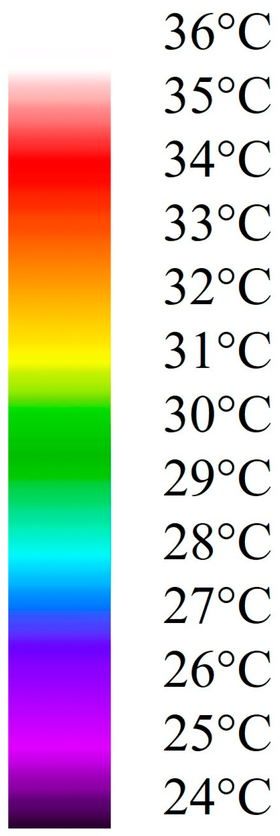
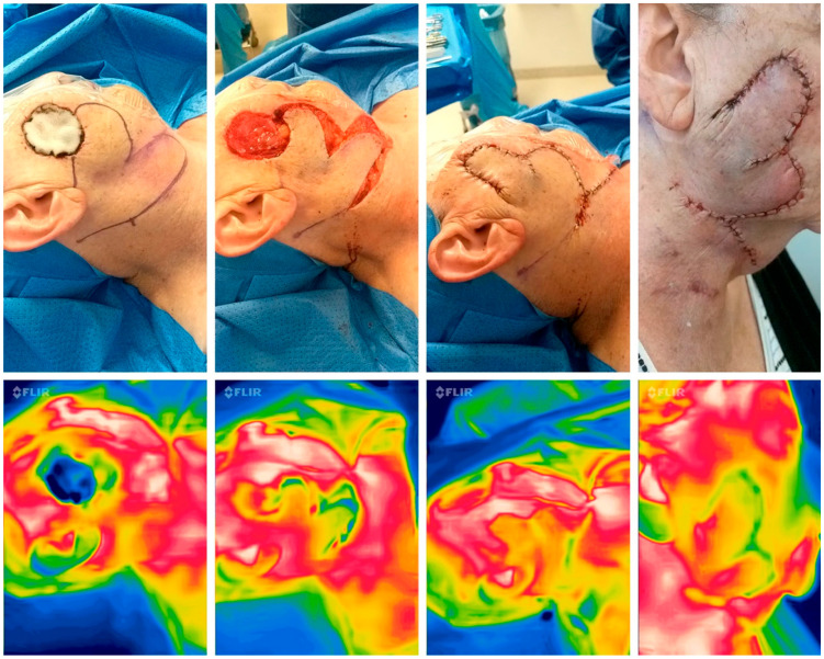
Methods: Thermal imaging was employed using the FLIR One System. An imaging protocol was established, defining points of interest (T1-T4) on pedicled skin flaps. Conducted over four months, the study integrated SBTI into reconstructive surgery for the face, head and neck defects post-tumor resections. SBTI's effectiveness was assessed with n = 11 pedicled flaps, capturing images at key stages and correlating them with clinical flap assessment. Thermal images were retrospectively graded by two surgeons, evaluating flap perfusion on a scale from 1 to 5, based on temperature differences (1 = ΔT < 2 °C, 2 = ΔT ≥ 2 °C, 3 = ΔT ≥ 4 °C, 4 = ΔT ≥ 6 °C, and 5 = ΔT ≥ 8 °C), with assessments averaged for consensus and compared with the clinical assessment control group.
Results: The study encountered challenges during implementation, leading to the exclusion of six patients. Patient data included 11 cases with n = 44 SBTI images. Intraoperative assessments consistently showed good perfusion. One postoperative dehiscence was noted, which retrospectively coincided with intraoperative SBTI grading, but not with clinical assessment. Statistical analysis indicated consistent outcomes following clinical and SBTI assessments. Thermal imaging accurately predicted flap viability, although it had limitations with small flaps.
Conclusion: SBTI proved effective, inexpensive, and noninvasive for assessing tissue perfusion, showing promise for predicting flap viability and intraoperative monitoring in head and neck surgery.
Keywords: pedicled flaps; perfusion; skin flaps; smartphone diagnostics; thermal imaging.
Conflict of interest statement
Lukas S. Fiedler, Lukas Adrian, Burkard M. Lippert and Tobias Meyer declared that they have no conflicts of interest related to this manuscript. No financial or personal relationships with other people or organizations could potentially influence their objectivity in conducting or reporting the research described in the manuscript. All potential sources of conflicts of interest have been disclosed by the journal’s guidelines. The authors declare any potential conflicts of interest related to this research. This encompasses financial interests, such as patent or stock ownership, board memberships, advisory roles, and consultancy or speaker fees from companies.
Figures
References
-
- Nischwitz S.P., Luze H., Schellnegger M., Gatterer S.J., Tuca A.C., Winter R., Kamolz L.P. Thermal, Hyperspectral, and Laser Doppler Imaging: Non-Invasive Tools for Detection of The Deep Inferior Epigastric Artery Perforators—A Prospective Comparison Study. J. Pers. Med. 2021;11:1005. doi: 10.3390/jpm11101005. - DOI - PMC - PubMed
-
- Veldhuizen I.J., Brouwer P., Aleisa A., Kurtansky N.R., Dusza S.W., Nehal K.S., Hoogbergen M.M., van der Hulst R., Lee E.H. Nasal skin reconstruction: Time to rethink the reconstructive ladder? J. Plast. Reconstr. Aesthet. Surg. 2022;75:1239–1245. doi: 10.1016/j.bjps.2021.11.028. - DOI - PMC - PubMed
-
- Larrabee W.F. Jr., Sherris D.A., Teixeira J., editors. Principles of Facial Reconstruction: A Subunit Approach to Cutaneous Repair. Thieme Medical Publishers, Inc.; New York, NY, USA: 2021. Soft Tissue Biomechanics and Physiology.
-
- Whitaker I.S., Rozen W.M., Chubb D., Acosta R., Kiil B.J., Birke-Sorensen H., Grinsell D., Ashton M.W. Postoperative monitoring of free flaps in autologous breast reconstruction: A multicenter comparison of 398 flaps using clinical monitoring, microdialysis, and the implantable Doppler probe. J. Reconstr. Microsurg. 2010;26:409–416. doi: 10.1055/s-0030-1249607. - DOI - PubMed
LinkOut - more resources
Full Text Sources
