

Histologic Evaluation of Early Papilla Healing after Augmentation with Injectable Hyaluronic Acid-A Proof of Concept
- PMID: 39064145
- PMCID: PMC11278162
- DOI: 10.3390/jcm13144102
Histologic Evaluation of Early Papilla Healing after Augmentation with Injectable Hyaluronic Acid-A Proof of Concept
Abstract
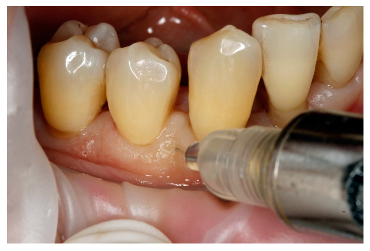
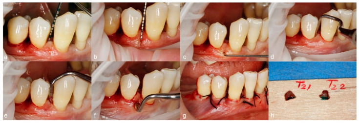
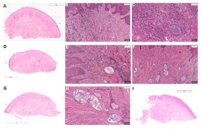
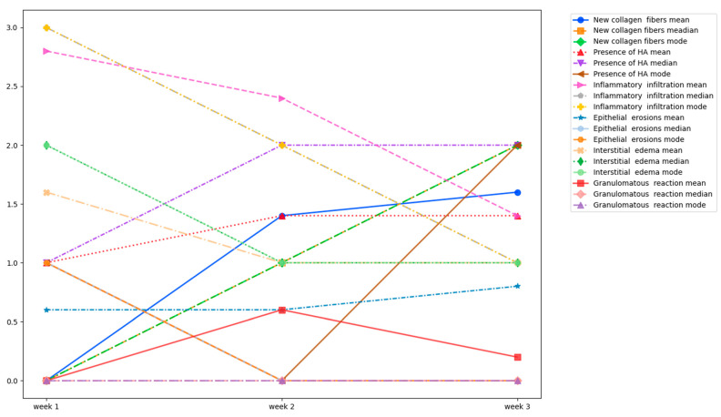
Objectives: This human histological study's purpose was to histologically evaluate papillae's healing after hyaluronic acid (HA) gel augmentation at three healing time points after one injection with hyaDENT BG®. Methods: Fifteen papillae from two patients with stage III, grade B periodontitis have been selected for this study. Every week for three weeks, five papillae were injected once with HA gel, and during the fourth week, the papillae were surgically removed as part of step 3 of the periodontal treatment. The histological analysis was performed on fifteen papillae, with five papillae corresponding to every timepoint of healing (weeks 1, 2, and 3). The primary outcome was considered to be the newly formed collagen fibers. The presence of residual HA, the integrity of epithelium or the presence of erosions/ulcerations, the presence and characteristics of inflammatory infiltrate, the presence of granulomatous reactions, and interstitial edema were considered to be secondary outcomes. Results: From the first to the third week, newly formed connective tissue begins to appear, while the observed HA pools (vesicles) content decreases. The density of inflammatory infiltrate was higher in the first week after injection, decreasing considerably by week 3; however, it was still visible throughout the healing time points. A granulomatous reaction was present in only three samples, while no signs of ulceration or necrosis could be observed; however, epithelial erosions could be observed on some samples after the first week. Conclusions: Papila augmentation with hyaluronic acid promotes new collagen formation from the second week of healing despite some foreign body granulomatous reactions.
Keywords: early wound healing; histologic analysis; hyaluronic acid; papilla augmentation.
Conflict of interest statement
The authors declare no conflicts of interest.
Figures
Similar articles
-
Evaluation of Interdental Papilla Regeneration Using Injectable Hyaluronic Acid: A Clinical Study.Cureus. 2024 Jul 14;16(7):e64510. doi: 10.7759/cureus.64510. eCollection 2024 Jul. Cureus. 2024. PMID: 39139319 Free PMC article.
-
Periodontal wound healing/regeneration of two-wall intrabony defects following reconstructive surgery with cross-linked hyaluronic acid-gel with or without a collagen matrix: a preclinical study in dogs.Quintessence Int. 2021 Mar 3;52(4):308-316. doi: 10.3290/j.qi.b937003. Quintessence Int. 2021. PMID: 33533237
-
Augmentation of glans penis using injectable hyaluronic acid gel.Int J Impot Res. 2003 Dec;15(6):456-60. doi: 10.1038/sj.ijir.3901058. Int J Impot Res. 2003. PMID: 14671667
-
Evaluation of the Effect of Hyaluronic Acid Injection on the Reconstruction of Reduced Interdental Papillae in Patients Referred to Shiraz School of Dentistry.J Dent (Shiraz). 2023 Sep;24(3):305-311. doi: 10.30476/dentjods.2022.94766.1808. J Dent (Shiraz). 2023. PMID: 37727351 Free PMC article.
-
Topical high-molecular-weight hyaluronan and a roofing barrier sheet equally inhibit postlaminectomy fibrosis.Spine J. 2005 Mar-Apr;5(2):180-90. doi: 10.1016/j.spinee.2004.06.019. Spine J. 2005. PMID: 15749618
References
-
- Al-Zarea B.K., Sghaireen M.G., Alomari W.M., Bheran H., Taher I. Black triangles causes and management: A review of literature. Br. J. Appl. Sci. Technol. 2015;6:1. doi: 10.9734/BJAST/2015/11287. - DOI
-
- Plato Palathingal J.M. Treatment of black triangle by using a sub-epithelial connective tissue graft. J. Clin. Diagn. Res. 2011;5:1688–1691.
Grants and funding
LinkOut - more resources
Full Text Sources
