

Uterine Mesenchymal Tumors: Updates on Pathology, Molecular Landscape, and Therapeutics
- PMID: 39064514
- PMCID: PMC11278911
- DOI: 10.3390/medicina60071085
Uterine Mesenchymal Tumors: Updates on Pathology, Molecular Landscape, and Therapeutics
Abstract
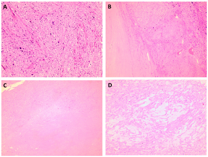
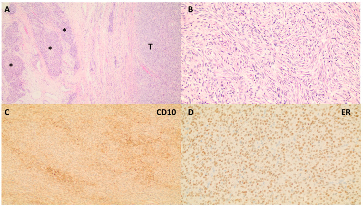
Background: Mesenchymal uterine tumors are a diverse group of neoplasms with varying biological potential. Many of these neoplasms can have overlapping morphologic similarities, which, in some instances, render their diagnosis and categorization thorough histomorphologic examination inconclusive. In the last decade, an exponential amount of molecular data aiming to more accurately characterize and, consequently, treat these tumors have accumulated. Objective: The goal of this narrative review is to provide a pathologic review, a genetic update, and to know the new therapeutic avenues of primary uterine mesenchymal neoplasms.
Keywords: gynecology; mesenchymal tumor; molecular update; pathology.
Conflict of interest statement
The author declares no conflicts of interest.
Figures


References
-
- WHO Classification of Tumours Editorial Board . Female Genital Tumours. 5th ed. Volume 4 International Agency for Research on Cancer; Lyon, France: 2020.
-
- Croce S., Devouassoux-Shisheboran M., Pautier P., Ray-Coquard I., Treilleux I., Neuville A., Arnould L., Just P.-A., Belda M.A.L.F., Averous G., et al. Uterine sarcomas and rare uterine mesenchymal tumors with malignant potential. Diagnostic guidelines of the French Sarcoma Group and the Rare Gynecological Tumors Group. Gynecol. Oncol. 2022;167:373–389. doi: 10.1016/j.ygyno.2022.07.031. - DOI - PubMed
-
- Croce S., Ribeiro A., Brulard C., Noel J.-C., Amant F., Stoeckle E., Devouassoux-Shisheborah M., Floquet A., Arnould L., Guyon F., et al. Uterine smooth muscle tumor analysis by comparative genomic hybridization: A useful diagnostic tool in challenging lesions. Mod. Pathol. 2015;28:1001–1010. doi: 10.1038/modpathol.2015.3. - DOI - PubMed
-
- Chiang S., Samore W., Zhang L., Sung Y.-S., Turashvili G., Murali R., Soslow R.A., Hensley M.L., Swanson D., Dickson B.C., et al. PGR Gene Fusions Identify a Molecular Subset of Uterine Epithelioid Leiomyosarcoma with Rhabdoid Features. Am. J. Surg. Pathol. 2019;43:810–818. doi: 10.1097/PAS.0000000000001239. - DOI - PMC - PubMed
Publication types
MeSH terms
LinkOut - more resources
Full Text Sources
Medical
