Review
. 2024 Jul 10;16(14):2195.
doi: 10.3390/nu16142195.

The "Sunshine Vitamin" and Its Antioxidant Benefits for Enhancing Muscle Function

Affiliations
Review

The "Sunshine Vitamin" and Its Antioxidant Benefits for Enhancing Muscle Function

Cristina Russo et al. Nutrients. .

Abstract

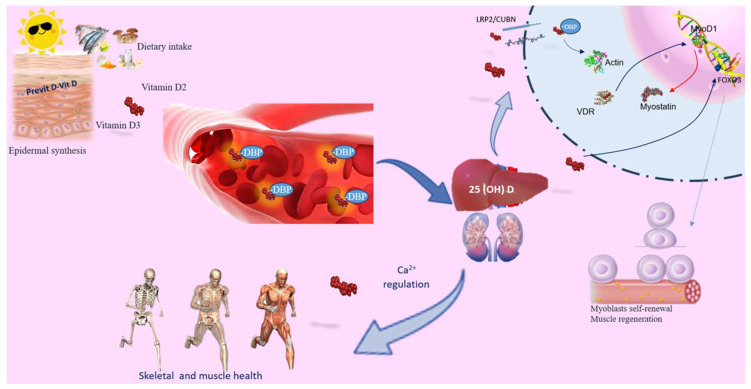
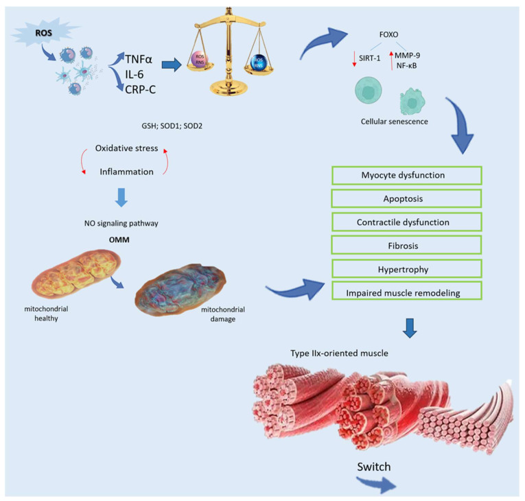
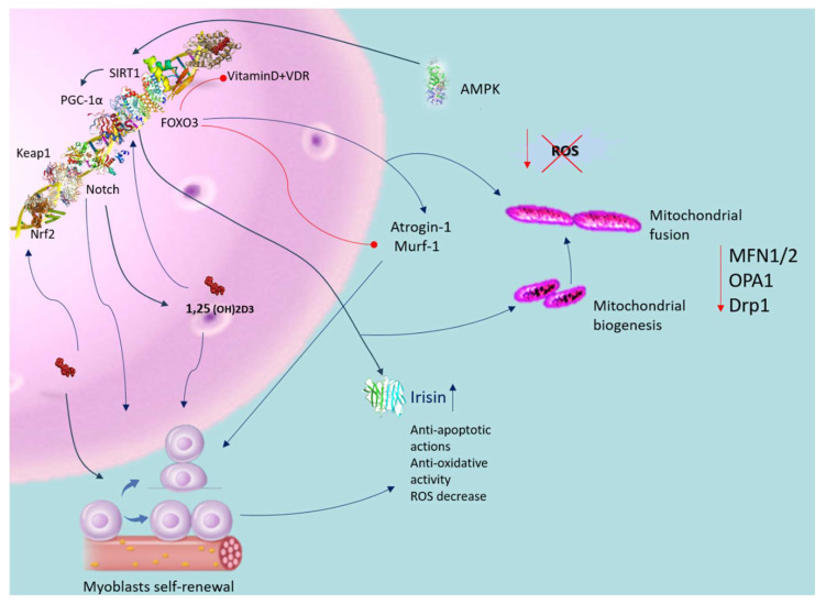
Pathological states marked by oxidative stress and systemic inflammation frequently compromise the functional capacity of muscular cells. This progressive decline in muscle mass and tone can significantly hamper the patient's motor abilities, impeding even the most basic physical tasks. Muscle dysfunction can lead to metabolic disorders and severe muscle wasting, which, in turn, can potentially progress to sarcopenia. The functionality of skeletal muscle is profoundly influenced by factors such as environmental, nutritional, physical, and genetic components. A well-balanced diet, rich in proteins and vitamins, alongside an active lifestyle, plays a crucial role in fortifying tissues and mitigating general weakness and pathological conditions. Vitamin D, exerting antioxidant effects, is essential for skeletal muscle. Epidemiological evidence underscores a global prevalence of vitamin D deficiency, which induces oxidative harm, mitochondrial dysfunction, reduced adenosine triphosphate production, and impaired muscle function. This review explores the intricate molecular mechanisms through which vitamin D modulates oxidative stress and its consequent effects on muscle function. The aim is to evaluate if vitamin D supplementation in conditions involving oxidative stress and inflammation could prevent decline and promote or maintain muscle function effectively.

Keywords: calcifediol; calcitriol; muscle homeostasis; muscular dysfunction; oxidative stress; public health.


Conflict of interest statement

The authors declare that they do not have any potential conflicts of interest.

Figures

Figure 1
Figure 1
Vitamin D’s role in skeletal muscle. Vitamin D can be acquired through skin synthesis or from diet. Both vitamin D3 and D2 undergo the same metabolic processes to produce their active forms. While the primary role of vitamin D is to regulate calcium levels and ensure skeletal and muscle health, it also serves as a powerful immunoregulator, influencing the inflammatory response, muscle damage, and aerobic capacity. Circulating 25(OH)D binds to the carrier protein DBP. PTH promotes renal Ca2+ retention and activates the synthesis of active vitamin D, which, in conjunction with the vitamin D receptor (VDR), facilitates Ca2+ and phosphate absorption. Vitamin D deficiency (VDD) or inadequate sun exposure can elevate PTH levels, leading to skeletal fragility. In skeletal muscle, the 25(OH)D-DBP complex is transported into target cells through the LRP2/CUBN transmembrane complex. Inside the cell, the D-DBP complex associates with cytoplasmic actin. 1,25(OH)2D triggers the expression of protein 1, affecting MyoD1 activation. Vitamin D also regulates the FOXO3 signaling pathways, enhancing myoblast self-renewal. VDR expression in skeletal muscle promotes muscle protein synthesis, is crucial for maintaining muscle mass, and aids in muscle regeneration. Abbreviations in alphabetical order: 1,25(OH)2D3—calcitriol; FOXO—forkhead family of transcription factors; LRP2/CUBN—megalin–cubilin transmembrane complex; MyoD1—myogenic determination factor 1; PTH—parathyroid hormone; DBP—vitamin D binding protein; vitamin D receptor (VDR).
Figure 2
Figure 2
Relationship between oxidative stress and weakness of muscle. ROS generated from localized inflammation activate immune cells, initiating a harmful cycle characterized by the release of pro-inflammatory mediators, such as TNFα, IL-6, and CRP. Despite the presence of an impaired antioxidant system, including decreased levels of GSH and SOD, oxidative damage remains unchecked. Furthermore, inflammation disrupts mitochondrial function in muscle through the NO signaling pathway, triggering cell death via OMM permeabilization. Oxidative stress accelerates cellular senescence by activating FOXO and diminishing SIRT-1, leading to heightened MMP-9 and NF-κB activity. This escalated oxidative stress precipitates myocyte dysfunction and apoptosis, resulting in contractile dysfunction, fibrosis, hypertrophy, and impaired muscle remodeling. Additionally, there is a transition towards a type-IIx-oriented muscle phenotype with compromised oxygen distribution and utilization, ultimately impairing functionality. Abbreviations in alphabetical order: CRP—C reactive protein; FOXO—forkhead family of transcription factors; GSH—glutathione peroxidase; IL-6—Interleukin 6; MMP-9—matrix metallopeptidase 9; NF-κB—nuclear factor kappa-light-chain-enhancer of activated B cells; NO—nitrogen monoxide; OMM—outer mitochondrial membrane; RNS—reactive nitrogen species; ROS—reactive oxygen species; SIRT-1—sirtuin-1; SOD—superoxide dismutase; TNFα—tumor receptor necrosis factor alpha.
Figure 3
Figure 3
Antioxidative role of vitamin D in muscle dysfunction. Blue arrow indicates activation; red arrow indicates inhibition. Vitamin D activates the VDR in satellite cells, enhancing their self-renewal, proliferation, and differentiation capabilities. Activation of the VDR also mitigates oxidative stress, promoting mitochondrial biogenesis and fusion while reducing oxidative damage and dysfunction. This process improves mitochondrial network structure through the regulation of MFN1/2, OPA1, and Drp1 expression. Vitamin D positively influences Sirt1 activity and enhances mitochondrial function. Supplementation with vitamin D activates Sirt1 and AMPK in skeletal muscle cells. Vitamin D further increases the expression of irisin precursors in muscle cells, contingent upon intact Sirt1 expression. Both AMPK and Sirt1 regulate PGC-1α activation and transcription, influencing irisin secretion in skeletal muscle cells. Vitamin D upregulates FOXO1 protein and suppresses atrogin-1 and MuRF1 expression. Additionally, VDS triggers VDR and induces the Nrf2-Keap1 antioxidant pathway. Abbreviations in alphabetical order: AMP—5′ AMP-activated protein kinase; 1,25(OH)2D3—calcitriol; Drp1—dynamin-related protein 1; IL-1β—interleukin-1 beta; MFN1/2—mitofusin; OPA—mitochondrial dynamin like GTPase; PGC-1α—peroxisome proliferator-activated receptor-gamma coactivator; PTH—parathyroid hormone; ROS—reactive oxygen species; SIRT-1—sirtuin-1; SOD—superoxide dis-mutase; TNFα—tumor receptor necrosis factor alpha; VDR—vitamin D receptor.

References

    1. Rai M., Demontis F. Systemic Nutrient and Stress Signaling via Myokines and Myometabolites. Annu. Rev. Physiol. 2016;78:85–107. doi: 10.1146/annurev-physiol-021115-105305. - DOI - PubMed
    1. Blaauw B., Schiaffino S., Reggiani C. Mechanisms modulating skeletal muscle phenotype. Compr. Physiol. 2013;3:1645–1687. doi: 10.1002/cphy.c130009. - DOI - PubMed
    1. Srikanthan P., Karlamangla A.S. Muscle mass index as a predictor of longevity in older adults. Am. J. Med. 2014;127:547–553. doi: 10.1016/j.amjmed.2014.02.007. - DOI - PMC - PubMed
    1. Casabona A., Valle M.S., Laudani L., Crimi C., Russo C., Malaguarnera L., Crimi N., Cioni M. Is the Power Spectrum of Electromyography Signal a Feasible Tool to Estimate Muscle Fiber Composition in Patients with COPD? J. Clin. Med. 2021;10:3815. doi: 10.3390/jcm10173815. - DOI - PMC - PubMed
    1. Derbré F., Gratas-Delamarche A., Gómez-Cabrera M.C., Viña J. Inactivity-induced oxidative stress: A central role in age-related sarcopenia? Eur. J. Sport Sci. 2014;1:S98–S108. doi: 10.1080/17461391.2011.654268. - DOI - PubMed

LinkOut - more resources