Review
. 2024 Jul 19;12(7):1481.
doi: 10.3390/microorganisms12071481.

Native Infective Endocarditis: A State-of-the-Art-Review

Affiliations
Review

Native Infective Endocarditis: A State-of-the-Art-Review

Francesco Nappi. Microorganisms. .

Abstract

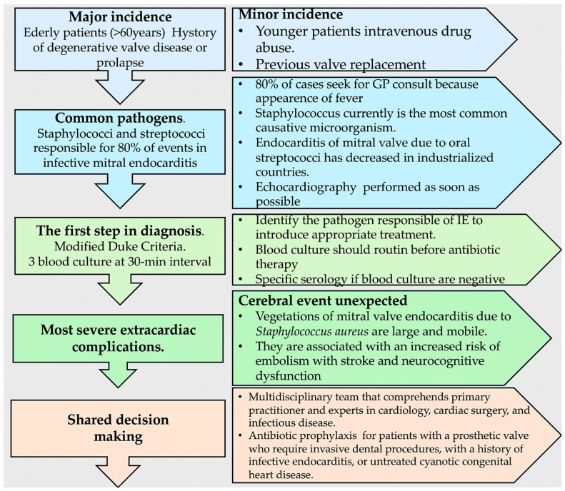
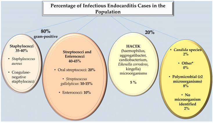
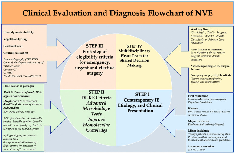
Native valve infective endocarditis (NVE) is a global phenomenon, defined by infection of a native heart valve and involving the endocardial surface. The causes and epidemiology of the disease have evolved in recent decades, with a doubling of the average patient age. A higher incidence was observed in patients with implanted cardiac devices that can result in right-sided infection of the tricuspid valve. The microbiology of the disease has also changed. Previously, staphylococci, which are most often associated with health-care contact and invasive procedures, were the most common cause of the disease. This has now been superseded by streptococci. While innovative diagnostic and therapeutic strategies have emerged, mortality rates have not improved and remain at 30%, which is higher than that for many cancer diagnoses. The lack of randomized trials and logistical constraints impede clinical management, and long-standing controversies such as the use of antibiotic prophylaxis persist. This state of the art review addresses clinical practice, controversies, and strategies to combat this potentially devastating disease. A multidisciplinary team will be established to provide care for patients with presumptive NVE. The composition of the team will include specialists in cardiology, cardiovascular surgery, and infectious disease. The prompt administration of combination antimicrobial therapy is essential for effective NVE treatment. Additionally, a meticulous evaluation of each patient is necessary in order to identify any indications for immediate valve surgery. With the intention of promoting a more comprehensive understanding of the procedural management of native infective endocarditis and to furnish clinicians with a reference, the current evidence for the utilization of distinct strategies for the diagnosis and treatment of NVE are presented.

Keywords: gram-positive bacteria; infective endocarditis; native valve endocarditis; vegetation.


Conflict of interest statement

The author declares no conflicts of interest.

Figures

Figure 4
Figure 4
Modified Duke Criteria. * Staphylococcus aureus, Viridans streptococci, Streptococcus gallolyticus, HACEK (Haemophilus species, Aggregatibacter (formerly Actinobacillus) species, Cardiobacterium species, Eikenella corrodens, and Kingella species), and community-acquired enterococci in the absence of a primary focus. Refs. [14,15,28].
Figure 5
Figure 5
Clinical evaluation and diagnosis flowchart. Refs. [4,5,6,7,8,9,10,11,12,13,14,15,16,18,19,20,21,22,23,24,25,26,27,28,29,30,31,32,33,34].
Figure 6
Figure 6
Sensitivity and specificities of echocardiography in the detection of abscesses. Refs. [27,28,31,32]. From Nappi et al. Bridging Molecular and Clinical Sciences to Achieve the Best Treatment of Enterococcus faecalis Endocarditis. Microorganisms 2023, 11, 2604. Ref. [42].
Figure 1
Figure 1
The following section reviews the echocardiographic findings in mitral valve endocarditis. (A) depicts a representative example of the normal mitral valve in two-dimensional transesophageal echocardiography, with the probe rotated to 120°. The thinner leaflets of the mitral valve are visible in the open position, in mid-diastole, with the aortic valve in the closed position. The left atrium is of an appropriate size, without any enlargement, and the left ventricular wall thickness is also within normal limits. (B) Mitral xenograft endocarditis. (C) Two-dimensional transesophageal echocardiography displays a vegetation on the atrial aspect of the anterior leaflet. (D) Three-dimensional reconstruction of the mitral valve (surgeon’s view) depicts the vegetation in C. Prosthetic valve endocarditis is evident. (E,F) The images presented are those obtained from the short-axis view (59 degrees of probe angle) and (G,H) the long-axis view (120 degrees). The images are presented in two different ways: with (A) and without color-Doppler analysis. The color doppler analysis was performed on a mechanical aortic prosthesis with a posterior semilunar abscess, which involved the aortomitral junction.
Figure 2
Figure 2
Percentage of Infectious Endocarditis Cases in the Population. * Fungal endocarditis, usually Candida or Aspergillus, is rare but often fatal, arising in patients who are immunosuppressed or after cardiac surgery, mostly on prosthetic valves. Includes small numbers of Enterobacteriaceae, Propionibacterium acnes, Coxiella burnetii, Bartonella quintana, Tropheryma whipplei, Gordonia bronchialis, Bacillus spp., Erysipelothrix rhusiopathiae, Neisseria elongata, Moraxella catarrhalis, Veillonella spp., Listeria monocytogenes, Acinetobacter ursingii, Campylobacter fetus, Francisella tularensis, and Pseudoonas aeruginosa, Lactobacillus spp., Corynebacterium spp., Catabacter hongkongensi. Refs. [1,2,3,4,5,6,7,8,9,10].
Figure 3
Figure 3
The diagnosis of culture-negative endocarditis and related testing to detect microorganisms in the red boxes are presented. The pink box illustrates the microorganisms and related clinical and epidemiologic clues (Box blue). The varying coloration of the red box signifies the type of testing. ⚘ In the event that a dash is present, it signifies that the test to detect the microorganism in question is not available or not applicable at this moment in time. HACEK stands for Haemophilus species, Aggregatibacter (formerly Actinobacillus) species, Cardiobacterium species, Eikenella corrodens, and Kingella species. It also encompasses PCR polymerase chain reaction and RT-PCR reverse-transcriptase PCR; §, the sensitivity of the method is significantly greater if the RT-PCR or broad-range 16S or 18S RNA PCR assay is conducted on valvular vegetation or abscess material, in comparison with the use of blood as a specimen; ☨ PCR assays that cover a broad range of targets often include the 16S and 18S ribosomal RNA genes; # The tests for Legionella pneumophila serotype 1, as indicated by sierologic tests and urinary antigen tests, are the only tests capable of detecting the aforementioned serotype; ## Serologic tests are employed solely for the purpose of detecting the presence of Mycoplasma pneumoniae; * In the event of an extracardial lesion, a biopsy of the affected tissue (e.g., small bowel and synovium, if present) is recommended. Refs. [2,4,6,8].
Figure 7
Figure 7
Clinical assessment of IE. To diagnose IE, a series of imaging techniques may be employed, including TTE, TEE, CT, and CT/MRI. These techniques are employed in a stepwise manner to either confirm or exclude the presence of an infection. The use of 18-FDG PET-CT or SPECT/CT has been demonstrated to have a high degree of specificity for the identification of NVEs. Abbreviations; CT, computed tomography; 18F- FDG PET/CT, positron emission CT with 18F-fluorodeoxyglucose; IE, infective endocarditis; MRI; magnetic resonance imaging; NVE; native endocarditis; TEE; transesophageal echocardiography; TTE, transthoracic echocardiography. * 2023 Duke-ISCVID IE Criteria. Ref. [28].
Figure 8
Figure 8
(A) This illustration demonstrates the efficacy of antibiotic treatment in NVEs for Viridans streptococci and Streptococcus gallolyticus at a penicillin MIC of ≤0.12 μg/mL. The duration of therapy once blood cultures have converted to negative is shown. Abbreviations; MIC, minimal inhibitory concentration; wk, week [27,46,47,48,49,50,51,52,53,54,55,56,57,58,59,60,61,62,63,64,65,66,67,68,69,70,71,72]. (B) This illustration demonstrates the efficacy of antibiotic treatment in NVEs for Viridans streptococci and Streptococcus gallolyticus at a penicillin MIC of >0.12 to <0.5 μg/mL and for enterococci. The duration of therapy once blood cultures have converted to negative is shown. Abbreviations; MIC, minimal inhibitory concentration, wk, week [27,46,47,48,49,50,51,52,53,54,55,56,57,58,59,60,61,62,63,64,65,66,67,68,69,70,71,72]. (C) This illustration demonstrates the efficacy of antibiotic treatment in NVEs for methicillin-susceptible Staphylococcus aureus, methicillin-resistant S. aureus and HACEK. The duration of therapy once blood cultures have converted to negative is shown. Abbreviations; MIC, hr, hour; wk, week. * HACEK denotes Haemophilus species, Aggregatibacter (formerly Actinobacillus) species, Cardiobacterium species, Eikenella corrodens, and Kingella species [27,46,47,48,49,50,51,52,53,54,55,56,57,58,59,60,61,62,63,64,65,66,67,68,69,70,71,72]. (D) This illustration demonstrates the efficacy of antibiotic treatment in NVEs for Abiotrophia defectiva, granulicatella species, Viridans streptococci, S. gallolyticus, at a penicillin MIC ≥0.5 μg/mL. The duration of therapy once blood cultures have converted to negative is shown. Abbreviations; MIC, minimal inhibitory concentration, wk, week. Refs. [27,46,47,48,49,50,51,52,53,54,55,56,57,58,59,60,61,62,63,64,65,66,67,68,69,70,71,72]. * 2023 ECC guidelines for IE.
Figure 8
Figure 8
(A) This illustration demonstrates the efficacy of antibiotic treatment in NVEs for Viridans streptococci and Streptococcus gallolyticus at a penicillin MIC of ≤0.12 μg/mL. The duration of therapy once blood cultures have converted to negative is shown. Abbreviations; MIC, minimal inhibitory concentration; wk, week [27,46,47,48,49,50,51,52,53,54,55,56,57,58,59,60,61,62,63,64,65,66,67,68,69,70,71,72]. (B) This illustration demonstrates the efficacy of antibiotic treatment in NVEs for Viridans streptococci and Streptococcus gallolyticus at a penicillin MIC of >0.12 to <0.5 μg/mL and for enterococci. The duration of therapy once blood cultures have converted to negative is shown. Abbreviations; MIC, minimal inhibitory concentration, wk, week [27,46,47,48,49,50,51,52,53,54,55,56,57,58,59,60,61,62,63,64,65,66,67,68,69,70,71,72]. (C) This illustration demonstrates the efficacy of antibiotic treatment in NVEs for methicillin-susceptible Staphylococcus aureus, methicillin-resistant S. aureus and HACEK. The duration of therapy once blood cultures have converted to negative is shown. Abbreviations; MIC, hr, hour; wk, week. * HACEK denotes Haemophilus species, Aggregatibacter (formerly Actinobacillus) species, Cardiobacterium species, Eikenella corrodens, and Kingella species [27,46,47,48,49,50,51,52,53,54,55,56,57,58,59,60,61,62,63,64,65,66,67,68,69,70,71,72]. (D) This illustration demonstrates the efficacy of antibiotic treatment in NVEs for Abiotrophia defectiva, granulicatella species, Viridans streptococci, S. gallolyticus, at a penicillin MIC ≥0.5 μg/mL. The duration of therapy once blood cultures have converted to negative is shown. Abbreviations; MIC, minimal inhibitory concentration, wk, week. Refs. [27,46,47,48,49,50,51,52,53,54,55,56,57,58,59,60,61,62,63,64,65,66,67,68,69,70,71,72]. * 2023 ECC guidelines for IE.
Figure 9
Figure 9
Indication for early surgery. Refs. [4,27,28,31,40,73,77].
Figure 10
Figure 10
Indications for surgery of isolated or complex valve endocarditis. This illustration presents a summary of the 2023 ESC Guidelines for the management of endocarditis, 2023 Duke-International Society for Cardiovascular Infectious Diseases Criteria for Infective Endocarditis: Updating the Modified Duke Criteria, JCS 2017 guideline on prevention and treatment of infective endocarditis and 2016 The American Association for Thoracic Surgery (AATS) consensus guidelines: surgical treatment of infective endocarditis: executive summary The guidelines are presented in the form of a flowchart, which depicts three different pathways (light blue, light green, and light grey box). according to the degree of urgency. The clinical presentation and imaging findings are described in each pathway. The algorithm used to inform decision-making regarding elective, urgent, or emergency surgery is presented in light blue, light green, or gray boxes, respectively, in the flow diagram. The decision regarding the surgical options (i.e., repair or replacement) is based on the clinical and anatomic findings on preoperative imaging. In the event that the IE is confined to a limited region of the valve leaflets, mitral valve repair should be considered. In cases of extensive anatomic involvement of the valve, surgical mitral valve replacement is the recommended surgical approach. The timing of surgery should be determined through a collaborative, interdisciplinary approach. In the context of emergency surgery, the infected valve must be treated within 24 h of the completion of the diagnostic workup. In the case of patients whose condition is urgent, surgery should be performed within a few days of the indication. Elective surgery should be delayed for a minimum of one to two weeks following the initiation of antibiotic therapy. Refs. [4,19,20,27,28,31,49,77]. * higher frequence of events. † For an indication of the necessity for emergency surgical intervention, please direct your attention to Figure 9.
Figure 11
Figure 11
Guidelines for Infective Endocarditis. Refs. [28,77]. * For an indication of the necessity for early surgical intervention, please direct your attention to Figure 9.
Figure 12
Figure 12
Clinical evaluation and diagnosis flowchart of native valve endocarditis.

Similar articles

Cited by

References

    1. Nappi F., Spadaccio C., Mihos C. Infective endocarditis in the 21st century. Ann. Transl. Med. 2020;8:1620. doi: 10.21037/atm-20-4867. - DOI - PMC - PubMed
    1. Cahill T.J., Prendergast B.D. Infective endocarditis. Lancet. 2016;387:882–893. doi: 10.1016/S0140-6736(15)00067-7. - DOI - PubMed
    1. Bin Abdulhak A.A., Baddour L.M., Erwin P.J., Hoen B., Chu V.H., Mensah G.A., Tleyjeh I.M. Global and regional burden of infective endocarditis, 1990 2010: A systematic review of the literature. Glob. Heart. 2014;9:131–143. doi: 10.1016/j.gheart.2014.01.002. - DOI - PubMed
    1. Nappi F., Spadaccio C., Dreyfus J., Attias D., Acar C., Bando K. Mitral endocarditis: A new management framework. J. Thorac. Cardiovasc. Surg. 2018;156:1486–1495. doi: 10.1016/j.jtcvs.2018.03.159. - DOI - PubMed
    1. Nappi F., Avtaar Singh S.S. Host-Bacterium Interaction Mechanisms in Staphylococcus aureus Endocarditis: A Systematic Review. Int. J. Mol. Sci. 2023;24:11068. doi: 10.3390/ijms241311068. - DOI - PMC - PubMed

LinkOut - more resources