

Hot-Melt Extrusion-Based Dexamethasone-PLGA Implants: Physicochemical, Physicomechanical, and Surface Morphological Properties and In Vitro Release Corrected for Drug Degradation
- PMID: 39065592
- PMCID: PMC11280434
- DOI: 10.3390/pharmaceutics16070895
Hot-Melt Extrusion-Based Dexamethasone-PLGA Implants: Physicochemical, Physicomechanical, and Surface Morphological Properties and In Vitro Release Corrected for Drug Degradation
Abstract
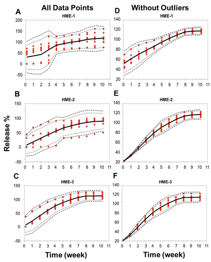
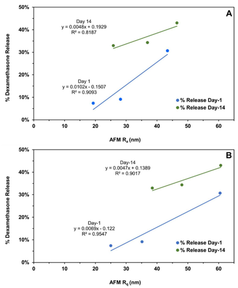
Developing bioequivalent (BE) generic products of complex dosage forms like intravitreal implants (IVIs) of corticosteroids such as dexamethasone prepared using hot-melt extrusion (HME), based on biodegradable poly (lactide-co-glycolide) (PLGA) polymers, can be challenging. A better understanding of the relationship between the physicochemical and physicomechanical properties of IVIs and their effect on drug release and ocular bioavailability is crucial to develop novel BE approaches. It is possible that the key physicochemical and physicomechanical properties of IVIs such as drug properties, implant surface roughness, mechanical strength and toughness, and implant erosion could vary for different compositions, resulting in changes in drug release. Therefore, this study investigated the hypothesis that biodegradable ophthalmic dexamethasone-loaded implants with 20% drug and 80% PLGA polymer(s) prepared using single-pass hot-melt extrusion (HME) differ in physicochemical and/or physicomechanical properties and drug release depending on their PLGA polymer composition. Acid end-capped PLGA was mixed with an ester end-capped PLGA to make three formulations: HME-1, HME-2, and HME-3, containing 100%, 80%, and 60% w/w of the acid end-capped PLGA. Further, this study compared the drug release between independent batches of each composition. In vitro release tests (IVRTs) indicated that HME-1 implants can be readily distinguished by their release profiles from HME-2 and HME-3, with the release being similar for HME-2 and HME-3. In the early stages, drug release generally correlated well with polymer composition and implant properties, with the release increasing with PLGA acid content (for day-1 release, R2 = 0.80) and/or elevated surface roughness (for day-1 and day-14 release, R2 ≥ 0.82). Further, implant mechanical strength and toughness correlated inversely with PLGA acid content and day-1 drug release. Drug release from independent batches was similar for each composition. The findings of this project could be helpful for developing generic PLGA polymer-based ocular implant products.
Keywords: LC-MS/MS; biodegradable; corticosteroid; dexamethasone; hot-melt extrusion (HME); implants; intravitreal; ocular drug delivery systems; poly-(lactide-co-glycolide) PLGA; sustained drug release.
Conflict of interest statement
The authors declare no conflicts of interest.
Figures


References
-
- FDA, 21 CFR §314.94 (a)(9)(iv) [(accessed on 21 June 2024)]; Available online: https://www.ecfr.gov/current/title-21/chapter-I/subchapter-D/part-314.
-
- FDA, 21 CFR 320.22 (B)(1) [(accessed on 21 June 2024)]; Available online: https://www.ecfr.gov/current/title-21/chapter-I/subchapter-D/part-320.
-
- RothenWeinhold A., Besseghir K., Gurny R. Analysis of the influence of polymer characteristics and core loading on the in vivo release of a somatostatin analogue. Eur. J. Pharm. Sci. 1997;5:303–313. doi: 10.1016/S0928-0987(97)00022-5. - DOI
Grants and funding
LinkOut - more resources
Full Text Sources
Research Materials
