. 2024 Jul 5;16(7):904.
doi: 10.3390/pharmaceutics16070904.

A New Casjensviridae Bacteriophage Isolated from Hospital Sewage for Inactivation of Biofilms of Carbapenem Resistant Klebsiella pneumoniae Clinical Isolates

Affiliations

A New Casjensviridae Bacteriophage Isolated from Hospital Sewage for Inactivation of Biofilms of Carbapenem Resistant Klebsiella pneumoniae Clinical Isolates

Sambuddha Chakraborty et al. Pharmaceutics. .

Abstract

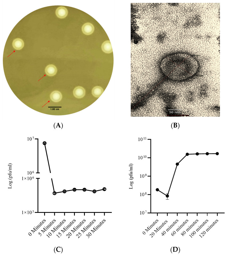
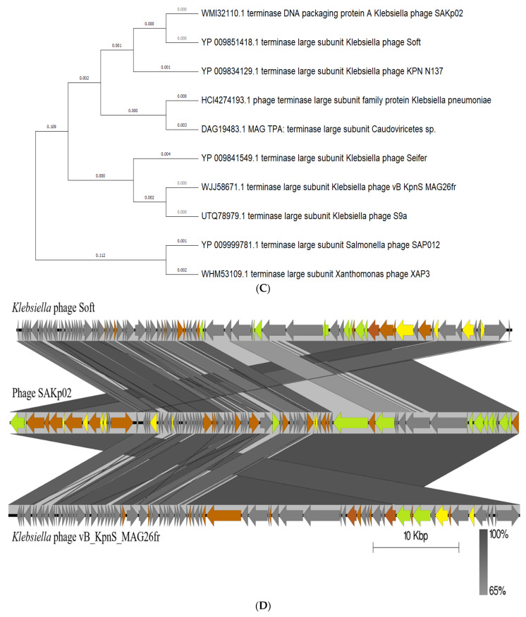
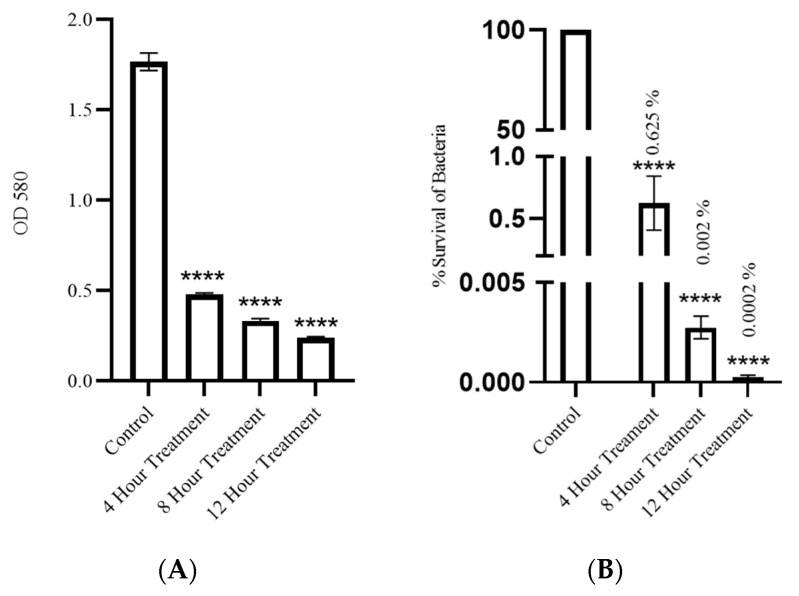
Klebsiella pneumoniae, a member of the ESKAPE pathogen group, is a prominent cause of hospital-acquired infections. The WHO has recognized carbapenem-resistant K. pneumoniae as a critical-one priority pathogen. These resilient superbugs have the ability to form biofilms and present a significant global threat. In the present study, we isolated and characterized a bacteriophage SAKp02, from hospital sewage, infectious to carbapenem-resistant K. pneumoniae patient isolates. SAKp02 could infect 43 of 72 clinical isolates, indicating a broad host spectrum. Whole genome analysis classified SAKp02 within the family Casjensviridae, with a 59,343 bp genome encoding 82 ORFs. Comparative genomic analysis revealed significant differences between SAKp02 and its closest viruses, indicating a distinct genetic makeup positioning it as a novel phage strain within the lineage. The SAKp02 genome comprises bacteriolytic enzymes, including holin, endolysin, and phage depolymerase, crucial for bacterial lysis and biofilm disruption. It reduced biofilm biomass by over threefold compared to the control and eradicated 99% of viable cells within a 4 h treatment period. Scanning electron microscopy corroborated the ability of the phage to dismantle biofilm matrices and lyse bacterial cells. Safe and effective treatments are warranted, and hence, the fully characterized lytic phages with therapeutic potential against drug-resistant clinical isolates of bacteria are needed. Our study is the first to report the antibacterial and antibiofilm activity of Casjensviridae phages, and our discovery of a novel K. pneumoniae phage broadens the arsenal against the bacteria.

Keywords: K. pneumoniae; alternative to antibiotics; antibiofilm strategy; antimicrobial resistance; biofilm bacteriophage.


Conflict of interest statement

The authors declare no conflicts of interest.

Figures

Figure 1
Figure 1
Plaque and structural morphology of phage SAKp02: (A) Plaque morphology of phage SAKp02 on the lawn of K. pneumoniae B3768 strain. Depolymerase activity is marked with an arrow; (B) TEM Image of phage. Adsorption rate and time of SAKp02 and single step growth curve: (C) SAKp02 took 5 min to adsorb to the host with a rate of 95% of total phage particles; (D) Phage SAKp02 has shown a 20 min latency period and a burst size of ~202 phage particles per host cell.
Figure 2
Figure 2
Killing kinetics and host range of phage SAKp02. (A) Phage SAKp02 could restrict the growth of clinical strain B3768 in comparison to the control (Bacteria + LB) at different MOI. (B) Endpoint OD of the control and phage treatment with different MOIs after 12 h post-phage treatment. (C) Phage SAKp02 could infect 43 K. pneumoniae clinical isolates among 72 tested strains. Data represented are means ± standard error of the mean of at least 3 independent experiments. **** p < 0.0001.
Figure 3
Figure 3
Stability studies of phage SAKp02; (A) At pH 1, 3, and 14, complete degradation of phage SAKp02 was observed. (B) At 65 °C, a significant~6-fold reduction was observed, and complete denaturation of phage particles at 100 °C was seen. (C) SAKp02 showed stability in PBS, chloroform, and 0.9% saline. Data represented are means ± standard error of the mean of at least 3 independent experiments. *p < 0.05, ** p < 0.005, *** p < 0.0005, **** p < 0.0001.
Figure 4
Figure 4
Overview of whole genome of phage SAKp02 and bioinformatic analysis. (A) The GC content and genome alignment of phage SAKp02 constructed by GC Content calculator. The blue skew indicates the GC content according to the nucleotide regions. Mean GC content is marked as a straight black line. Calculation shows that the GC content of SAKp02 is 56.1%. (B) The genome map of phage SAKp02 generated by the genomevx. ORFs coding for hypothetical proteins are denoted in grey. Genes with predicted functions are colored; orange-colored genes are indicated as phage enzyme associated with replication process at different stages of viral life cycle, green colored CDSs are denoted as structural proteins of the virus, and yellow colored CDSs are proteins associated with regulatory activities. (C) Neighbor-joining phylogenetic tree of terminase large subunits from bacteriophage SAKp02 and the related phages. Sequences were downloaded from NCBI. (D) EasyFigure was used to create a linear representation and comparative analysis, visualizing alignments phage Soft (top DNA), SAKp02 (middle DNA), and phage vB_KpnS_MAG26fr (bottom DNA). Arrows denote the transcription direction of predicted ORFs and are categorized as grey (hypothetical proteins), green (structural proteins), orange (viral enzymes), and yellow (regulatory proteins). Shaded regions indicate well-conserved segments with 65–100% amino acid similarity, with darker grey regions showing higher identity. The scale is in 10 kbp increments. (E) A heatmap generated by VIRIDIC illustrates the intergenomic similarities on the right half, using color-coding to provide a quick visual comparison of the 10 phage genomes most closely related to SAKp02 (NC-048805.1: Phage soft, OR290970.1: SAKp02, OP558002.1: Klebsiella phage vB_KpnS_MAG26fr).
Figure 4
Figure 4
Overview of whole genome of phage SAKp02 and bioinformatic analysis. (A) The GC content and genome alignment of phage SAKp02 constructed by GC Content calculator. The blue skew indicates the GC content according to the nucleotide regions. Mean GC content is marked as a straight black line. Calculation shows that the GC content of SAKp02 is 56.1%. (B) The genome map of phage SAKp02 generated by the genomevx. ORFs coding for hypothetical proteins are denoted in grey. Genes with predicted functions are colored; orange-colored genes are indicated as phage enzyme associated with replication process at different stages of viral life cycle, green colored CDSs are denoted as structural proteins of the virus, and yellow colored CDSs are proteins associated with regulatory activities. (C) Neighbor-joining phylogenetic tree of terminase large subunits from bacteriophage SAKp02 and the related phages. Sequences were downloaded from NCBI. (D) EasyFigure was used to create a linear representation and comparative analysis, visualizing alignments phage Soft (top DNA), SAKp02 (middle DNA), and phage vB_KpnS_MAG26fr (bottom DNA). Arrows denote the transcription direction of predicted ORFs and are categorized as grey (hypothetical proteins), green (structural proteins), orange (viral enzymes), and yellow (regulatory proteins). Shaded regions indicate well-conserved segments with 65–100% amino acid similarity, with darker grey regions showing higher identity. The scale is in 10 kbp increments. (E) A heatmap generated by VIRIDIC illustrates the intergenomic similarities on the right half, using color-coding to provide a quick visual comparison of the 10 phage genomes most closely related to SAKp02 (NC-048805.1: Phage soft, OR290970.1: SAKp02, OP558002.1: Klebsiella phage vB_KpnS_MAG26fr).
Figure 5
Figure 5
Assessment of antibiofilm activity of SAKp02. (A) The scanning electron microscopy (SEM) analysis (sample coating, gold sputter coating; mounting, carbon tape; accelerating voltage, 5.00 kV; working distance, 5.1–5.3 mm; magnification, 5.00 K X; imaging, secondary electron imaging; detector, secondary electron detector; contrast, topographic contrast) revealed that K. pneumoniae exhibited a compact bacterial community covered by an extracellular polymeric substance (EPS) layer (white arrows). This observation indicates strong biofilm formation by K. pneumoniae MDR clinical strains. (B) Four-hour treatment with SAKp02 could effectively degrade the EPS layers and reduce the density of the bacteria. (C,D). After 8 and 12 h of treatment with phage SAKp02, there was a noticeable degradation of the biofilm matrix and bacterial cell. Cellular debris (red arrow) on the slides indicates a lytic phage infection.
Figure 6
Figure 6
Evaluation of antibiofilm efficacy of SAKp02. (A) Crystal violet assay indicated an effective demolition of biofilm biomass after 4 h of phage treatment; 12 h of phage treatment showed the lowest biofilm biomass; a 7-fold decrease in OD compared to control is seen. (B) Effective lysis of biofilm viable cells was seen with SAKP02 infection; only <0.001 percent of bacteria survived after 12 h of phage treatment. Data represented are means ± standard error of the mean of at least 3 independent experiments. **** p <0.0001.

References

    1. Podschun R., Ullmann U. Klebsiella spp. as nosocomial pathogens: Epidemiology, taxonomy, typing methods, and pathogenicity factors. Clin. Microbiol. Rev. 1998;11:589–603. doi: 10.1128/CMR.11.4.589. - DOI - PMC - PubMed
    1. Luo K., Tang J., Qu Y., Yang X., Zhang L., Chen Z., Kuang L., Su M., Mu D. Nosocomial infection by Klebsiella pneumoniae among neonates: A molecular epidemiological study. J. Hosp. Infect. 2021;108:174–180. doi: 10.1016/j.jhin.2020.11.028. - DOI - PubMed
    1. Mohd Asri N.A., Ahmad S., Mohamud R., Mohd Hanafi N., Mohd Zaidi N.F., Irekeola A.A., Shueb R.H., Yee L.C., Mohd Noor N., Mustafa F.H., et al. Global Prevalence of Nosocomial Multidrug-Resistant Klebsiella pneumoniae: A Systematic Review and Meta-Analysis. Antibiotics. 2021;10:1508. doi: 10.3390/antibiotics10121508. - DOI - PMC - PubMed
    1. Mancuso G., Midiri A., Gerace E., Biondo C. Bacterial Antibiotic Resistance: The Most Critical Pathogens. Pathogens. 2021;10:1310. doi: 10.3390/pathogens10101310. - DOI - PMC - PubMed
    1. Chang D., Sharma L., Dela Cruz C.S., Zhang D. Clinical Epidemiology, Risk Factors, and Control Strategies of Klebsiella pneumoniae Infection. Front. Microbiol. 2021;12:750662. doi: 10.3389/fmicb.2021.750662. - DOI - PMC - PubMed

LinkOut - more resources