. 2024 Jun 28;16(7):1049.
doi: 10.3390/v16071049.

Protein C Pretreatment Protects Endothelial Cells from SARS-CoV-2-Induced Activation

Affiliations

Protein C Pretreatment Protects Endothelial Cells from SARS-CoV-2-Induced Activation

Bruna Rafaela Dos Santos Silva et al. Viruses. .

Abstract

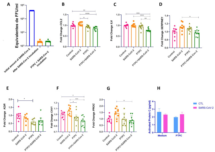
SARS-CoV-2 can induce vascular dysfunction and thrombotic events in patients with severe COVID-19; however, the cellular and molecular mechanisms behind these effects remain largely unknown. In this study, we used a combination of experimental and in silico approaches to investigate the role of PC in vascular and thrombotic events in COVID-19. Single-cell RNA-sequencing data from patients with COVID-19 and healthy subjects were obtained from the publicly available Gene Expression Omnibus (GEO) repository. In addition, HUVECs were treated with inactive protein C before exposure to SARS-CoV-2 infection or a severe COVID-19 serum. An RT-qPCR array containing 84 related genes was used, and the candidate genes obtained were evaluated. Activated protein C levels were measured using an ELISA kit. We identified at the single-cell level the expression of several pro-inflammatory and pro-coagulation genes in endothelial cells from the patients with COVID-19. Furthermore, we demonstrated that exposure to SARS-CoV-2 promoted transcriptional changes in HUVECs that were partly reversed by the activated protein C pretreatment. We also observed that the serum of severe COVID-19 had a significant amount of activated protein C that could protect endothelial cells from serum-induced activation. In conclusion, activated protein C protects endothelial cells from pro-inflammatory and pro-coagulant effects during exposure to the SARS-CoV-2 virus.

Keywords: SARS-CoV-2; bioinformatics; blood coagulation disorders; endothelial cell; inflammation.


Conflict of interest statement

The authors declare no conflicts of interest.

Figures

Figure 1
Figure 1
Overview of the study design. (A). Public data from single-cell RNA sequencing (scRNA-seq) studies on human pulmonary tissues were retrieved from the Gene Expression Omnibus (GEO) and European Genome-phenome Archive (EGA) under accession numbers GSE171668 and EGAS00001004344. (B). HUVEC of different passages pretreated or not with purified inactive human protein C for four hours at a concentration of 0.8 ng/μL. The upper panel shows HUVECs exposed to SARS-CoV-2 infection. Cells were grown in 12-well plates, with three wells per group—experimental N of 3. After incubation, the medium was collected for an ELISA test, and cell lysate was collected for subsequent RNA extraction and analysis by quantitative real-time PCR (RT-qPCR). The lower panel shows HUVECs incubated for 4 h with serum from patients with severe COVID-19. Cells were grown in 12-well plates, with two wells per group—experimental N of 4. After incubation, the medium was collected for an ELISA test, and the cell lysate was collected for subsequent RNA extraction and analysis by RT-qPCR.
Figure 2
Figure 2
Gene expression of endothelial damage and the PC pathway in pulmonary endothelial cells of healthy patients and patients with coronavirus disease 2019 (COVID-19) based on single-cell transcriptomics. (A). Heatmap showing genes expressed among healthy pulmonary endothelial subtypes. (B). Heatmap showing genes expressed among COVID-19 pulmonary endothelial subtypes. (C). Dot plot representation of genes expressed in healthy versus COVID-19 pulmonary endothelial cells.
Figure 3
Figure 3
PC pretreatment reduces the expression of genes related to inflammation and coagulation in HUVECs exposed to SARS-CoV-2 infection. (A). Viral quantification of SARS-CoV-2 using real-time PCR on the medium collected after viral incubation. (BG). HUVECs of different passages pretreated or not with purified inactive human protein C for 4 h at a concentration of 0.8 ng/μL and then exposed or not to SARS-CoV-2 infection. Cells were grown in 12-well plates, with three wells per group—experimental N of 3. The cells were washed with PBS before PC treatment, and a medium without FBS was used for the incubation. After the viral inoculum was removed, the medium was replaced with DMEM or MEM without FBS, and the cell lysate was collected for subsequent extraction and analysis of RNA by quantitative real-time PCR (RT-qPCR). One-way ANOVA followed by Tukey’s multiple comparison test was used for statistical analyses. * p ≤ 0.05, ** p < 0.01, *** p < 0.001, and **** p < 0.0001 compared with the groups indicated in the figure. (H). Activated protein C (aPC) levels were determined with an ELISA assay after incubation with SARS-CoV-2. The medium was collected after 24 h for the ELISA test. The two-way ANOVA followed by Tukey’s multiple-comparison test was used for statistical analyses. * p ≤ 0.05 compared with the indicated groups. Data are presented as mean ± s.e.m. PTPC: Pretreatment with PC. CTL: control—only cell culture medium was added.
Figure 4
Figure 4
aPC present in severe COVID-19 serum may protect ECs from increased expression of inflammatory and pro-coagulation genes. (AG). HUVECs of different passages pretreated or not with purified inactive human protein C for 4 h at a concentration of 0.8 ng/μL and then incubated with serum from a patient with severe COVID-19 or healthy serum at a concentration of 5%. Cells were grown in 12-well plates, with two wells per group—experimental N of 4. Cells were washed with PBS before PC treatment, and a medium without FBS was used for incubation. After serum incubation for 4 h, cell lysate was collected for subsequent extraction and analysis of RNA by quantitative real-time PCR (RT-qPCR). Two-way ANOVA followed by Tukey’s multiple comparison test was used for statistical analyses. * p ≤ 0.05, ** p < 0.01, *** p < 0.001 and **** p < 0.0001 compared with the groups indicated in the figure. Data are presented as mean  ±  s.e.m., with individual data points indicated. (HJ). Activated protein C (aPC) levels were determined after serum incubation with an ELISA assay. The medium was collected after 4 h for the ELISA test. Two-way ANOVA followed by Tukey’s multiple-comparison test was used for statistical analyses. * p ≤ 0.05 compared with the indicated groups. Data are presented as mean ± s.e.m. PTPC: Pretreatment with PC. CTL: control—only cell culture medium was added.
Figure 5
Figure 5
Panels comparing endothelial activation induced by SARS-CoV-2 and the cytoprotective effect of aPC. Left panel: The crucial role of endothelial cells in the pathogenesis of COVID-19 is related to the EC response to SARS-CoV-2 viral infection by detecting the adjacent infection and mounting a pro-inflammatory response to SARS-CoV-2 by stimulating circulating proteins. In addition, it is possible that even if direct infection in the ECs by SARS-CoV-2 does not occur, exposure to the virus is sufficient to promote transcriptional changes that lead to upregulation of pro-inflammatory and pro-coagulation genes and downregulation of cytoprotective signaling via Apc/PAR-1. Under stress conditions, serum aPC levels naturally rise in response to tissue damage; however, these levels may subsequently fall, allowing the already-known correlation of initially high serum aPC levels being associated with a worse prognosis in COVID-19. Right panel: The inactive PC might be stored in the EC (completing the PC reserve), and as soon as the cell is exposed to the virus, this protein promptly becomes activated and protects the vascular bed; consequently, its cytoprotective, anti-inflammatory, and anti-clotting signaling occurs more rapidly, thus actually protecting the cell from deleterious viral effects. PAR-1: Protease-activated receptor type 1; PC: Protein C; aPC: activated protein C; CCL2:C-C Motif Chemokine Ligand 2; ITGAM: Integrin Subunit Alpha M; VCAM1: vascular cell adhesion molecule 1; VWF: Von Willebrand factor.

References

    1. Xu Z., Shi L., Wang Y., Zhang J., Huang L., Zhang C., Liu S., Zhao P., Liu H., Zhu L. Pathological findings of COVID-19 associated with acute respiratory distress syndrome. Lancet Respir. Med. 2020;8:420–422. doi: 10.1016/S2213-2600(20)30076-X. - DOI - PMC - PubMed
    1. Yang X., Yu Y., Xu J., Shu H., Liu H., Wu Y., Zhang L., Yu Z., Fang M., Yu T. Clinical course and outcomes of critically ill patients with SARS-CoV-2 pneumonia in Wuhan, China: A single-centered, retrospective, observational study. Lancet Respir. Med. 2020;8:475–481. doi: 10.1016/S2213-2600(20)30079-5. - DOI - PMC - PubMed
    1. Charfeddine S., Ibn Hadj Amor H., Jdidi J., Torjmen S., Kraiem S., Hammami R., Bahloul A., Kallel N., Moussa N., Touil I. Long COVID-19 syndrome: Is it related to microcirculation and endothelial dysfunction? Insights from TUN-EndCOV study. Front. Cardiovasc. Med. 2021;8:745758. doi: 10.3389/fcvm.2021.745758. - DOI - PMC - PubMed
    1. Østergaard L. SARS-CoV-2 related microvascular damage and symptoms during and after COVID-19: Consequences of capillary transit-time changes, tissue hypoxia and inflammation. Physiol. Rep. 2021;9:e14726. doi: 10.14814/phy2.14726. - DOI - PMC - PubMed
    1. Wang M., Hao H., Leeper N.J., Zhu L. Thrombotic regulation from the endothelial cell perspectives. Arterioscler. Thromb. Vasc. Biol. 2018;38:e90–e95. doi: 10.1161/ATVBAHA.118.310367. - DOI - PMC - PubMed

Publication types

LinkOut - more resources