. 2024 Jul 16;16(7):1145.
doi: 10.3390/v16071145.

Chimeric Viruses Enable Study of Antibody Responses to Human Rotaviruses in Mice

Affiliations

Chimeric Viruses Enable Study of Antibody Responses to Human Rotaviruses in Mice

Sarah Woodyear et al. Viruses. .

Abstract

The leading cause of gastroenteritis in children under the age of five is rotavirus infection, accounting for 37% of diarrhoeal deaths in infants and young children globally. Oral rotavirus vaccines have been widely incorporated into national immunisation programs, but whilst these vaccines have excellent efficacy in high-income countries, they protect less than 50% of vaccinated individuals in low- and middle-income countries. In order to facilitate the development of improved vaccine strategies, a greater understanding of the immune response to existing vaccines is urgently needed. However, the use of mouse models to study immune responses to human rotavirus strains is currently limited as rotaviruses are highly species-specific and replication of human rotaviruses is minimal in mice. To enable characterisation of immune responses to human rotavirus in mice, we have generated chimeric viruses that combat the issue of rotavirus host range restriction. Using reverse genetics, the rotavirus outer capsid proteins (VP4 and VP7) from either human or murine rotavirus strains were encoded in a murine rotavirus backbone. Neonatal mice were infected with chimeric viruses and monitored daily for development of diarrhoea. Stool samples were collected to quantify viral shedding, and antibody responses were comprehensively evaluated. We demonstrated that chimeric rotaviruses were able to efficiently replicate in mice. Moreover, the chimeric rotavirus containing human rotavirus outer capsid proteins elicited a robust antibody response to human rotavirus antigens, whilst the control chimeric murine rotavirus did not. This chimeric human rotavirus therefore provides a new strategy for studying human-rotavirus-specific immunity to the outer capsid, and could be used to investigate factors causing variability in rotavirus vaccine efficacy. This small animal platform therefore has the potential to test the efficacy of new vaccines and antibody-based therapeutics.

Keywords: antibody; reverse genetics; rotavirus; vaccine.


Conflict of interest statement

The authors declare no conflicts of interest. The funders had no role in the design of the study, collection, analyses, interpretation of data, manuscript writing, or in the decision to publish the results.

Figures

Figure 1
Figure 1
Schematic diagram of segmented dsRNA genome of chimeric rotaviruses. Reverse genetics was used to generate chimeric viruses encoding either human or murine outer capsid proteins.
Figure 2
Figure 2
Virus shedding and clinical disease induced by oral infection of neonatal mice with chimeric rotaviruses compared to a human rotavirus strain. (A) Viral load detected in stool by qPCR (dotted line for lower limit of quantification). (B) Diarrhoea observed in neonatal mice infected at seven days old. Numbers in brackets in the key indicate the number of pups per litter.
Figure 3
Figure 3
Analysis of serum antibody responses in mice infected with chimeric rotaviruses. Mice were infected with chimeric rotaviruses (murine outer capsid as black circles, human outer capsid as orange squares) or Rotarix control (shown as purple triangles) at seven days old and serum samples were collected for antibody analysis at the timepoints shown in the schematic diagram (A). For all graphs, each point corresponds to an individual mouse; note that some samples were not available for all assays due to limited sample volumes. (B) Analysis of serum from 21-day-old mice by sandwich ELISA with primate lysate. (C) Longitudinal samples analysed by sandwich ELISA with primate rotavirus. (D) Western blot of SA11- and Rotarix-infected cell lysates, probed with sera from mice infected with either murine outer capsid rotavirus or human outer capsid rotavirus. (E) Samples from 10-week-old mice analysed by sandwich ELISA with human rotavirus (Rotarix strain). (F) Extracellular neutralisation of human rotavirus by serum samples from 10-week-old mice as quantified by fluorescent focus assay. (G) Intracellular neutralisation of human rotavirus by serum samples from 10-week-old mice as quantified by fluorescent focus assay. Dashed horizontal lines in Figure (AC) represent the positive threshold based on the OD450 of serum from uninfected control mice. Statistical significance was determined by one-way ANOVA (B), repeated measures ANOVA (C), or unpaired two-tailed t-tests (* p < 0.05; ** p < 0.01; **** p < 0.0001). Tukey’s adjusted pair-wise comparisons (B) and Bonferroni-corrected pair-wise comparisons (C) are shown.
Figure 4
Figure 4
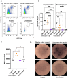
Analysis of B cell responses in mice infected with chimeric (murine outer capsid as black circles, human outer capsid as orange squares) or human Rotarix control (shown as purple triangles). Seven-day-old mice were infected with the panel of viruses, and B cell analysis was performed 14 days later. (A) Representative flow plots of germinal centre (GL7 + FAS+) B cells identified in Peyer’s patches (PPs) by flow cytometry. Numbers represent the percentage of total B220 + cells. (B) Quantification of germinal centres in PPs and MLNs by flow cytometry. (C) Quantification of spot forming unit (SFU) area by B cell ELISpot for Rotarix-specific B cells. (D) Representative images of B cell ELISpot wells. Statistical significance was determined by one-way ANOVA. Tukey’s adjusted pair-wise comparisons are shown for p < 0.05 (* p < 0.05; ** p < 0.01; *** p < 0.001; **** p < 0.0001).

References

    1. Ruiz-Palacios G.M., Pérez-Schael I., Velázquez F.R., Abate H., Breuer T., Clemens S.C., Cheuvart B., Espinoza F., Gillard P., Innis B.L., et al. Safety and Efficacy of an Attenuated Vaccine against Severe Rotavirus Gastroenteritis. N. Engl. J. Med. 2006;354:11–22. doi: 10.1056/NEJMoa052434. - DOI - PubMed
    1. Vesikari T., Matson D.O., Dennehy P., Van Damme P., Santosham M., Rodriguez Z., Dallas M.J., Heyse J.F., Goveia M.G., Black S.B., et al. Safety and Efficacy of a Pentavalent Human–Bovine (WC3) Reassortant Rotavirus Vaccine. N. Engl. J. Med. 2006;354:23–33. doi: 10.1056/NEJMoa052664. - DOI - PubMed
    1. Cates J.E., Tate J.E., Parashar U. Rotavirus vaccines: Progress and new developments. Expert Opin. Biol. Ther. 2022;22:423–432. doi: 10.1080/14712598.2021.1977279. - DOI - PMC - PubMed
    1. Desselberger U. Differences of Rotavirus Vaccine Effectiveness by Country: Likely Causes and Contributing Factors. Pathogens. 2017;6:65. doi: 10.3390/pathogens6040065. - DOI - PMC - PubMed
    1. Caddy S., Papa G., Borodavka A., Desselberger U. Rotavirus research: 2014–2020. Virus Res. 2021;304:198499. doi: 10.1016/j.virusres.2021.198499. - DOI - PubMed

Publication types

MeSH terms

LinkOut - more resources