Review
. 2024 Jul 3;34(7):bhae294.
doi: 10.1093/cercor/bhae294.

Dysfunction of motor cortices in Parkinson's disease

Affiliations
Review

Dysfunction of motor cortices in Parkinson's disease

Hong-Yuan Chu et al. Cereb Cortex. .

Abstract

The cerebral cortex has long been thought to be involved in the pathophysiology of motor symptoms of Parkinson's disease. The impaired cortical function is believed to be a direct and immediate effect of pathologically patterned basal ganglia output, mediated to the cerebral cortex by way of the ventral motor thalamus. However, recent studies in humans with Parkinson's disease and in animal models of the disease have provided strong evidence suggesting that the involvement of the cerebral cortex is much broader than merely serving as a passive conduit for subcortical disturbances. In the present review, we discuss Parkinson's disease-related changes in frontal cortical motor regions, focusing on neuropathology, plasticity, changes in neurotransmission, and altered network interactions. We will also examine recent studies exploring the cortical circuits as potential targets for neuromodulation to treat Parkinson's disease.

Keywords: Parkinson’s disease; basal ganglia; cerebral cortex; dopamine; pathophysiology.


Figures

Fig. 1
Fig. 1
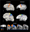
Regional maps of the localization of motor cortices. A–D) The lateral and medial surface of the human and macaque brains. Bottom) The localization of M1 and M2 cortices on the lateral surface and in coronal sections through the rostrocaudal axis of the mouse brain. The inset shows the abbreviations: CCZ: caudal cingulate zone, M1: primary motor cortex, M2: secondary motor cortex, PMC: pre-motor cortex, pSMA: presupplementary motor area, SMA: supplementary motor area. The lateral and sagittal views of the macaque and mouse brains are from: Scalable Brain Atlas - Neuroanatomy at your fingertips (incf.org). The coronal sections of the mouse brain are from the Allen Brain Atlas website.
Fig. 2
Fig. 2
Changes in cortical dopaminergic and glutamatergic innervation patterns in parkinsonian monkeys. A–D) TH-immunostained axonal profiles in the M1 of a control monkey (A) compared with a motor asymptomatic (B), mildly symptomatic (C), and symptomatic (D) MPTP-treated rhesus monkey. D’) Average optical density measurements of TH immunostaining in M1 in the 4 groups of monkeys used in this study. From left to right, the bars represent values from each animal in the order depicted in A–D) (see Masilamoni et al. 2022 for more details). E, F) vGluT2-immunostained profiles in M1 of a control and an MPTP-treated parkinsonian monkey (E), along with the corresponding optical density measurements in layer II–III and Vb (F). Note the significant reduction in vGluT2 labeling in layer Vb of the MPTP-treated monkey (see Villalba et al. 2021 for more details). G, H) Confocal images of vGluT2-immunolabled terminal profiles in layer V of M1 from a control and a 6-OHDA-treated mouse (G) and summarized results (see Chen et al. 2023 for details). A–F) are from Villalba et al. 2021, while F) and G) are from Chen et al. 2023. Both parts of this figure are used with permission.
Fig. 3
Fig. 3
Dendritic spine pathology and myelination changes in parkinsonian mice. A–D) Spine dynamics in M1 in MPTP-treated mice. Neurons were identified by expression of yellow fluorescent protein (Thy1-YFP-H line) using trans-cranial two-photon laser scanning microscopy to study the growth or pruning of dendritic spines over time in controls and parkinsonian mice. A) illustrates the timeline of treatment and imaging. B) depicts imaged spines at 0, 4, and 8 days in control and MPTP-treated mice. Arrows: spines eliminated; arrowheads: spines formed; asterisks: filopodia. Scale bar: 2 mm. C, D) Bar graphs showing the spines eliminated (C) or formed (D) at different time points (as shown in A). See Guo et al. (2015) for more details. E, F) Myelin and axonal pathologies in white matter underlying the primary motor cortex in advanced PD cases. E) shows immunofluorescence labeling of axons (NFL, left panel), alpha-synuclein-containing neurites (P-αsyn, middle panel), and myelin basic protein (MBP, right panel). The white arrowheads indicate segments of axons in which P-αsyn displaced the longitudinal NFL and MBP labeling, suggesting myelination damage of some axonal profiles. The blue arrowheads point at P-αsyn+/NFL+/MBP+ axonal segments. F) shows negative Spearman rank correlations between the levels of myelin proteins (MBP and myelin proteolipid protein-PLP) and α-synuclein in PD cases. Gray zones indicate 95% confidence intervals that are automatically calculated using the predicted values for the line of best fit (see (Fu et al. 2022) for more details). Figure 2A–D reproduced from Guo et al. (2015), and E) and F) appeared in Fu et al. (2022). Both components are used with permission.
Fig. 4
Fig. 4
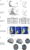
Examples of recordings of electrophysiologic and magnetoencephalographic recordings of abnormal M1 activity in the parkinsonian state. A) In vivo single-cell recordings in M1 of parkinsonian NHPs (A1) and in brain slices of parkinsonian mice (A2) demonstrate that corticostriatal neurons show no changes in firing rates (primates) (A1, top) or the spiking response to current injections (their excitability) (A2, top), while PT neurons fire at lower rates (A1, bottom), and are less excitable (A2, bottom). B) In vivo multielectrode recordings in M1 in normal and parkinsonian NHPs demonstrate that cortical neurons show a greater tendency to discharge in synchronized bursts. C) The images show an analysis of recordings of ECoGs in parkinsonian patients, made while the patients underwent DBS lead placement procedures. Shown is the (color-coded) modulation index, a measure of the coupling between the phase of low-frequency oscillations and the amplitude of high-frequency oscillations. Parkinsonian patients show strong phase-amplitude coupling between beta- and gamma oscillatory ECoG activity that is ameliorated by DBS. D) The figure shows an oscillatory network associated with parkinsonian resting tremor, as identified with MEG and dynamic imaging of coherent sources analysis. As demonstrated, M1 (circle B) is coupled to other cortical areas, including the lateral premotor cortex (circle A) and the SMA (circle C), with diencephalic areas (circle D), and with the cerebellum (circle E). Part A1 of the figure reproduced from Pasquereau and Turner (2011), part A2 reproduced from Chen et al. (2023), part B comes from Goldberg et al. (2002), and part C from Hemptinne et al. (2015) and part D from Timmermann et al. (2007). All figures are used with permission. Part B is covered by copyright 2002, Society for Neuroscience.
Fig. 5
Fig. 5
Examples of imaging studies in patients with parkinsonism. Left and middle panels: Increased FDG uptake in sensorimotor cortex of PD patients compared to healthy controls. Right panel: A significant positive correlation between sensorimotor cortical FDG uptake and UPDRS III score in PD patients. From Zang et al. (2022) and used with permission.
Fig. 6
Fig. 6
Spectrogram of LFPs in simulated M1. A) Control state. B) Parkinsonian state (modified from Doherty et al. 2024).
Fig. 7
Fig. 7
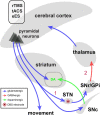
Simplified circuit diagram showing potential mechanisms of cortical neuromodulation for PD treatment. Cortical pyramidal neurons and local circuits can be functionally modulated by electric stimulation of basal ganglia structures (e.g. the STN) via 1) antidromic and 2) orthodromic circuits, by the less invasive direct rTMS, tACS modalities, or by epidural electric stimulation (eES) of the motor cortex. Concentric circles inside the STN indicate the DBS electrode.

Similar articles

Cited by

References

    1. Abbasi O, Hirschmann J, Storzer L, Ozkurt TE, Elben S, Vesper J, Wojtecki L, Schmitz G, Schnitzler A, Butz M. Unilateral deep brain stimulation suppresses alpha and beta oscillations in sensorimotor cortices. NeuroImage. 2018:174:201–207. 10.1016/j.neuroimage.2018.03.026. - DOI - PubMed
    1. Albin RL, Young AB, Penney JB. The functional anatomy of basal ganglia disorders. Trends Neurosci. 1989:12(10):366–375. 10.1016/0166-2236(89)90074-X. - DOI - PubMed
    1. Albin RL, Koeppe RA, Bohnen NI, Wernette K, Kilbourn MA, Frey KA. Spared caudal brainstem SERT binding in early Parkinson's disease. J Cereb Blood Flow Metab. 2008:28(3):441–444. 10.1038/sj.jcbfm.9600599. - DOI - PubMed
    1. Albin RL, Zee S, Laar T, Sarter M, Lustig C, Muller M, Bohnen NI. Cholinergic systems, attentional-motor integration, and cognitive control in Parkinson's disease. Prog Brain Res. 2022:269:345–371. 10.1016/bs.pbr.2022.01.011. - DOI - PMC - PubMed
    1. Andersen KB, Hansen AK, Sommerauer M, Fedorova TD, Knudsen K, Vang K, Van Den Berge N, Kinnerup M, Nahimi A, Pavese N, et al. . Altered sensorimotor cortex noradrenergic function in idiopathic REM sleep behaviour disorder - a PET study. Parkinsonism Relat Disord. 2020:75:63–69. 10.1016/j.parkreldis.2020.05.013. - DOI - PubMed

Publication types

MeSH terms