. 2024 Oct 1;201(2):254-262.
doi: 10.1093/toxsci/kfae092.

Copper-mediated neurotoxicity and genetic vulnerability in the background of neurodegenerative diseases in C. elegans

Affiliations

Copper-mediated neurotoxicity and genetic vulnerability in the background of neurodegenerative diseases in C. elegans

Ann-Kathrin Weishaupt et al. Toxicol Sci. .

Abstract

The mechanisms associated with neurodegenerative diseases, such as Alzheimer's disease (AD) and Parkinson's disease (PD), have yet to be fully characterized, and genetic as well as environmental factors in their disease etiology are underappreciated. Although mutations in genes such as PARKIN and LRRK2 have been linked to PD, the idiopathic component of the disease suggests a contribution of environmental risk factors, including metals, such as copper (Cu). Cu overexposure has been reported to cause oxidative stress and neurotoxicity, but its role in neurodegenerative diseases is rarely studied. Using Caenorhabditis elegans (C. elegans) as a model organism for neurotoxicity, we assessed the effects of Cu oversupply in AD and PD models. Our findings reveal that although copper treatment did not induce neurodegeneration in wild-type worms or the AD model, it significantly exacerbated neurodegeneration in the PD-associated mutants PARKIN and LRRK2. These results suggest that genetic predisposition for PD enhances the sensitivity to copper toxicity, highlighting the multifactorial nature of neurodegenerative diseases. Furthermore, our study provides insight into the mechanisms underlying Cu-induced neurotoxicity in PD models, including disruptions in dopamine levels, altered dopamine-dependent behavior and degraded dopaminergic neurons. Overall, our novel findings contribute to a better understanding of the complex interactions between genetic susceptibility, environmental factors, and neurodegenerative disease pathogenesis, emphasizing the importance of a tightly regulated Cu homeostasis in the etiology of PD. Copper oversupply exacerbated neurodegeneration in Caenorhabditis elegans models of Parkinson's disease, highlighting the genetic susceptibility and emphasizing the crucial role of tightly regulated copper homeostasis in Parkinson's disease pathogenesis.

Keywords: C. elegans; copper toxicity; genetic predisposition; neurodegenerative diseases.


Figures

Fig. 1.
Fig. 1.
a) Total Cu levels [ng/µg protein] and b) survival [%] following 24 h of Cu incubation in wild-type worms, pdr-1Δ, g2019s, and CL2006 mutants. Data presented are mean values of n = 4 experiments ± SEM. Statistical analysis using 2-way ANOVA test with Tukey’s multiple comparison. Significance level with α  =  0.05: *: P ≤ 0.05; ***: P ≤ 0.001 compared with untreated control.
Fig. 2.
Fig. 2.
Relative levels of a) reduced GSH and b) oxidized GSSG normalized to untreated wild-type control [%] following 24 h Cu treatment quantified by LC-MS/MS. Data presented are mean values of n ≥ 4 independent experiments + SEM. Statistical analysis using 2-way ANOVA test with Tukey’s multiple comparison. Significance level with α = 0.05: *: P ≤ 0.05; **: P ≤ 0.01 and ***: P ≤ 0.001 compared with untreated control and §: P ≤ 0.05 and §§§: P ≤ 0.001 compared with wild-type in the same condition.
Fig. 3.
Fig. 3.
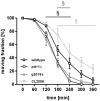
Aldicarb-induced paralysis assay in untreated worms. Plotted is the fraction of moving worms [%] against the assay procedure time [min]. Data presented are mean values of n = 4 independent and blinded experiments ± SEM. Statistical analysis using 2-way ANOVA test with Tukey’s multiple comparison. Significance level with α = §: P ≤ 0.05 compared with wild-type in same condition.
Fig. 4.
Fig. 4.
Neurotransmitter levels of a) DA, b) SRT, c) GABA and d) ACh in ng per mg protein following Cu treatment. Data presented are mean values of n ≥ 4 independent experiments + SEM. Statistical analysis using 2-way ANOVA test with Tukey’s multiple comparison. Significance level with α = 0.05: *: P ≤ 0.05 compared with untreated control and §§: P ≤ 0.01 and §§§: P ≤ 0.001 compared with wild-type in the same condition.
Fig. 5.
Fig. 5.
Basal slowing response following Cu treatment. The difference between body bends per 20 s on uncoated plates and body bends per 20 s on coated plates (Δ body bends/20 s) was assessed. cat-2Δ worms, which are restricted in DA synthesis, are used as a positive control (C+). Data presented are mean values of n = 3 independent experiments + SEM. Statistical analysis using 2-way ANOVA test with Tukey’s multiple comparison. Significance level with α  =  0.05: *: P ≤ 0.05 and ***: P ≤ 0.001 compared with untreated control and §§: P ≤ 0.01 and §§§: P ≤ 0.001 compared with wild-type in the same condition.
Fig. 6.
Fig. 6.
a) Schematic overview of the dopaminergic neurons (ADE and CEP) in C. elegans’ head region which can be visualized by fluorescence microscopy in the BY200 strain. b) Exemplary images show different events of the neurons morphology marked by arrows: no alterations, irregularities (kinks and bends of dendrites, shrunken soma) and severe damage (loss of dendrites or soma). c) 6-OHDA (25 mM) was used as a positive control in wild-type worms. d) Exemplary fluorescence images of wild-type worms (BY200), pdr-1Δ and g2019s (which were each crossed with strain BY200). Images display either untreated worms or supplemented with 2 mM Cu for 24 h. e) Overview of the number of events [%] counted in N = 5–10 worms per group. Data presented are mean values of n = 3 independent experiments.

References

    1. Alvarez J, Alvarez-Illera P, Santo-Domingo J, Fonteriz RI, Montero M. 2022. Modeling Alzheimer’s disease in Caenorhabditis elegans. Biomedicines. 10(2):288. 10.3390/biomedicines10020288. - DOI - PMC - PubMed
    1. An Y, Li S, Huang X, Chen X, Shan H, Zhang M. 2022. The role of copper homeostasis in brain disease. Int J Mol Sci. 23(22):13850. 10.3390/ijms232213850. - DOI - PMC - PubMed
    1. Bagheri S, Squitti R, Haertlé T, Siotto M, Saboury AA. 2017. Role of copper in the onset of Alzheimer’s disease compared to other metals. Front Aging Neurosci. 9:446. 10.3389/fnagi.2017.00446. - DOI - PMC - PubMed
    1. Bloem BR, Okun MS, Klein C. 2021. Parkinson’s disease. Lancet. 397(10291):2284–2303. 10.1016/S0140-6736(21)00218-X. - DOI - PubMed
    1. Boll M-C, Alcaraz-Zubeldia M, Montes S, Rios C. 2008. Free copper, ferroxidase and SOD1 activities, lipid peroxidation and NO(x) content in the CSF. A different marker profile in four neurodegenerative diseases. Neurochem Res. 33(9):1717–1723. 10.1007/s11064-008-9610-3. - DOI - PubMed

MeSH terms

Substances

LinkOut - more resources