. 2024 Oct 17;31(10):1815-1826.e5.
doi: 10.1016/j.chembiol.2024.06.016. Epub 2024 Jul 26.

Dissecting the neuroprotective interaction between the BH4 domain of BCL-w and the IP3 receptor

Affiliations

Dissecting the neuroprotective interaction between the BH4 domain of BCL-w and the IP3 receptor

Sophia X Tang et al. Cell Chem Biol. .

Abstract

BCL-w is a BCL-2 family protein that promotes cell survival in tissue- and disease-specific contexts. The canonical anti-apoptotic functionality of BCL-w is mediated by a surface groove that traps the BCL-2 homology 3 (BH3) α-helices of pro-apoptotic members, blocking cell death. A distinct N-terminal portion of BCL-w, termed the BCL-2 homology 4 (BH4) domain, selectively protects axons from paclitaxel-induced degeneration by modulating IP3 receptors, a noncanonical BCL-2 family target. Given the potential of BCL-w BH4 mimetics to prevent or mitigate chemotherapy-induced peripheral neuropathy, we sought to characterize the interaction between BCL-w BH4 and the IP3 receptor, combining "staple" and alanine scanning approaches with molecular dynamics simulations. We generated and identified stapled BCL-w BH4 peptides with optimized IP3 receptor binding and neuroprotective activities. Point mutagenesis further revealed the sequence determinants for BCL-w BH4 specificity, providing a blueprint for therapeutic targeting of IP3 receptors to achieve neuroprotection.

Keywords: BCL-2 family; BCL-w; BH4 domain; IP3 receptor; axon degeneration; chemotherapy induced peripheral neuropathy; neuroprotection; paclitaxel; stapled peptide.


Conflict of interest statement

Declaration of interests M.F.P.-M., G.H.B., R.A.S., and L.D.W. are named co-inventors on international patent application WO 2018/039545 (and associated patent applications and granted national patents) related to this work.

Figures

Figure 1.
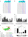
Figure 1.. Binding activities of a stapled BCL-w BH4 library to endogenous and recombinant IP3R1
(A) The BH4 domain of BCL-w (cyan) is composed of the α1 helix and a portion of the unstructured loop between α1 and α2, with amino acids 11–28 of the helical portion subjected to all-hydrocarbon stapling. (B) Structure of the IP3 receptor type 1 (IP3R1) depicting its structural and functional domains. Domain 3, which has been implicated as a cytosolic surface site of BCL-2 and BCL-XL BH4 interaction, is highlighted in pink. (C and D) Compositions of i, i+4 (C) and i, i+7 (D) staple scanning libraries of the BCL-w BH4 domain sequence. The stapling amino acids are indicated as X for S-pentenylalanine and 8 for R-octenylalanine, and colored in blue. (E) Helical wheel depiction of the helical portion of the BCL-w BH4 domain, highlighting regions of hydrophobicity (yellow), positive (blue) and negative (red) charge, and hydrophilic residues (green). (F and G) Cerebellar lysates were incubated with biotinylated and i, i+4 (F) or i, i+7 (G) stapled BCL-w BH4 peptides followed by streptavidin pull-down, gel electrophoresis, and anti-IP3R1 western blot. Band intensities were quantified by ImageJ analysis and normalized to the band of highest intensity for plotting. Error bars are mean ± SEM for experiments performed in triplicate with three independent preparations of protein and peptides. (H) The influence of stapling at a particular amino acid position of the BCL-w BH4 peptide is depicted by red, yellow, or green color, whereby an adverse “hit” is scored if a staple that begins or ends at a particular residue results in 25% or less endogenous IP3R1 pull-down activity. Residues with 2–3, 1, or no adverse “hits” are colored red, yellow, and green respectively, with stick representations shown for red and yellow hits. (I and J) Recombinant IP3R1 D3 protein was incubated with biotinylated and i, i+4 (H) or i, i+7 (I) stapled BCL-w BH4 peptides followed by streptavidin pull-down, gel electrophoresis, and anti-GST western blot. Band intensities were quantified by ImageJ analysis and normalized to the band of highest intensity for plotting. Error bars are mean ± SEM for experiments performed in triplicate with three independent preparations of protein and peptides. (K) The influence of stapling at a particular amino acid position of the BCL-w BH4 peptide is depicted by red, yellow, or green color, whereby an adverse “hit” is scored if a staple that begins or ends at a particular residue results in 25% or less recombinant IP3R1 D3 pull-down activity. Residues with 2–3, 1, or no adverse “hits” are colored red, yellow, and green respectively, with stick representations shown for red and yellow hits. See also Figures S1–S3, Videos S1–S7, and Data S1
Figure 2.
Figure 2.. Molecular Dynamics Simulations of the Interaction between IP3R1 D3 and the BH4 Domain of BCL-w
(A) Sample conformer of the BCL-w BH4 domain based on a molecular dynamics simulation of amino acid residues 11–28. (B) Clustering of coarse-grained simulation trajectories that capture binding of the BCL-w BH4 peptide to the ARM2 region of IP3R1 (amino acids 1200–1566). (C) A low energy conformer of the BCL-w BH4 helix (cyan) at a surface groove (gold) of the IP3R1 ARM2 domain (navy blue). (D-E) Contact probabilities of BCL-w BH4 (D) and IP3R1 ARM2 domain (E) residues. Data are aggregated over 80 ns of an all-atom, equilibrium, molecular dynamics simulation and aggregated for three different trajectories. (F) Hydrophobic interactions at the BCL-w BH4/IP3R1 helix-in-groove interface. (G) Hydrophilic and electrostatic interactions at the BCL-w BH4/IP3R1 helix-in-groove interface. (H) Relative location of the BCL-w BH4 binding site (gold) at a surface groove of the ARM2 domain, within the cytosolic portion of IP3R1 (grey). See also Videos S8–9.
Figure 3.
Figure 3.. Point Mutagenesis Reveals Key Binding Determinants for BCL-w BH4/IP3R1-ARM2 Interaction
(A and B) Sequence compositions of the alanine scanning libraries of the i, i+4 stapled SAH-BCL-w BH4-12 (A) and i, i+7 SAH-BCL-w BH4-27 (B) peptides. Alanine residues are highlighted in pink and the stapling amino acids S-pentenyl alanine and R-octenyl alanine are labeled X and 8, respectively and colored in blue. (C and D) Recombinant GST-IP3R1 D3 protein was incubated with biotinylated and i, i+4 (C) or i, i+7 (D) stapled BCL-w BH4 peptides followed by streptavidin pull-down, gel electrophoresis, and anti-GST western blot. Band intensities were quantified by ImageJ analysis and normalized to the band of highest intensity for plotting. Error bars are mean ± SEM for experiments performed in triplicate with three independent preparations of protein and peptides. (E) The influence of alanine mutagenesis at a particular amino acid position of the BCL-w BH4 peptide is depicted by red (at or below a 25% binding threshold in one or both datasets), yellow (at or below a 50% binding threshold in one or both datasets), or green color (above 50% binding activity across both datasets). (F) Sequence compositions of BCL-w and BCL-2 BH4 peptides bearing the indicated staples and point mutations. Gold, hydrophobic; navy blue, positively charged; red, negatively charged; green, hydrophilic; royal blue X, stapling amino acid. (G) Cerebellar lysates were incubated with the indicated peptides followed by streptavidin pull-down, gel electrophoresis, and anti-IP3R1 western blot. Band intensities were quantified by ImageJ analysis and normalized to the band of highest intensity for plotting. Error bars are mean ± SEM for experiments performed in triplicate with three independent preparations of protein and peptides. Statistical significance was calculated by one-way ANOVA with Bonferroni’s multiple comparisons test, where p is *** < 0.001. See also Figure S4 and Data S1
Figure 4.
Figure 4.. Sequence-specific BH4 Protection from Paclitaxel-induced Axonal Degeneration
(A) Quantitation of axonal degeneration in response to paclitaxel (30 nM, 24 h), with or without 2 h pretreatment with the indicated SAH-BCL-w BH4 peptides (10 nM). The axonal degeneration index reflects the ratio of the area of fragmented axons to total axon area of E15 DRG neurons grown in compartmentalized cultures. Data are mean ± SEM for five independent experiments (27–29 images each) with individual data points plotted. Statistical significance was calculated by one-way ANOVA with Bonferroni’s multiple comparisons test, where p is *** < 0.001, or ns (not significant) for p > 0.05. (B) Binarized images of Tuj1 immunostained axons treated in compartmentalized cultures as indicated. Scale bar, 20 μm. (C) Quantitation of axonal degeneration in response to paclitaxel (30 nM, 24 h), with or without 2 h pretreatment with the indicated SAH-BCL-w or SAH-BCL-2 BH4 peptides (10 nM). Data are mean ± SEM for four independent experiments (16–18 images each) with individual data points plotted. Statistical significance was calculated by one-way ANOVA with Bonferroni’s multiple comparisons test, where p is ** < 0.01, *** < 0.001, or ns (not significant) for p > 0.05. (D) Binarized images of Tuj1 immunostained axons treated in compartmentalized cultures as indicated. Scale bar, 20 μm. See also Figure S5 and Data S1

References

    1. Danial NN, and Korsmeyer SJ (2004). Cell death: critical control points. Cell 116, 205–219. 10.1016/s0092-8674(04)00046-7. - DOI - PubMed
    1. Sattler M, Liang H, Nettesheim D, Meadows RP, Harlan JE, Eberstadt M, Yoon HS, Shuker SB, Chang BS, Minn AJ, et al. (1997). Structure of Bcl-xL-Bak peptide complex: recognition between regulators of apoptosis. Science 275, 983–986. 10.1126/science.275.5302.983. - DOI - PubMed
    1. Cheng EH, Wei MC, Weiler S, Flavell RA, Mak TW, Lindsten T, and Korsmeyer SJ (2001). BCL-2, BCL-X(L) sequester BH3 domain-only molecules preventing BAX- and BAK-mediated mitochondrial apoptosis. Mol Cell 8, 705–711. 10.1016/s1097-2765(01)00320-3. - DOI - PubMed
    1. Gavathiotis E, Suzuki M, Davis ML, Pitter K, Bird GH, Katz SG, Tu HC, Kim H, Cheng EH, Tjandra N, and Walensky LD (2008). BAX activation is initiated at a novel interaction site. Nature 455, 1076–1081. 10.1038/nature07396. - DOI - PMC - PubMed
    1. Denisov AY, Madiraju MS, Chen G, Khadir A, Beauparlant P, Attardo G, Shore GC, and Gehring K (2003). Solution structure of human BCL-w: modulation of ligand binding by the C-terminal helix. J Biol Chem 278, 21124–21128. 10.1074/jbc.M301798200. - DOI - PubMed

MeSH terms

Substances

LinkOut - more resources