. 2025 Jun:72:615-632.
doi: 10.1016/j.jare.2024.07.022. Epub 2024 Jul 25.

A novel drug prejudice scaffold-imidazopyridine-conjugate can promote cell death in a colorectal cancer model by binding to β-catenin and suppressing the Wnt signaling pathway

Affiliations

A novel drug prejudice scaffold-imidazopyridine-conjugate can promote cell death in a colorectal cancer model by binding to β-catenin and suppressing the Wnt signaling pathway

Min Hee Yang et al. J Adv Res. 2025 Jun.

Abstract

Introduction: Globally, colorectal cancer (CRC) is the third most common type of cancer, and its treatment frequently includes the utilization of drugs based on antibodies and small molecules. The development of CRC has been linked to various signaling pathways, with the Wnt/β-catenin pathway identified as a key target for intervention.

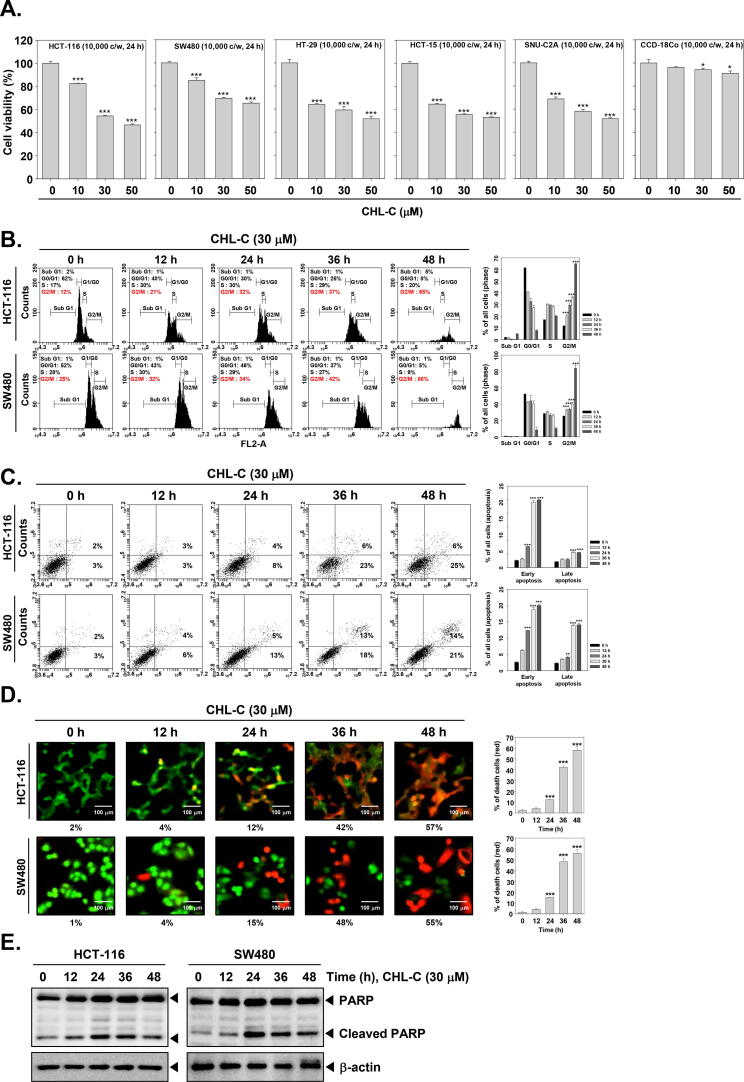
Objectives: We have explored the impact of imidazopyridine-tethered chalcone-C (CHL-C) in CRC models.

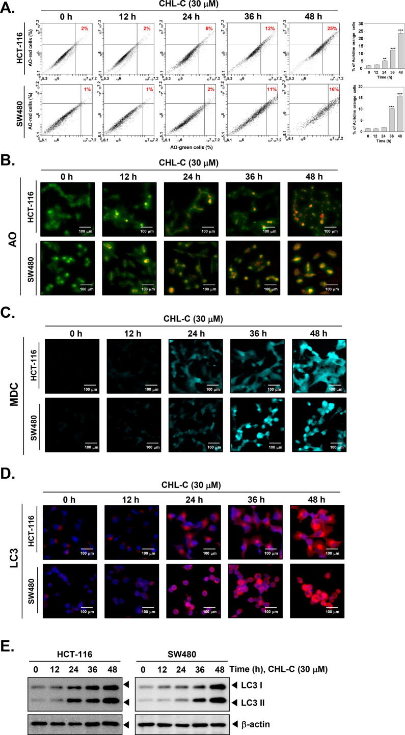
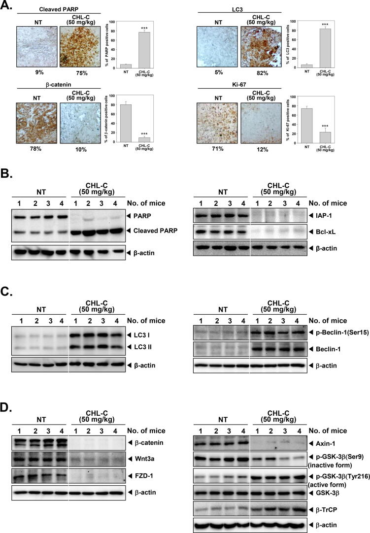
Methods: To determine the influence of CHL-C on apoptosis and autophagy, Western blot analysis, annexin V assay, cell cycle analysis, acridine orange staining, and immunocytochemistry were performed. Next, the activation of the Wnt/β-catenin signaling pathway and the anti-cancer effects of CHL-C in vivo were examined in an orthotopic HCT-116 mouse model.

Results: We describe the synthesis and biological assessment of the CHL series as inhibitors of the viability of HCT-116, SW480, HT-29, HCT-15, and SNU-C2A CRC cell lines. Further biological evaluations showed that CHL-C induced apoptosis and autophagy in down-regulated β-catenin, Wnt3a, FZD-1, Axin-1, and p-GSK-3β (Ser9), and up-regulated p-GSK3β (Tyr216) and β-TrCP. In-depth analysis using structure-based bioinformatics showed that CHL-C strongly binds to β-catenin, with a binding affinity comparable to that of ICG-001, a well-known β-catenin inhibitor. Additionally, our in vivo research showed that CHL-C markedly inhibited tumor growth and triggered the activation of both apoptosis and autophagy in tumor tissues.

Conclusion: CHL-C is capable of inducing apoptosis and autophagy by influencing the Wnt/β-catenin signaling pathway.

Keywords: Apoptosis; Autophagy; CHL-C; Colorectal cancer; Wnt/ β −catenin.


Conflict of interest statement

Declaration of competing interest The authors declare that they have no known competing financial interests or personal relationships that could have appeared to influence the work reported in this paper.

Figures

None
Graphical abstract
Scheme 1
Scheme 1
Synthesis of imidazopyridine tethered chalcones.
Fig. 1
Fig. 1
Reported inhibitors of the Wnt/β-catenin pathway and currently synthesized chalcones.
Fig. 2
Fig. 2
CHL-C induces apoptosis in human colorectal carcinoma cells. (A) Human colorectal carcinoma HCT-116, SW480, HT-29, HCT-15, SNU-C2A, and CCD-18Co cells were treated with CHL-C (0, 10, 30, 50 µM) for 24 h and cell viability was examined through MTT assay. Data represents mean ± SD. ***p < 0.001 vs. non-treated (NT) cells. (B-D) HCT-116 and SW480 cells were treated with 30 µM for CHL-C (0, 12, 24, 36, 48 h). Apoptotic cells were analyzed using cell cycle analysis, Annexin V assay, and live and dead assay. (E) The cells were treated as described above in panel B, and Western blot analysis for PARP and LC3 was carried out. β-actin was used as a loading control. The results shown are representative of the three independent experiments.
Fig. 3
Fig. 3
CHL-C induces autophagy in HCT-116 and SW480 cells. HCT-116 and SW480 cells were treated with CHL-C (30 µM) for 12, 24, 36, 48 h. (A and B) After CHL-C treatment, cells were stained with acridine orange and detected by flow cytometer and confocal microscope. Data represents mean ± SD. **p < 0.01 vs. non-treated (NT) cells, ***p < 0.001 vs. non-treated (NT) cells. (C) The cells were stained with Monodansylcadaverine (MDC) and detected using a confocal microscope. (D) Immunocytochemistry for LC3 distribution was performed. (E) Whole-cell lysates were prepared, and Western blotting for LC3 was performed. The results shown are representative of the three independent experiments. (For interpretation of the references to colour in this figure legend, the reader is referred to the web version of this article.)
Fig. 4
Fig. 4
CHL-C down-regulates the expression of various tumorigenic proteins and autophagy markers. (A and B) HCT-116 and SW480 cells were treated with CHL-C (30 μM) for indicated time intervals, and Western blotting for caspase-9, caspase-3, IAP-1, Bcl-2, and Bcl-xL was carried out. (C) Cells were pretreated with Z-DEVD-FMK (30 μM) for 30 min, and exposed to CHL-C (30 μM) for 48 h. Then, the annexin V assay was performed. (D) The cells were treated as described above in panel A, and Western blotting for various autophagy-related markers was carried out. (E) Cells were pretreated with 3-MA (3 mM) for 30 min, and exposed to CHL-C (30 μM) for 48 h. Then, an acridine orange assay was carried out. (For interpretation of the references to colour in this figure legend, the reader is referred to the web version of this article.)
Fig. 5
Fig. 5
In silico analysis of CHL-C. (A) Cartoon representation of docked CHL-C compound to the binding pocket of β-catenin. (B and C) Key interactions of compound CHL-C and ICG-001 with β-catenin. (D) Surface image of β-catenin along with docked compounds CHL-C (Black) & ICG-001 (Yellow) and its enlarged view for better visualization. (For interpretation of the references to colour in this figure legend, the reader is referred to the web version of this article.)
Fig. 6
Fig. 6
CHL-C induces cell death through β-catenin down-regulation. (A) Human colorectal carcinoma cells were treated with CHL-C in a time-dependent manner. The expression of Wnt/β-catenin signaling cascades-related proteins was analyzed using Western blotting. (B) HCT-116 and SW480 cells were treated as described above in panel A. Then, GSK3 β activity was analyzed using GSK3 β kinase enzyme system. (C and D) HCT-116 and SW480 cells were transfected with β-catenin or scrambled siRNA (50 nM) for 24 h. After that, cells were treated with CHL-C (30 µM) for 48 h. Western blotting for various proteins was carried out. (E) β-catenin or scrambled siRNA-transfected cells were treated with CHL-C (30 µM) for 48 h. Apoptotic cell death was analyzed using live and dead assays. (F) Cells were transfected and treated as described above in panel B, and immunocytochemistry for LC3 was carried out.
Fig. 7
Fig. 7
CHL-C attenuates tumorigenesis in preclinical model. (A) HCT-116-Luc cells-derived tumor tissues are orthotopically placed into the liver with a subsequent intraperitoneal administration of 0.1 % DMSO or CHL-C (50 mg) thrice a week, for three weeks. (B) The graph represents the body weight of mice. (C and D) Tumor burden was measured in vehicle-treated or CHL-C-treated tumor-bearing mice.
Fig. 8
Fig. 8
CHL-C alters the expression of oncogenic markers in tissues. (A) Immunohistochemistry was performed for cleaved PARP, LC3, β−catenin, and Ki-67. Data represents mean ± SD. ***p < 0.001 vs. non-treated (NT) cells. (B) The expression of apoptosis-related markers is analyzed through Western blotting. (C) Western blotting for LC3, p-Beclin-1(Ser15), and Beclin-1 was done. (D) The level of Wnt/ β−catenin signaling cascade-related proteins is analyzed using western blot analysis.
Fig. 9
Fig. 9
(A) The plot illustrates the stable connection between ligand CHL-C and β-catenin over 150 ns. The average deviation (RMSD) is 3.5 Å for the protein and 5.1 Å for the ligand. (B) The protein–ligand complex demonstrates overall stability with most residues below 2.5 Å RMSF, except for significant fluctuations (exceeding 3 Å) in the loop region. (C) Bar graph showing the interaction, contact, and fraction folds of CHL-C with residues of β-catenin.

References

    1. Siegel R.L., Wagle N.S., Cercek A., Smith R.A., Jemal A. Colorectal cancer statistics, 2023. CA Cancer J Clin. 2023;73(3):233–254. - PubMed
    1. Hani U., Honnavalli Y.K., Begum M.Y., Yasmin S., Osmani R.A.M., Ansari M.Y. Colorectal cancer: A comprehensive review based on the novel drug delivery systems approach and its management. J Drug Deliv Sci Tec. 2021:63.
    1. Pricci M., Girardi B., Giorgio F., Losurdo G., Ierardi E., Di Leo A. Curcumin and colorectal cancer: From basic to clinical evidences. Int J Mol Sci. 2020;21(7) - PMC - PubMed
    1. Blondy S., David V., Verdier M., Mathonnet M., Perraud A., Christou N. 5-Fluorouracil resistance mechanisms in colorectal cancer: From classical pathways to promising processes. Cancer Sci. 2020;111(9):3142–3154. - PMC - PubMed
    1. Cho Y.H., Ro E.J., Yoon J.S., Mizutani T., Kang D.W., Park J.C., et al. 5-FU promotes stemness of colorectal cancer via p53-mediated WNT/beta-catenin pathway activation. Nat Commun. 2020;11(1):5321. - PMC - PubMed

MeSH terms

LinkOut - more resources