. 2024 Jul 27;15(1):6338.
doi: 10.1038/s41467-024-50286-0.

Multi-compartmental diversification of neutralizing antibody lineages dissected in SARS-CoV-2 spike-immunized macaques

Affiliations

Multi-compartmental diversification of neutralizing antibody lineages dissected in SARS-CoV-2 spike-immunized macaques

Marco Mandolesi et al. Nat Commun. .

Abstract

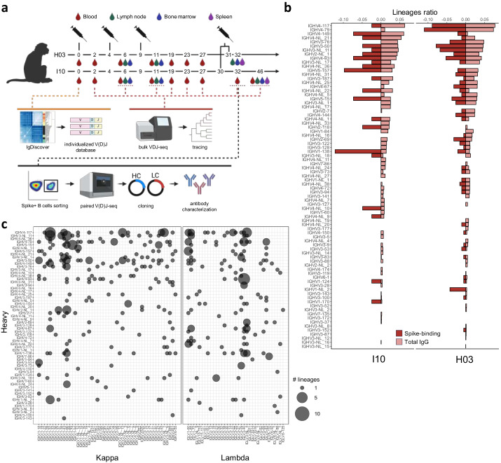
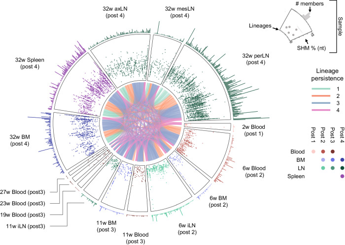
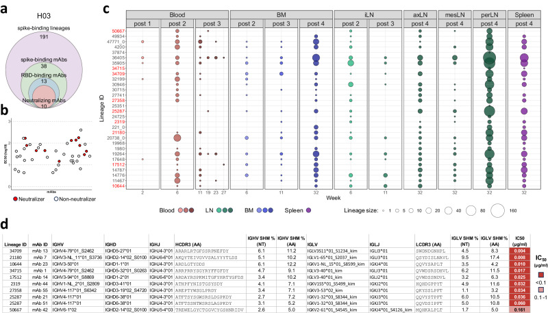
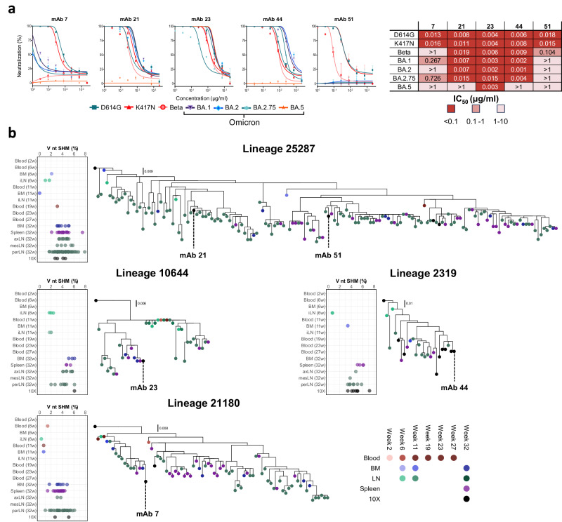
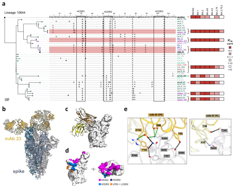
The continued evolution of SARS-CoV-2 underscores the need to understand qualitative aspects of the humoral immune response elicited by spike immunization. Here, we combine monoclonal antibody (mAb) isolation with deep B cell receptor (BCR) repertoire sequencing of rhesus macaques immunized with prefusion-stabilized spike glycoprotein. Longitudinal tracing of spike-sorted B cell lineages in multiple immune compartments demonstrates increasing somatic hypermutation and broad dissemination of vaccine-elicited B cells in draining and non-draining lymphoid compartments, including the bone marrow, spleen and, most notably, periaortic lymph nodes. Phylogenetic analysis of spike-specific monoclonal antibody lineages identified through deep repertoire sequencing delineates extensive intra-clonal diversification that shaped neutralizing activity. Structural analysis of the spike in complex with a broadly neutralizing mAb provides a molecular basis for the observed differences in neutralization breadth between clonally related antibodies. Our findings highlight that immunization leads to extensive intra-clonal B cell evolution where members of the same lineage can both retain the original epitope specificity and evolve to recognize additional spike variants not previously encountered.


Conflict of interest statement

D.J.S. consults for AstraZeneca AB on matters related to monoclonal antibody therapeutics for Covid-19. The remaining authors declare no competing interests.

Figures

Fig. 1
Fig. 1. Study design and immunoglobulin gene usage of vaccine-induced spike-binding B cells.
a Study overview. Two Chinese-origin rhesus macaques, H03 and I10, were inoculated four times with a pre-fusion stabilized spike ectodomain adjuvated with Matrix-M at weeks 0, 4, 9. Additional boosting was performed at week 31 for H03 and week 30 for I10. Samples (blood, LNs, BM and spleen) were collected as indicated and used to produce bulk IgG HC libraries. Samples from week 0 were used for IgM, IgK and IgL library production and immunoglobulin genotyping with IgDiscover. Samples from week 32 were used to sort spike-binding B cells, which were processed for 10X single-cell V(D)J sequencing and mAb cloning. Figure created with BioRender.com. b Comparison of IGHV usage in the total IgG repertoire (light red) and the spike-sorted repertoire (dark red). The y-axis shows IGHV genes ranked in descending order based on frequency usage in the bulk IgG repertoires. Frequencies are displayed on the x-axis and calculated based on the number of total IgG (n = 48,528 and n = 51,904 for H03 and I10, respectively) vs spike-sorted (n = 191 and n = 41 for H03 and I10, respectively) for each IGHV gene. Source data are provided as a Source Data file. c Combined IGHV (y-axis), IGKV (x-axis left) and IGLV (x-axis right) genes identified in single-cell sequence data from both animals. Each combination is represented as a circle with the size directly proportional to the number of lineages computed with each IGHV and IGKV or IGLV combination. Source data are provided as a Source Data file.
Fig. 2
Fig. 2. Multicompartmental longitudinal analysis of spike-elicited B cell lineage evolution.
Each sector represents a sample from H03. Starting from degree 0, the samples are ordered clockwise by sample time point. The base of each sector represents a curved x-axis with each lineage as a point. Each sector is divided into three levels. The outer level shows the number of members of that lineage, the mid-level shows each member’s SHM, and the inner common level connects lineages identified in multiple samples. The inner level is color-coded based on lineage persistence as determined by their identification at multiple time points (post 1, post 2, post 3, and post 4) in the traced data. The mid and outer sectors are color-coded with red, blue, green, and purple for blood, BM, LN, and spleen, respectively, with darker shades for increasing weeks. Source data are provided as a Source Data file.
Fig. 3
Fig. 3. Functional analysis of spike-binding mAbs and their lineage tracing in bulk IgG repertoires.
a Summary of the number of spike-binding lineages and cloned spike-binding, RBD-binding and neutralizing mAbs isolated from H03. b ELISA EC50 values based on binding to the ancestral prefusion D614G spike protein with neutralizing mAbs highlighted in red and non-neutralizing mAbs in white. Binding assays were performed in triplicates for each mAb. Source data are provided as a Source Data file. c Dissemination of spike-binding mAb lineages where the bubble size is directly proportional to the number of sequences identified for each lineage in each library. The x-axis of each section shows the sample compartment, grouped by time point with the sampling week indicated. Source data are provided as a Source Data file. d Summary table displaying V, D and J allele assignments, V gene SHM levels, CDR3 amino acid identity and IC50 neutralizing titers against the D614G variant for each neutralizing mAb. Neutralization assays were performed in triplicates for each mAb. The table displays only one of the most likely possible D allele assignments.
Fig. 4
Fig. 4. Neutralization breadth and phylogenic analysis of neutralizing Ab lineages.
a Neutralization curves (left) and IC50 values (right) against D614, K417N, Beta and Omicron subvariants BA.1, BA.2, BA.2.75, and BA.5 for the five broadest mAbs. For each curve, the plot displays single data as dots alongside mean values and standard deviation from triplicates. Source data are provided as a Source Data file. b Phylogenetic trees of the broadly neutralizing mAbs with the origin of the traced members displayed in red, blue, green, and purple for blood, BM, LN, and spleen, respectively, with darker shades for increasing weeks. Lineage members obtained from the 10x single-cell paired V(D)J sequencing are displayed in black. A summary of V gene SHM levels at the nucleotide level for each lineage member is displayed to the right of each tree with the compartment of origin and time point of detection shown in temporal order using the same color scheme.
Fig. 5
Fig. 5. Phylogenetic and cryo-EM analysis of mAb 23 in complex with spike.
a mAb 23 phylogenetic tree, with amino acid alignment of each member. The tree is rooted to its germline, each member is color coded in red, blue, green, and purple for blood, BM, LN and spleen, respectively, with darker shades for increasing weeks. Positions of SHM are indicated and the HCDRs are highlighted by black squares. IC50 neutralizing titers against D614G, BA.1, BA.2, BA.5, BA.4.6, BA.2.75, and BA.2.75.2 for expressed members of the lineage are shown to the right in a red color scale. Neutralization assays were performed in triplicates for each member. Source data are provided as a Source Data file. b Cryo-EM structure of the mAb 23 Fab interacting with the D614G prefusion spike. c Closer-up representation of mAb 23 interacting with the RBD. d Contact residues mapped on the RBD. hACE2 is colored purple, HCDR2 in dark blue, HCDR3 in light blue and LC in orange. e mAb 23 and RBD residues interaction. mAb23 is colored in yellow while the RBD residues are shown in white/gray. Ionic bonds are represented as dotted lines in green and hydrogen bonds in black.

References

    1. Karlsson Hedestam, G. B. et al. The challenges of eliciting neutralizing antibodies to HIV-1 and to influenza virus. Nat. Rev. Microbiol.6, 143–155 (2008). 10.1038/nrmicro1819 - DOI - PubMed
    1. Bq, O. et al. Enhanced neutralization resistance of SARS-CoV-2 Enhanced neutralization resistance of SARS-CoV-2 Omicron subvariants. Cell Host Microbe10.1016/j.chom.2022.11.012 (2022). - PMC - PubMed
    1. Tuekprakhon, A. et al. Antibody escape of SARS-CoV-2 Omicron BA.4 and BA.5 from vaccine and BA.1 serum. Cell185, 2422–2433.e13 (2022). 10.1016/j.cell.2022.06.005 - DOI - PMC - PubMed
    1. Hachmann, N. P. et al. Neutralization escape by SARS-CoV-2 omicron subvariants BA.2.12.1, BA.4, and BA.5. N. Engl. J. Med.387, 86–88 (2022). 10.1056/NEJMc2206576 - DOI - PMC - PubMed
    1. Shrestha, L. B., Foster, C., Rawlinson, W., Tedla, N. & Bull, R. A. Evolution of the SARS-CoV-2 omicron variants BA.1 to BA.5: implications for immune escape and transmission. Rev. Med. Virol.32, e2381 (2022). 10.1002/rmv.2381 - DOI - PMC - PubMed

MeSH terms

LinkOut - more resources