. 2024 Jul 27;14(1):17332.
doi: 10.1038/s41598-024-67697-0.

MASLD does not affect fertility and senolytics fail to prevent MASLD progression in male mice

Affiliations

MASLD does not affect fertility and senolytics fail to prevent MASLD progression in male mice

Jessica D Hense et al. Sci Rep. .

Abstract

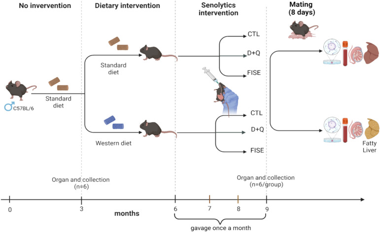
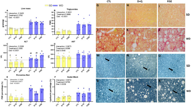
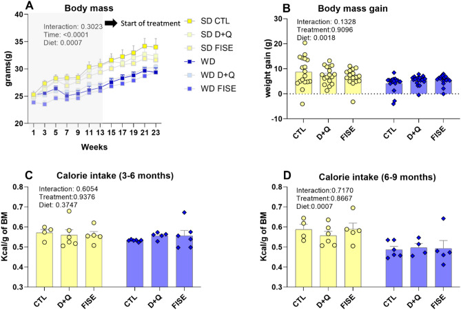
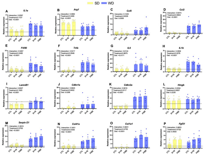
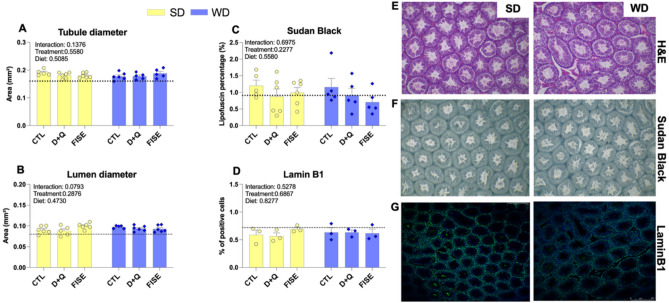
Senescent cells have been linked to the pathogenesis of metabolic dysfunction-associated steatotic liver disease (MASLD). However, the effectiveness of senolytic drugs in reducing liver damage in mice with MASLD is not clear. Additionally, MASLD has been reported to adversely affect male reproductive function. Therefore, this study aimed to evaluate the protective effect of senolytic drugs on liver damage and fertility in male mice with MASLD. Three-month-old male mice were fed a standard diet (SD) or a choline-deficient western diet (WD) until 9 months of age. At 6 months of age mice were randomized within dietary treatment groups into senolytic (dasatinib + quercetin [D + Q]; fisetin [FIS]) or vehicle control treatment groups. We found that mice fed choline-deficient WD had liver damage characteristic of MASLD, with increased liver size, triglycerides accumulation, fibrosis, along increased liver cellular senescence and liver and systemic inflammation. Senolytics were not able to reduce liver damage, senescence and systemic inflammation, suggesting limited efficacy in controlling WD-induced liver damage. Sperm quality and fertility remained unchanged in mice developing MASLD or receiving senolytics. Our data suggest that liver damage and senescence in mice developing MASLD is not reversible by the use of senolytics. Additionally, neither MASLD nor senolytics affected fertility in male mice.

Keywords: Aging; Dasatinib; Fisetin; Quercetin; Senescence.


Conflict of interest statement

The authors declare no competing interests.

Figures

Figure 1
Figure 1
Experimental design. Mice were assigned to a standard or Western diet for the first 3 months of the study. After that mice were assigned to a control group (CTL), dasatinib and quercetin (D + Q) and fisetin (FISE) groups for 3 months. Six mice from each group were euthanized at 3 and 6 months of age. The remaining mice were bred with young control females for a period of 8 days.
Figure 2
Figure 2
Liver mass (A), liver triglycerides (B), serum ALT (C) and AST (D), liver fibrosis (PSR; E), liver senescence (F), representative histological images stained with picrosirius red (G–L) and lipofuscin staining (M–R) in male mice fed with standard diet (SD) or western diet (WD) and treated with placebo (CTL), dasatinib and quercetin (D + Q) and fisetin (FIS). The dotted line represents measures in 3-month-old control male mice. Representative images are presented at × 400 magnification. All data are presented as mean ± SEM. P values ≤ 0.05 were considered significant. Different letters indicate statistical difference at P < 0.05 between individual groups when a significant interaction effect was observed.
Figure 3
Figure 3
Body mass (A), body mass gain (B), calorie intake from 3 to 6 months of age (C), calorie intake from 6 to 9 months of age (D) in male mice fed with standard diet (SD) or western diet (WD) and treated with placebo (CTL), dasatinib and quercetin (D + Q) or fisetin (FISE). All data are presented as mean ± SEM. P values ≤ 0.05 were considered significant. Different letters indicate statistical difference at P < 0.05 between individual groups when a significant interaction effect was observed.
Figure 4
Figure 4
Gene expression in the liver of male mice fed with standard diet (SD) or western diet (WD) and treated with placebo (CTL), dasatinib and quercetin (D + Q) and fisetin (FIS). Interleukin 1 alpha (Il1αA), Arginase-1 (Arg1B), C–C Motif Chemokine Ligand 5 (Ccl5C), C–C Motif Chemokine Ligand 2 (Ccl2D), Adhesion G Protein-Coupled Receptor E1 (f4/80E), Tumor Necrosis Factor alpha (TnfαF), Interleukin 6 (Il6G), Interleukin 1 Beta (Il1βH), Lamin B1 (LmnB1I), Cyclin Dependent Kinase Inhibitor 1A (Cdkn1a/p21J), Cyclin Dependent Kinase Inhibitor 2A (Cdkn2a/p16—K), High Mobility Group Box 1 (Hmgb1L), Serpin Family E Member 1 (Serpine1M), Collagen Type IV Alpha 1 Chain (Col4a1N), Collagen Type I Alpha 1 Chain (Col1a1O), Fibroblast Growth Factor 21 (Fgf21P). The dotted line represents measures in 3-month-old control male mice. All data are presented as mean ± SEM. P values ≤ 0.05 were considered significant. Different letters indicate statistical difference at P < 0.05 between individual groups when a significant interaction effect was observed.
Figure 5
Figure 5
Serum inflammatory markers in male mice fed with standard diet (SD) or western diet (WD) and treated with placebo (CTL), dasatinib and quercetin (D + Q) and fisetin (FIS). The dotted line represents measures in 3-month-old control male mice. All data are presented as mean ± SEM. P values ≤ 0.05 were considered significant. Different letters indicate statistical difference at P < 0.05 between individual groups when a significant interaction effect was observed.
Figure 6
Figure 6
Testicular gene expression in male mice fed with standard diet (SD) or western diet (WD) and treated with placebo (CTL), dasatinib and quercetin (D + Q) and fisetin (FIS). Interleukin 1 alpha (Il1αA), Arginase-1 (Arg1B), C–C Motif Chemokine Ligand 5 (Ccl5C), C–C Motif Chemokine Ligand 2 (Ccl2D), Adhesion G Protein-Coupled Receptor E1 (f4/80E), Tumor Necrosis Factor alpha (Tnf αF), Interleukin 6 (Il6G), Interleukin 1 Beta (Il1βH), Lamin B1 (LmnB1I), Cyclin Dependent Kinase Inhibitor 1A (Cdkn1a/p21J), Cyclin Dependent Kinase Inhibitor 2A (Cdkn2a/p16—K), High Mobility Group Box 1 (Hmgb1L), Serpin Family E Member 1 (Serpine1M). The dotted line represents measures in 3-month-old control male mice. All data are presented as mean ± SEM. P values ≤ 0.05 were considered significant. Different letters indicate statistical difference at P < 0.05 between individual groups when a significant interaction effect was observed.
Figure 7
Figure 7
Seminiferous tubule diameter (A), lumen diameter (B), lipofuscin (C) and lamin B1 staining (D) and representative images of sections with lipofuscin (F), lamin B1 (G), and H&E staining (E) of seminiferous tubules with a × 10 magnification in mice fed a standard diet (SD) or western diet (WD) and treated with placebo (CTL), dasatinib and quercetin (D + Q) and fisetin (FIS). The dotted line represents measures in 3-month-old control male mice. Representative images are presented at × 400 magnification. All data are presented as mean ± SEM. P values ≤ 0.05 were considered significant. Different letters indicate statistical difference at P < 0.05 between individual groups when a significant interaction effect was observed.

References

    1. Pouwels, S. et al. Non-alcoholic fatty liver disease (NAFLD): A review of pathophysiology, clinical management and effects of weight loss. BMC Endocr. Disord.22, 63. 10.1186/s12902-022-00980-1 (2022). 10.1186/s12902-022-00980-1 - DOI - PMC - PubMed
    1. Schierwagen, R. et al. Seven weeks of Western diet in apolipoprotein-E-deficient mice induce metabolic syndrome and non-alcoholic steatohepatitis with liver fibrosis. Sci. Rep.5, 12931. 10.1038/srep12931 (2015). 10.1038/srep12931 - DOI - PMC - PubMed
    1. Castellanos-Tapia, L. et al. Mediterranean-like mix of fatty acids induces cellular protection on lipid-overloaded hepatocytes from western diet fed mice. Ann. Hepatol.19, 489–496. 10.1016/j.aohep.2020.06.005 (2020). 10.1016/j.aohep.2020.06.005 - DOI - PubMed
    1. Dave, A. et al. Consumption of grapes modulates gene expression, reduces non-alcoholic fatty liver disease, and extends longevity in female C57BL/6J mice provided with a high-fat western-pattern diet. Foods11, 984. 10.3390/foods11131984 (2022). 10.3390/foods11131984 - DOI - PMC - PubMed
    1. Lian, C. Y., Zhai, Z. Z., Li, Z. F. & Wang, L. High fat diet-triggered non-alcoholic fatty liver disease: A review of proposed mechanisms. Chem. Biol. Interact.330, 109199. 10.1016/j.cbi.2020.109199 (2020). 10.1016/j.cbi.2020.109199 - DOI - PubMed

LinkOut - more resources