. 2024 Jul 28;15(7):535.
doi: 10.1038/s41419-024-06926-w.

Glucocorticoid receptor controls atopic dermatitis inflammation via functional interactions with P63 and autocrine signaling in epidermal keratinocytes

Affiliations

Glucocorticoid receptor controls atopic dermatitis inflammation via functional interactions with P63 and autocrine signaling in epidermal keratinocytes

Lisa M Sevilla et al. Cell Death Dis. .

Abstract

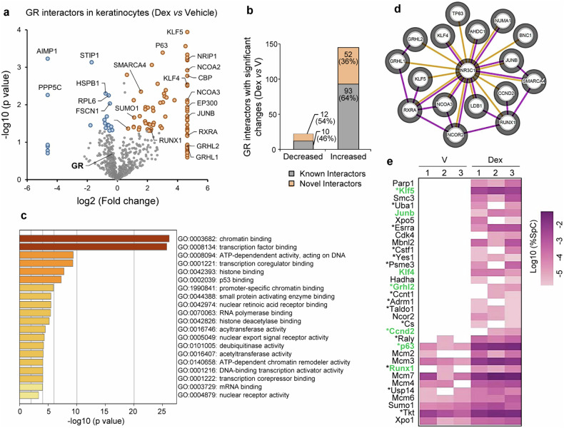
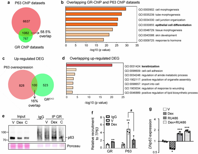
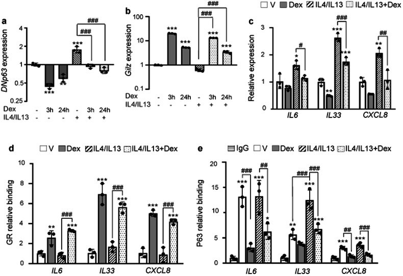
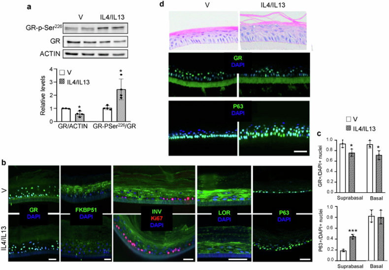
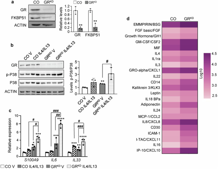
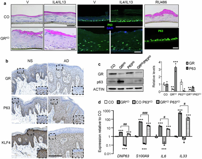
Atopic dermatitis (AD), a prevalent chronic inflammatory disease with multifactorial etiology, features epidermal barrier defects and immune overactivation. Synthetic glucocorticoids (GCs) are widely prescribed for treating AD due to their anti-inflammatory actions; however, mechanisms are incompletely understood. Defective local GC signaling due to decreased production of endogenous ligand and/or GC receptor (GR) levels was reported in prevalent inflammatory skin disorders; whether this is a consequence or contributing factor to AD pathology is unclear. To identify the chromatin-bound cell-type-specific GR protein interactome in keratinocytes, we used rapid immunoprecipitation of endogenous proteins and mass spectrometry identifying 145 interactors that increased upon dexamethasone treatment. GR-interacting proteins were enriched in p53/p63 signaling, including epidermal transcription factors with critical roles in AD pathology. Previous analyses indicating mirrored AD-like phenotypes between P63 overexpression and GR loss in epidermis, and our data show an intricate relationship between these transcription factors in human keratinocytes, identifying TP63 as a direct GR target. Dexamethasone treatment counteracted transcriptional up-regulation of inflammatory markers by IL4/IL13, known to mimic AD, causing opposite shifts in GR and P63 genomic binding. Indeed, IL4/IL13 decreased GR and increased P63 levels in cultured keratinocytes and human epidermal equivalents (HEE), consistent with GR down-regulation and increased P63 expression in AD lesions vs normal skin. Moreover, GR knockdown (GRKD) resulted in constitutive increases in P63, phospho-P38 and S100A9, IL6, and IL33. Also, GRKD culture supernatants showed increased autocrine production of TH2-/TH1-/TH17-TH22-associated factors including IL4, CXCL10, CXCL11, and CXCL8. GRKD HEEs showed AD-like features including hyperplasia and abnormal differentiation, resembling phenotypes observed with GR antagonist or IL4/IL13 treatment. The simultaneous GR/P63 knockdown partially reversed constitutive up-regulation of inflammatory genes in GRKD. In summary, our data support a causative role for GR loss in AD pathogenesis via functional interactions with P63 and autocrine signaling in epidermal keratinocytes.


Conflict of interest statement

The authors declare no competing interests.

Figures

Fig. 1
Fig. 1. Identification of chromatin-bound GR-interacting proteins in dexamethasone (Dex)-treated keratinocytes.
Mouse keratinocytes were treated with Dex (100 nM) or vehicle (V) for 1 h, and GR-bound chromatin fragments isolated for mass spectrometry. a Volcano plot shows GR interactors; loss (blue) or gain (orange) of interactions upon Dex treatment are indicated. Note that for visualization purposes, fold changes that could not be computed (present in one group and completely absent in the other) were attributed the maximum value. b The graph shows significantly changed GR interactions in Dex vs vehicle-treated cells. c Functional categorization of 145 significant Dex-induced interactors (Metascape, Molecular Function). d Network of GR-interacting proteins involved in skin/keratinocyte function (identified by IPA) was built using the STRING database using confidence level of 0.4 (default setting) and with no additional interactors. The orange edges represent novel GR interactors, while the purple edges represent previously reported interactions and/or predicted protein-protein networks (from databases or experimentally determined). e Heatmap of GR interacting proteins involved in p53/p63 signaling (IPA).
Fig. 2
Fig. 2. GR/p63 interaction in keratinocytes.
a Venn diagram of ChIP-seq datasets for P63 bound to active enhancers (human) and GR (mouse). Note that almost 60% of GR bound genes are also bound by P63. b Functional categorization of overlapping GR and P63 bound genes in (a) for biological process with Metascape. c Venn diagram of up-regulated differentially expressed genes (DEG) in GR epidermal KO (GREKO) epidermis and p63-overexpressing epidermis. d Functional categorization of overlapping genes in (c) with Metascape, biological process. e p63 immunoblot of nuclear GR immunoprecipitations (IP) in mouse keratinocytes treated 1 h with vehicle (V), 100 nM dexamethasone (Dex), or 100 nM corticosterone (C). f Chromatin immunoprecipitation shows Dex-dependent modulation of GR and P63 recruitment to TP63 in N/TERT-2G (Dex, 1 μM, 1 h). g RT-QPCR for DNP63 evaluating response to Dex (1 μM, 3 h) in N/TERT-2G that were pre-incubated 16 h with V or 10μM RU486. Statistical significance using Student’s t-test (f, GR) or one-way ANOVA (f, P63; g) with post hoc Tukey multiple comparison test: *, #p < 0.05; **p < 0.01; ***p < 0.001. Asterisks: significant differences relative to vehicle (f, GR; g) or IgG (f, P63); hash: significant differences between groups indicated by brackets. N = 3 for all experiments.
Fig. 3
Fig. 3. GR and P63 show inverse correlation in IL4/IL13-treated keratinocytes.
Gene expression of DNP63 (a) and TSC22D3/GILZ (b) after treatment with Dex (1 μM, 3 and 24 h), IL4/IL13 (50 ng/ml, 24 h), or both. c RT-QPCR evaluating indicated genes following 24 h incubation with vehicle (V), Dex (1 μM), and/or IL4/IL13 (50 ng/ml). ChIP-QPCR shows Dex (1 μM, 1 h) and IL4/IL13 (50 ng/mL, 16 h)-dependent modulation of GR (d) and P63 (e) recruitment to IL6, IL33 and CXCL8. Statistical significance using one-way ANOVA with post hoc Tukey multiple comparison test: *, #p < 0.05; **, ##p < 0.01; ***, ###p < 0.001. Asterisks: significant differences relative to vehicle (ad) or IgG (e); hashes: significant differences between groups indicated by brackets. N = 3 for all experiments.
Fig. 4
Fig. 4. IL4/IL13 treatment results in reduction of GR expression and function in human keratinocytes.
a Primary human keratinocytes were treated 72 h with vehicle or 30 ng/ml IL4/IL13 prior to immunoblotting (upper); quantitation (lower). N = 4. b Immunofluorescence of sections of human epidermal equivalents (HEEs) with primary keratinocytes treated with vehicle (V) or IL4/IL13 (30 ng/ml, 72 h) with indicated antibodies. c Graphs showing ratios of GR+ (top) or P63+ nuclei (bottom) relative to total DAPI stained nuclei in HEEs treated with vehicle (V) or IL4/IL13. Statistical significance determined using Student’s t-test, *p < 0.05; ***p < 0.001. N = 3. d Sections of HEEs with N/TERT-2G cells treated with vehicle (V) or IL4/IL13 (50 ng/ml, 72 h). Top panels: hematoxylin & eosin. Lower panels: immunofluorescence with indicated antibodies. Bars: 50 μm.
Fig. 5
Fig. 5. Keratinocyte GR loss-of-function results in enhanced inflammation in human keratinocytes.
a Immunoblot (left) and quantitation (right) of control (CO) or GR-knockdown (GRKD) keratinocyte lysates with indicated antibodies. b, c CO or GRKD keratinocytes were treated 24 h with vehicle (V) or 50 ng/ml IL4/IL13. Immunoblotting (b) with P38 and phospho(p)-P38 antibodies (left); quantitation (right). RT-QPCR (c) for expression of indicated genes. Mean and SD are shown in graphs. Statistical significance using Student’s t-test (a) or 2-way ANOVA and post hoc Tukey multiple comparison test (b, c) indicated as: *, #p < 0.05; **, ##p < 0.01; ***, ###p < 0.001. Asterisks: statistically significant differences relative to CO V; hashes: comparisons between groups indicated by brackets. N = 3 for all experiments. d Heatmap shows relative levels of secreted factors in CO or GRKD primary keratinocyte supernatants (vehicle-treated) using a multiplex antibody array. Data are average of three independent experiments and represent factors with statistical significance using Student’s t-test; p < 0.05.
Fig. 6
Fig. 6. Atopic dermatitis inflammation upon GR loss-of-function can be partially rescued by simultaneous GR/P63 knockdown.
a Sections of GR-knockdown (GRKD) and control (CO) HEEs treated with vehicle (V) or 50 ng/ml IL4/IL13 (72 h) or 5μM RU486 (13 d) stained with hematoxylin & eosin (left panels and upper panel RU486) or P63-specific antibodies. Bars: 50 μm. N = 3. b Sections from normal (NS) or lesional (AD) skin. N = 2 for each condition. Bar: 300 μm. c, d CO or GRKD N/TERT-2G were transiently transfected with negative control or P63-specific (P63KD) siRNAs. Western blot (left) and quantification (right) show knockdown of GR and P63 (c). RT-QPCR for indicated genes (d). N = 3. Mean and SD are shown in graphs. Statistical significance using 2-way ANOVA (d) with post hoc Tukey multiple comparison test: *, #p < 0.05; **, ##p < 0.01; ***, ###p < 0.001. Asterisks: significant differences relative to CO; hashes: significant differences between groups indicated by brackets (d).

Similar articles

Cited by

References

    1. Bieber T, Paller AS, Kabashima K, Feely M, Rueda MJ, Ross Terres JA, et al. Atopic dermatitis: pathomechanisms and lessons learned from novel systemic therapeutic options. J Eur Acad Dermatol Venereol. 2022;36:1432–49. https://pubmed.ncbi.nlm.nih.gov/35575442/. 10.1111/jdv.18225 - DOI - PubMed
    1. Meesters LD, Niehues H, Johnston L, Smits JPH, Zeeuwen PLJM, Brown SJ, et al. Keratinocyte signaling in atopic dermatitis: Investigations in organotypic skin models toward clinical application. J Allergy Clin Immunol. 2023;151:1231–5. https://pubmed.ncbi.nlm.nih.gov/36841264/. 10.1016/j.jaci.2023.02.012 - DOI - PubMed
    1. Strehl C, Ehlers L, Gaber T, Buttgereit F. Glucocorticoids-all-rounders tackling the versatile players of the immune system. Front Immunol. 2019;10:1744. https://pubmed.ncbi.nlm.nih.gov/31396235/. 10.3389/fimmu.2019.01744 - DOI - PMC - PubMed
    1. Timmermans S, Souffriau J, Libert C. A general introduction to glucocorticoid biology. Front Immunol. 2019;10:1545. 10.3389/fimmu.2019.01545 - DOI - PMC - PubMed
    1. Vettorazzi S, Nalbantoglu D, Gebhardt JCM, Tuckermann J. A guide to changing paradigms of glucocorticoid receptor function-a model system for genome regulation and physiology. FEBS J. 2022;289:5718–43. https://pubmed.ncbi.nlm.nih.gov/34213830/. 10.1111/febs.16100 - DOI - PubMed

MeSH terms

LinkOut - more resources