Case Reports
. 2024 Jul 28;15(1):6359.
doi: 10.1038/s41467-024-50686-2.

Helical superstructures between amyloid and collagen in cardiac fibrils from a patient with AL amyloidosis

Affiliations
Case Reports

Helical superstructures between amyloid and collagen in cardiac fibrils from a patient with AL amyloidosis

Tim Schulte et al. Nat Commun. .

Abstract

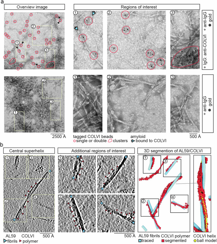
Systemic light chain (LC) amyloidosis (AL) is a disease where organs are damaged by an overload of a misfolded patient-specific antibody-derived LC, secreted by an abnormal B cell clone. The high LC concentration in the blood leads to amyloid deposition at organ sites. Indeed, cryogenic electron microscopy (cryo-EM) has revealed unique amyloid folds for heart-derived fibrils taken from different patients. Here, we present the cryo-EM structure of heart-derived AL amyloid (AL59) from another patient with severe cardiac involvement. The double-layered structure displays a u-shaped core that is closed by a β-arc lid and extended by a straight tail. Noteworthy, the fibril harbours an extended constant domain fragment, thus ruling out the variable domain as sole amyloid building block. Surprisingly, the fibrils were abundantly concatenated with a proteinaceous polymer, here identified as collagen VI (COLVI) by immuno-electron microscopy (IEM) and mass-spectrometry. Cryogenic electron tomography (cryo-ET) showed how COLVI wraps around the amyloid forming a helical superstructure, likely stabilizing and protecting the fibrils from clearance. Thus, here we report structural evidence of interactions between amyloid and collagen, potentially signifying a distinct pathophysiological mechanism of amyloid deposits.


Conflict of interest statement

The authors declare no competing interests.

Figures

Fig. 1
Fig. 1. AL59 extract comprises additional polymer decorating the amyloid fibrils.
a Cryo-EM micrographs and b Cryo-ET 2D projections reveal undecorated and polymer-decorated AL59 fibrils, highlighted by yellow and red arrows, respectively. The remarkably shaped polymer beads are encircled in yellow and red for the unbound and amyloid-associated forms, respectively. A thin yellow line indicated the estimated crossover-length. The image scale-bar is valid for both panels. See Supplementary Fig. 1.
Fig. 2
Fig. 2. The structure of glycosylated AL59 exhibits a meandering double layer comprising a central β-arc, u-shaped core and a terminal straight segment.
a Cross-sectional view of the reconstructed map, visualised according to the depicted σ-colour scale. The polypeptide backbone and side-chains of the models are shown as black cartoon and sticks, respectively. b The polypeptide backbone is shown as cartoon, coloured on a rainbow-spectrum for residue numbers from 1 to 120. The three kinks are highlighted as pink straight lines. The u-shaped core is highlighted by a black outline. The terminal and β-arc segments are labelled. c Side-view of the deposited model comprising five subunits. The height difference of the polypeptide layer is indicated for the central layer, highlighted as thick ribbon. d ~5 Å cross-sectional slices through the reconstructed AL59 map are shown applying a black/white (0/0.09) linear scale (left). The structural heterogeneity panel depicts two additional alternative conformations of AL59 amyloid core (highlighted by pink arrow) that emerged during 3D classifications.
Fig. 3
Fig. 3. AL59 adopts a fold related to λ3-FOR005, but harbours an extended constant domain (CL) fragment in its amyloid core.
a Sequence alignment of AL59 belonging to the λ3 gene to the four other ex vivo LC amyloid structures λ3-FOR005 [https://www.rcsb.org/structure/6Z1O], λ6-AL55 [https://www.rcsb.org/structure/6HUD], λ1-FOR001 [https://www.rcsb.org/structure/7NSL] and λ1-FOR006 [https://www.rcsb.org/structure/6IC3]. β-strands and strict β-turns are indicated by numbered β and non-numbered TT symbols, respectively. Strict sequence identity is indicated by a red box with white character, similarities within and across groups are indicated by red characters and blue frames, respectively. Secondary structure elements of λ3-AL59 and λ3-FOR005 are shown above. CDR segments of λ3-AL59 and λ3-FOR005 are labelled and highlighted in yellow and orange, respectively. The CL-derived fragment in the amyloid core of AL59 is highlighted in light pink. b The AI-generated native AL59 structure (left) is compared to the amyloid structure (right). VL and CL domains are coloured white and light pink, respectively. The CDRs are coloured yellow and labelled. The non-amyloidogenic CL part is semi-transparent. Modified N- and C-termini of LC peptide fragments detected by LC-MS/MS are shown as red and blue Cα-spheres, respectively. The residue-level line-plot depicts the domain boundaries and modified N- and C-termini of LC fragments detected by LC-MS/MS. c The superimposed λ3-AL59 and λ3-FOR005 structures are shown as white and black cartoons, respectively.
Fig. 4
Fig. 4. Collagen VI is the abundant polymer concatenating with AL59.
a The 2D class average reveals two half-beads linked by an intra-bead fibre. Each half-bead entails head, body and tail substructures. Inter-bead fibres appear blurry, likely due to high flexibility. b Two views of the half-bead map that is visualised according to depicted σ-colour scale. The main features (darker blue) are comparable to a map at a threshold of 1.5 σ. See Supplementary Figs. 4 and 5.
Fig. 5
Fig. 5. The fibril-decorating polymer is COLVI and follows the helical pitch of AL59.
a Immuno-electron microscopy (IEM) revealed AL59 fibrils, free polymer and dense fibril clusters tagged with gold particles. Top panel: > 50 gold particles, highlighted in red, were counted in this image of the extract stained with anti-COLVI IgG and gold-conjugated anti-IgG antibodies 70R-CR009x (Fitzgerald) followed by a 12 nm gold-conjugated anti-IgG antibody, respectively. Three magnified sections emphasise (1) three gold particles within ~150 Å distance to AL59 fibrils, (2) three single gold particles and three duplets tagging free COLVI polymer as well as (3) 11 gold particles within a dense amyloid fibril cluster. bottom panel: No gold particles were found in the sample treated only with the gold-conjugated anti-IgG antibodies. Three magnified sections are shown for direct comparison to the top panel. See Supplementary Figs. 6 and 7. b Left: An overview cryo-ET slice with a thickness of 0.925 nm and four additional regions of interest (highlighted boxes) reveal amyloid fibrils (blue arrowheads) decorated with COLVI polymers (red arrowheads). The height level of the overview slice highlights the central AL59/COLVI interaction. The four boxes are shown at different height levels and higher magnification. Right: 3D renderings of the overview cryo-ET slice and the highlighted central AL59/COLVI interaction show traced AL59 fibril tubes (blue) decorated with segmented COLVI densities (red). A helical ball model (yellow) with helical twist and rise of AL59, but three times larger helical radius, is shown within the COLVI density to demonstrate the helical imprinting of AL59 on COLVI.

References

    1. Merlini, G. et al. Systemic immunoglobulin light chain amyloidosis. Nat. Rev. Dis. Prim.4, 1–19 (2018). - PubMed
    1. Merlini, G. AL amyloidosis: from molecular mechanisms to targeted therapies. Hematol. Am. Soc. Hematol. Educ. Program2017, 1–12 (2017).10.1182/asheducation-2017.1.1 - DOI - PMC - PubMed
    1. Haupt, C. The AL amyloid fibril: looking for a link between fibril formation and structure. Hemato2, 505–514 (2021).10.3390/hemato2030032 - DOI
    1. Del Pozo-Yauner, L. et al. Role of the mechanisms for antibody repertoire diversification in monoclonal light chain deposition disorders: when a friend becomes foe. Front. Immunol.14, 1203425 (2023). 10.3389/fimmu.2023.1203425 - DOI - PMC - PubMed
    1. Bodi, K. et al. AL-Base: a visual platform analysis tool for the study of amyloidogenic immunoglobulin light chain sequences. Amyloid16, 1–8 (2009). 10.1080/13506120802676781 - DOI - PMC - PubMed

Publication types