. 2024 Aug 28;44(8):BSR20240542.
doi: 10.1042/BSR20240542.

Characterization of human aquaporin ion channels in a yeast expression system as a tool for novel ion channel discovery

Affiliations

Characterization of human aquaporin ion channels in a yeast expression system as a tool for novel ion channel discovery

Saeed Nourmohammadi et al. Biosci Rep. .

Abstract

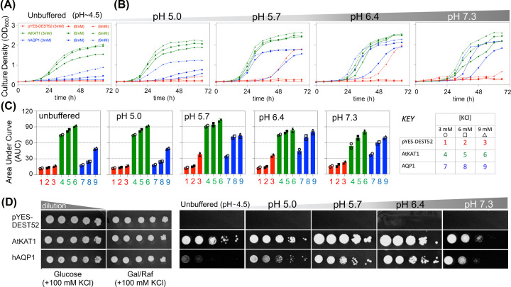
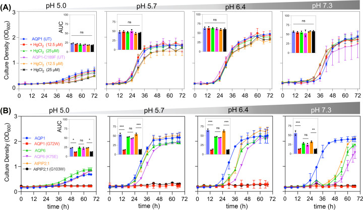
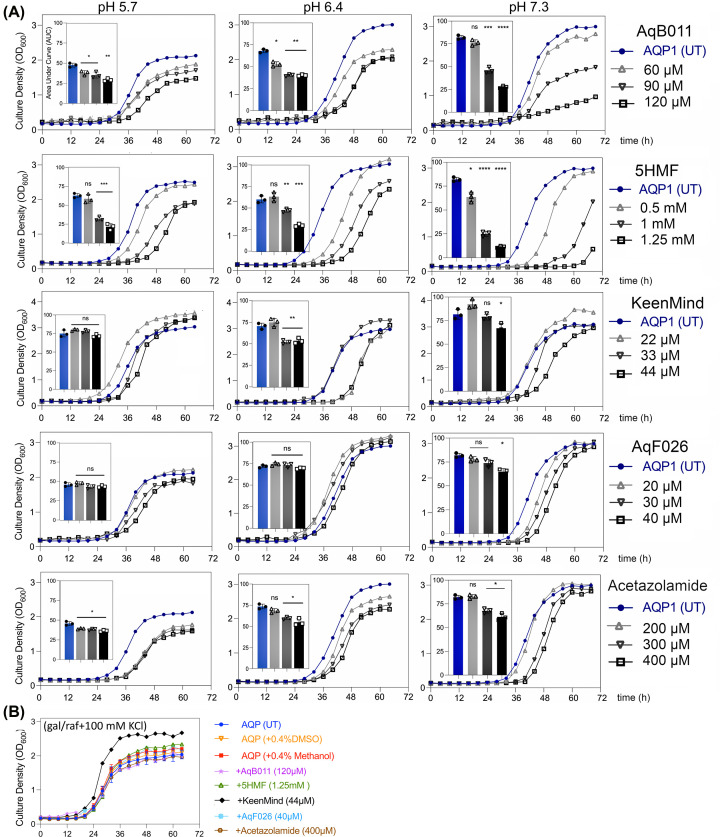
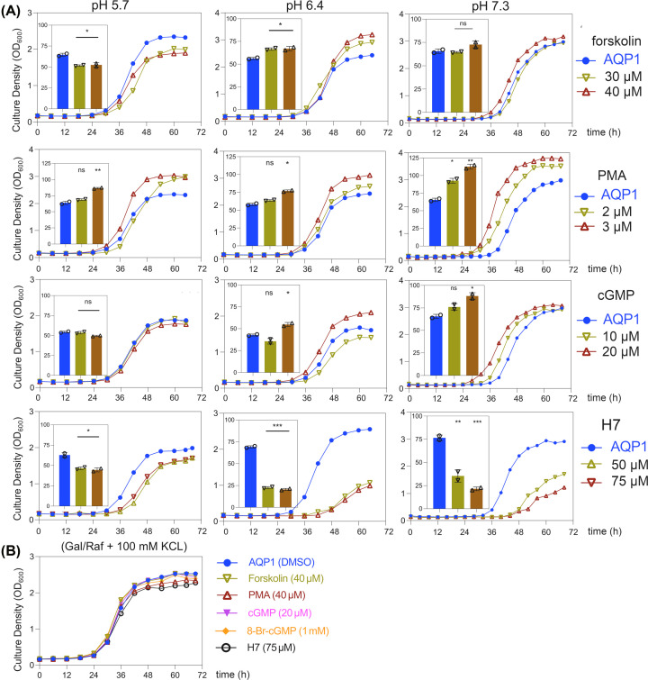
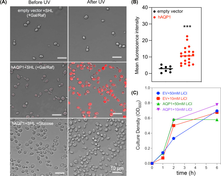
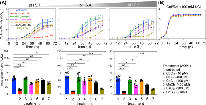
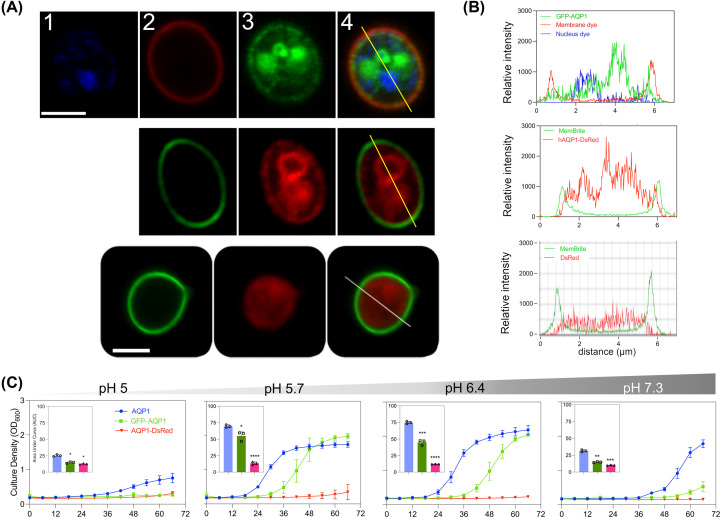
Aquaporin (AQP) channels found in all domains of life are transmembrane proteins which mediate passive transport of water, glycerol, signaling molecules, metabolites, and charged solutes. Discovery of new classes of ion-conducting AQP channels has been slow, likely reflecting time- and labor-intensive methods required for traditional electrophysiology. Work here defines a sensitive mass-throughput system for detecting AQP ion channels, identified by rescue of cell growth in the K+-transport-defective yeast strain CY162 following genetic complementation with heterologously expressed cation-permeable channels, using the well characterized human AQP1 channel for proof of concept. Results showed AQP1 conferred transmembrane permeability to cations which rescued survival in CY162 yeast. Comprehensive testing showed that growth response properties fully recapitulated AQP1 pharmacological agonist and antagonist profiles for activation, inhibition, dose-dependence, and structure-function relationships, demonstrating validity of the yeast screening tool for AQP channel identification and drug discovery efforts. This method also provided new information on divalent cation blockers of AQP1, pH sensitivity of antagonists, and ion permeability of human AQP6. Site-directed mutagenesis of AQP1 channel regulatory domains confirmed that yeast growth rescue was mediated by the introduced channels. Optical monitoring with a lithium-sensitive photoswitchable probe in living cells independently demonstrated monovalent cation permeability of AQP1 channels in yeast plasma membrane. Ion channel properties of AQP1 expressed in yeast were consistent with those of AQP1 expressed in Xenopus laevis oocyte and K+-transport defective Escherichia coli. Outcomes here establish a powerful new approach for efficient screening of phylogenetically diverse AQPs for yet untested functions as cation channels.

Keywords: Saccharomyces cerevisiae; aquaporins; drug discovery and design; high-throughput screening; ion channels.


Conflict of interest statement

The authors declare no conflict of interest; the research was conducted in the absence of any commercial or financial relationships that could be construed as a potential conflict of interest.

Figures

Figure 1
Figure 1. Complementation of growth in K+-uptake deficient CY162 yeast by transfection with hAQP1
K+ uptake enabled by heterologous hAQP1 expression yielded concentration-dependent rescue of growth in CY162 yeast cells. hAQP1-expressing yeast were grown in liquid culture (200 µl/well) in (A) unbuffered (∼pH 4.5) inducing medium or (B) in inducing media adjusted to pH 5.0, 5.7, 6.4, or 7.3. Growth was monitored in 96 well plates by optical density at 600 nm (OD600) as a function of time in media supplemented with KCl at 3 mM (circle), 6 mM (square), or 9 mM (triangle symbols). Mock transfected yeast (pYES-DEST52, red) was the negative control; CY162 yeast expressing the K+ channel AtKAT1 (green) was the positive control. (C) Growth responses quantified as calculated Area Under Curve (AUC) values were compiled in histograms showing mean ± SD for two independent experiments with three replicates each. (D) Phenotypic assays on solid media showed colony growth of hAQP1 and control lines, aliquoted from stock (OD600 = 1.0) using 10-fold serial dilutions on low-salt yeast nitrogen base medium supplemented with 6 mM KCl, incubated for 5 days. Yeast expressing hAQP1 or AtKAT1 showed strong growth in standard media (right panels); supplementation with high (100 mM) KCl allowed growth of negative control as well (left panels).
Figure 2
Figure 2. Rescue of yeast growth by AQP channel activity assessed by mercury sensitivity and effects of site directed mutations
(A) hAQP1-mediated water permeability is not required for compensation of the CY162 yeast growth defect, as indicated by lack of sensitivity to the water pore blocker mercuric chloride (HgCl2) at 12.5 and 25 µM. Yeast expressing the Hg2+-insensitive hAQP1 mutant C189F showed growth not different from that in wild-type, with or without HgCl2 at any pH. (B) Three different classes of characterized AQP ion channels (mammalian AQP1 and AQP6, and plant AtPIP2;1) rescued CY162 growth at pH 5.7 and above in 6 mM KCl. Two mutations resulting in dysfunctional channels (hAQP1 G72W; AtPIP2;1 G103W) failed to rescue growth; whereas the mutant hAQP6 K75E which retains ion channel functionality supported growth not different from hAQP6 wild-type. Results compiled from two independent experiments (two replicates each) are quantified as AUC histograms (insets; mean ± SD). Statistical significance (ANOVA with post-hoc Bonferroni tests) is indicated as *P<0.05, **P<0.01, ***P<0.001; or ns (not significant).
Figure 3
Figure 3. Assessment of pharmacological modulators of hAQP1 channel activity on yeast growth
(A) Agents were tested for CY162 cell growth in media with 6 mM KCl at pH 5.7, 6.4, or 7.3, monitored for 68 h. AUC data histograms (insets) show results (mean ± SD) of two independent experiments each done in triplicate. Statistical significance compared with untreated AQP1 (unpaired t-test) is shown as ****P<0.0005, ***P<0.005, **P<0.05, *P<0.5, and ns, not significant. Vehicle controls matched to drug treatments as ‘untreated’ (UT) groups were: DMSO for AqB011, AqF026 and 5HMF; methanol for KeenMind; and water for acetazolamide. Data are compiled from two independent experiments with three replicates each; error bars show mean ± SD. (B) In permissive medium with 100 mM KCl, yeast expressing hAQP1 and drug treatments at the highest doses grew well on standard medium, showing absence of general toxicity.
Figure 4
Figure 4. Regulation of hAQP1-induced growth rescue by modulators of intracellular signaling pathways
(A) Growth responses for CY162 cells expressing hAQP1, grown in YNB inducing medium with 6 mM KCl at pH 5.7, 6.4 or 7.3. Vehicle (0.4% v/v DMSO) or water (UT) served as the matched control conditions for treatments with direct ligand (cGMP) and protein kinase modulating agents (forskolin, PMA, cGMP, and H7) as indicated. AUC summary data are shown in inset histograms. Statistical significance (unpaired T test) was determined by comparison of each treatment with matched vehicle or water controls, and shown as *P<0.05, **P<0.01, ***P<0.001; or ns (not significant). (B) Equivalent growth rates in permissive media (100 mM KCl) confirmed no toxic effects on growth for any of the treatments at maximal doses. All growth curves were compiled from two independent experiments, with three technical replicates each.
Figure 5
Figure 5. Optical confirmation of monovalent cation permeation in hAQP1-expressing yeast using a photoswitchable lithium sensor SHL
(A) CY162 cells expressing pYES-DEST52 empty vector (EV) (upper row) or human (h)AQP1 (middle and lower rows) imaged by confocal microscopy with the Li+ sensor in the OFF (before UV) and ON (after UV) configurations, showing cation entry into cells induced to express AQP1 (middle row), not in non-AQP1 expressing controls (upper and lower rows). Li+ -bound SHL was detected using Ex 514 nm and Em 550-650 nm (see Methods for details). Controls were empty vector (upper low), and non-induced AQP1-transfected cells supplemented with 100 mM KCl (lower row). Scale bars are 10 µM. (B) Scatter plot of cellular fluorescence intensities for 8 empty vector control and 20 AQP1-expressing yeast cells which were induced in gal/raf with 6 mM KCl (as shown in A, middle row). Statistical significance determined by unpaired Student’s t-test shows ***P<0.0001. (C) Lack of effect of lithium exposure on yeast cell growth responses in hAQP1-expressing or empty vector CY162 cells induced in gal/raf at pH 7.3.
Figure 6
Figure 6. Effects of extracellular divalent cations on growth of hAQP1-expressing yeast
(A) Effect of divalent cations on the growth of CY162 cells expressing hAQP1 at different pH values all with 6 mM KCl. Curves are shown for untreated hAQP1 (blue); Cd2+ (10 µM; red), Ni2+ (400 µM; green), Co2+ (400 µM; pink), Mn2+ (400 µM; orange), Ba2+ (200 µM; black) and Ca2+ (2 mM; sage). Growth rates compiled as AUC are shown below the curve plots as corresponding histograms, for data from least two independent experiments with two replicates each; error bars show mean ± SD. Statistical significance determined by ANOVA with post-hoc Bonferroni tests *P<0.05, **P<0.01, ***P<0.001; or ns (not significant). (B) Yeast expressing hAQP1 in permissive media with 100 mM KCl showed equivalent growth across treatments with or without divalent cations, indicating block was not due to general toxicity.
Figure 7
Figure 7. Fusion-protein tagging of hAQP1 influences subcellular localization and cation-dependent growth rescue in yeast
(A) Live cell confocal microscopy images of CY162 cells expressing GFP tagged (upper row) or DsRED tagged (middle row) hAQP fusion proteins, or cells expressing fluorescent tag alone (lower row). Hoechst nuclear stain (1); plasma membrane dye (2; red upper row; green middle row); tagged GFP-hAQP1 (3; upper row) and tagged hAQP1-DsRed (middle row); cytoplasmic tag alone (lower row); and merged images (4). Scale bars 5 µm. Cells were induced in gal/raf with 6 mM KCl for ∼70 hours before imaging. (B) Lines in panel A show cross sections taken through cell centers to measure signal intensities for tagged hAQP1 or DsRed alone, with membrane dye and nuclear dye levels as indicated, plotted as a function of X-Y distance to determine overlap between fusion protein and plasma membrane localization. (C) Growth responses of CY162 cells transformed with hAQP1 (blue), GFP-hAQP1 (green), or hAQP1-DsRed (red) in media with 6 mM KCl at ∼70 hours, with pH (5.0-7.3). Compiled AUC values from three independent experiments (2 replicates each) were assessed by unpaired Student’s t-test showing *P<0.05, **P<0.01, ***P<0.001, ****P<0.0001; or ns (not significant).

Similar articles

Cited by

References

    1. Zampighi G.A., Hall J.E. and Kreman M. (1985) Purified lens junctional protein forms channels in planar lipid films. Proc. Natl. Acad. Sci. 82, 8468–8472 10.1073/pnas.82.24.8468 - DOI - PMC - PubMed
    1. Tyerman S.D., McGaughey S.A., Qiu J., Yool A.J. and Byrt C.S. (2021) Adaptable and multifunctional ion-conducting aquaporins. Annu. Rev. Plant Biol. 72, 703–736 10.1146/annurev-arplant-081720-013608 - DOI - PubMed
    1. Henderson S.W., Nourmohammadi S., Ramesh S.A. and Yool A.J. (2022) Aquaporin ion conductance properties defined by membrane environment, protein structure, and cell physiology. Biophys. Rev. 1–18 10.1007/s12551-021-00925-3 - DOI - PMC - PubMed
    1. Kourghi M., Pei J.V., De Ieso M.L., Nourmohammadi S., Chow P.H. and Yool A.J. (2018) Fundamental structural and functional properties of Aquaporin ion channels found across the kingdoms of life. Clin. Exp. Pharmacol. Physiol. 45, 401–409 10.1111/1440-1681.12900 - DOI - PubMed
    1. Wagner K., Unger L., Salman M.M., Kitchen P., Bill R.M. and Yool A.J. (2022) Signaling Mechanisms and Pharmacological Modulators Governing Diverse Aquaporin Functions in Human Health and Disease. Int. J. Mol. Sci. 23, 1388 10.3390/ijms23031388 - DOI - PMC - PubMed

Publication types

LinkOut - more resources