[Preprint]. 2024 Jul 18:rs.3.rs-4578315.
doi: 10.21203/rs.3.rs-4578315/v1.

Activation of a GPCR, ORL1 receptor: A novel therapy to prevent heart failure progression

Affiliations

Activation of a GPCR, ORL1 receptor: A novel therapy to prevent heart failure progression

Saliha Pathan et al. Res Sq. .

Update in

Abstract

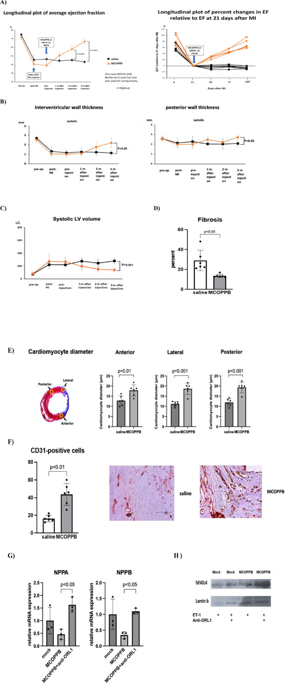
Purpose: The number of ischemic heart failure (HF) patients is growing dramatically worldwide. However, there are at present no preventive treatments for HF. Our previous study showed that Gata4 overexpression improved cardiac function after myocardial infarction in the rat heart. We also found that Gata4 overexpression significantly increased a Pnoc gene expression, an endogenous ligand for cell membrane receptor, ORL1. We hypothesized that an activation of ORL1 receptor would suppress HF in a rat ischemic heart model.

Method: Adult Sprague Dawley rats (8 weeks old, 6 males and 6 females) underwent left anterior descending coronary artery ligation. Three weeks later, normal saline or MCOPPB (ORL1 activator, 2.5mg/kg/day) intraperitoneal injection was started, and continued 5 days a week, for 3 months. Echocardiography was performed six times, pre-operative, 3 days after coronary artery ligation, pre-MCOPPB or saline injection, and 1, 2, and 3 months after saline or MCOPPB injection started. Animals were euthanized after 3 months follow up and the heart was harvested for histological analysis.

Results: ORL1 activator, MCOPPB, significantly improved cardiac function after myocardial infarction in rat (Ejection fraction, MCOPPB vs saline at euthanasia, 67 ± 3 vs 43 ± 2, p < 0.001). MCOPPB also decreased fibrosis and induced angiogenesis.

Conclusion: ORL1 activator, MCOPPB, may be a novel treatment for preventing HF progression.

Keywords: MCOPPB; ORL1 receptor activation; Prevention of ischemic heart failure progression.


Conflict of interest statement

Declarations Conflicts of interest/Competing interests: none

Figures

Figure 1
Figure 1. Volcano plot of RNAseq analysis.
A) Cardiac fibroblasts were treated with lentivirus encoding Gata4 or GFP (each 20MOI) for 14 days (n=3). PNOC gene was significantly upregulated by Gata4. B) Chromatography assay for MCOPPB detection in the heart. Rats received MCOPPB 2.5mg/kg/day or saline intraperitoneally for 4 days (n=3/group) and were euthanized immediately after the last injection. The heart tissues were examined with Mass spectrometry. MCOPPB was detected in the heart tissue of all MCOPPB received rats but no MCOPPB in the saline received rats. Representative chromatography of MCOPPB received rat is shown. C) Schematic showing the experimental design of MCOPPB study. Twelve adult Sprague Dawley rats were enrolled. Three weeks after the coronary artery ligation, they were treated with saline or MCOPPB for 3 months; six rats (3 males and 3 females) for saline, and another 6 rats (3 males and 3 females) for MCOPPB. Echocardiography was performed six times, 1: Pre-op, 2: Post-op, 3: Pre-treatment, 4: Four weeks after treatment started, 5: Eight weeks after treatment started, and 6: twelve weeks after treatment started. D) Fibrosis analysis. The excised heart was cut transversally and sectioned with 2 (2- to 3-mm thick) slices obtained, One, immediately cephalad and another one immediately caudad to the transverse centerline of the infarct region, which was readily identifiable by gross inspection. To assess the extent of fibrosis, 14 sections per animal (at a 120-μm interval between each section) were stained with Masson’s trichrome. E) Three regions where cardiomyocyte diameter was measured: The slide demonstrating the greatest area of fibrosis, as identified by Masson’s Trichrome staining, was selected for each animal. In each slide, 20 longitudinally oriented cardiomyocytes from each 3 regions, anterior, lateral, and posterior, were examined, and the diameters were defined. The mean value of 20 measurements represented 1 sample from each position in each animal.
Figure 2
Figure 2. Cardiac function was improved with MCOPPB administration.
A) Echocardiography results show longitudinal plot of changes in average ejection fraction (graph on left) and individual relative percent changes in ejection fraction (graph on right) associated with saline (black line) and MCOPPB (orange line) administration, started at day 21 after myocardial infarction induction. Ejection fraction significantly improved in the MCOPPB group (MCOPPB vs saline at 2 months follow-up, 58±3 vs 45±2, p<0.001, MCOPPB vs saline at euthanasia, 67±3 vs 43±2, p<0.001). Two-tailed ANOVA with Bonferroni post hoc test was used. n=6/group. B) Systolic wall thickness increased in MCOPPB group. LV wall thickness was measured by echocardiography with short axis M-mode image, at interventricular wall (left), and posterior wall (right). Both end-systolic interventricular septum (IVS) (left) and end-systolic left ventricular posterior wall (LVPW) (right) were significantly greater in MCOPPB group compared to saline group at euthanasia (end-systolic IVS, MCOPPB vs saline, 2.2±0.5mm vs 1.1±0.3mm, p<0.01, end-systolic LVPW, MCOPPB vs saline, 2.7±0.2mm vs 2.1±0.3mm, p<0.05). Two-tailed ANOVA with Bonferroni post hoc test was applied. n=6/group. Orange line: MCOPPB group, black line : saline group. C) End-systolic volume was significantly decreased in MCOPPB-received group at euthanasia. End-systolic volume was calculated with VenoLAB software: (7.0/(2.4+LVIDs))xLVIDs3, LVIDs=left ventricular internal diameter end systole). End-systolic volume was significantly decreased in MCOPPB-received group at euthanasia (MCOPPB vs saline, 140±30μL vs 280±44 μL, p<0.001). Orange line: MCOPPB group, Black line: Saline group. Two-tailed ANOVA with Bonferroni post-hoc test was used. n=6/group. D) MCOPPB decreased extent of left ventricular wall fibrosis. The percent of left ventricular wall fibrosis was determined by Masson-Trichrome staining of the sections of myocardial tissue harvested 3 months after the administration of MCOPPB or saline (Figure 1D). Fibrosis area significantly decreased in MCOPPB group (% fibrosis area, MCOPPB vs saline, 14±2 vs 29±10, p<0.05). Two-tailed t-test was used. F) MCOPPB increased angiogenesis in infarcted heart. Myocardial angiogenesis was analyzed by immunohistochemical staining with CD31 antibody. The number of capillaries was counted with two slides per animal, most positive cells found in peri-infarct region (x200 magnification). MCOPPB group had significantly higher vessel counts in border zone (44±12 vs 16±4, p<0.01). The graph shows the number of vessels/field. n=6/group. Two-tailed t test was used. Photo-images are two representative slides from saline and MCOPPB groups. Scale bar=150μm. G) NPPA and NPPB were downregulated by MCOPPB. Rat neonatal cardiomyocytes were treated with nociception agonist, MCOPPB (0.5mM), ET-1 (100nM) and anti-ORL1(ORL1 antagonist, [Nphe1]Nociceptin(1–13)NH2]) (10mM). qPCR shows that NPPA and NPPB, which are downstream transcription genes of NFAT signaling pathway and related to pathological hypertrophy, were downregulated by MCOPPB. Significantly, those downregulations by MCOPPB were diminished by adding ORL1 antagonist. Two-tailed ANOVA with Bonferroni post hoc test was used. Primers for NPPA: forward, 5’-CGTATACAGTGCGGTGTCCAAC-3’. reverse, 5’-CATCTTCTCCTCCAGGTGGTCTAG-3’. Primers for NPPB: forward, 5’-AAGTCCTAGCCAGTCTCCAGAACA −3’. Reverse, 5’-TTGAGAGCTGTCTCTGAGCCATT-3’. H) ORL1 inhibitor increased nuclear dephosphorylated NFAT. Rat neonatal cardiomyocytes were treated with MCOPPB (0.5 μM), ET-1 (100nM) and anti-ORL1 (10 μM). Nucler fractions of cells were extracted using NE-PER nuclear reagents kit and analyzed by western blotting with NFATc4 antibody(Santa Cruz Biotechnology- SC 271597). Lamin b (Santa Cruz Biotechnology- SC 374015) was used as a loading control. NFATc4 expression was significantly increased by adding ORL1 antagonist.

References

    1. Bian J, Popovic ZB, Benejam C, Kiedrowski M, Rodriguez LL, Penn MS. Effect of cell-based intercellular delivery of transcription factor Gata4 on ischemic cardiomyopathy. Circ Res. 2007;100:1622–1633. DOI: 10.1161/01.RES.000026978.75877.68. - DOI - PubMed
    1. Rysä J, Tenhunen O, Serpi R, Soini Y, Nemer M, Leskinen H, Ruskoaho H. Gata-4 is an angiogenic survival factor of the infarcted heart. Circ Heart Fail. 2010;3:440–450. DOI: 10.1161/CIRCHEARTFAILURE.109.889642. - DOI - PubMed
    1. Mathison M, Singh VP, Sanagasetti D, Yang L, Pinnamaneni JP, Yang J, Rosengart TK. Cardiac reprogramming factor Gata4 reduces postinfarct cardiac fibrosis through direct repression of the profibrotic mediator snail. J Thorac Cardiovasc Surg. 2017;154:1601–1610. DOI: 10.1016/j.jtcvs.2017.06.035. - DOI - PMC - PubMed
    1. Acosta AJ, Rysä J, Szabo Z, Moilanen A-M, Serpi R, Ruskoaho H. Phosphorylation of GATA4 at serine 105 is required for left ventricular remodelling process in angiotensin II-induced hypertension in rats. Basic Clin Pharmacol Toxicol. 2020;127:178–195. DOI: 10.1111/bcpt.13398. - DOI - PMC - PubMed
    1. Välimäki M, Ruskoaho HJ. Targeting GATA4 for cardiac repair. IUBMB Life. 2020;72:68–79. DOI: 10.1002/jub.2150. - DOI - PMC - PubMed

Publication types

LinkOut - more resources