

This is a preprint.
Structural Characterization of Disulfide-Linked p53-Derived Peptide Dimers
- PMID: 39070635
- PMCID: PMC11275974
- DOI: 10.21203/rs.3.rs-4644285/v1
Structural Characterization of Disulfide-Linked p53-Derived Peptide Dimers
Abstract
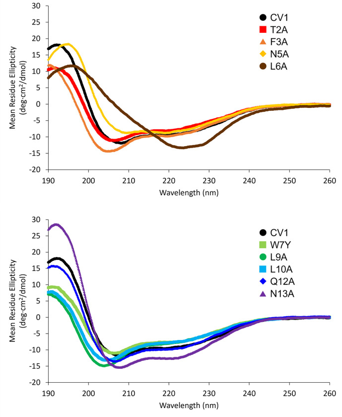
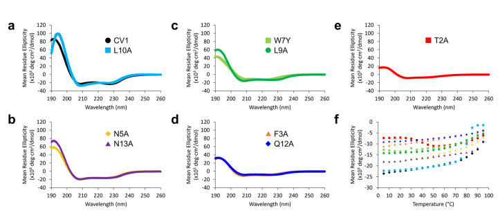
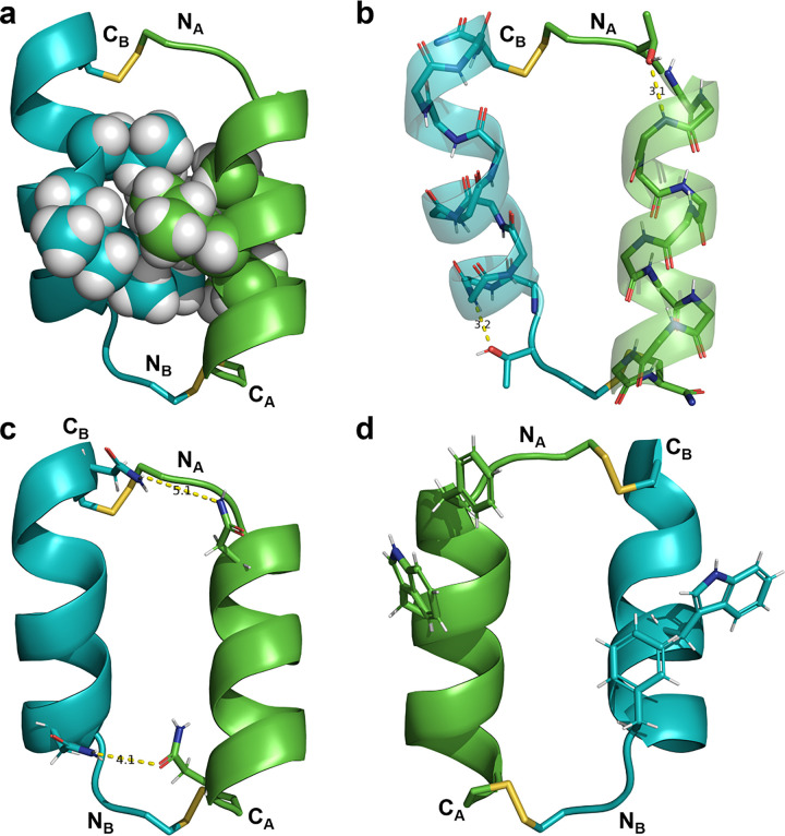
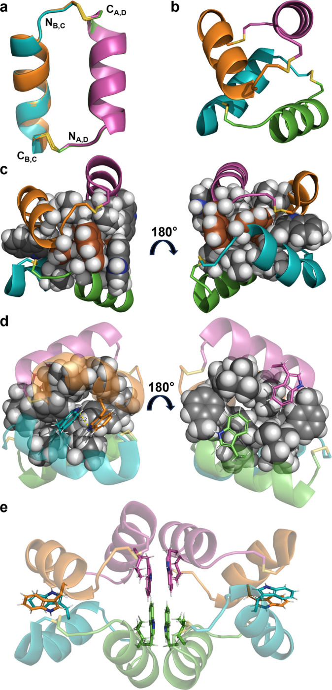
Disulfide bonds provide a convenient method for chemoselective alteration of peptide and protein structure and function. We previously reported that mild oxidation of a p53-derived bisthiol peptide (CTFANLWRLLAQNC) under dilute non-denaturing conditions led to unexpected disulfide-linked dimers as the exclusive product. The dimers were antiparallel, significantly α-helical, resistant to protease degradation, and easily reduced back to the original bisthiol peptide. Here we examine the intrinsic factors influencing peptide dimerization using a combination of amino acid substitution, circular dichroism (CD) spectroscopy, and X-ray crystallography. CD analysis of peptide variants suggests critical roles for Leu6 and Leu10 in the formation of stable disulfide-linked dimers. The 1.0 Å resolution crystal structure of the peptide dimer supports these data, revealing a leucine-rich LxxLL dimer interface with canonical knobs-into-holes packing. Two levels of higher-order oligomerization are also observed in the crystal: an antiparallel "dimer of dimers" mediated by Phe3 and Trp7 residues in the asymmetric unit and a tetramer of dimers mediated by Trp7 and Leu10. In CD spectra of Trp-containing peptide variants, minima at 227 nm provide evidence for the dimer of dimers in dilute aqueous solution. Importantly, and in contrast to the original dimer model, the canonical leucine-rich core and robust dimerization of most peptide variants suggests a tunable molecular architecture to target various proteins and evaluate how folding and oligomerization impact various properties, such as cell permeability.
Keywords: Dimerization; Disulfide; Folding; Structure.
Conflict of interest statement
The authors declare no competing financial interest.
Figures
References
-
- Adams P. D., Afonine P. V., Bunkoczi G., Chen V. B., Davis I. W., Echols N., Headd J. J., Hung L.-W., Kapral G. J., Grosse-Kunstleve R. W., McCoy A. J., Moriarty N. W., Oeffner R., Read R. J., Richardson D. C., Richardson J. S., Terwilliger T. C., & Zwart P. H. (2010). PHENIX: a comprehensive Python-based system for macromolecular structure solution. Acta Crystallographica Section D, 66(2), 213–221. 10.1107/S0907444909052925 - DOI - PMC - PubMed
-
- Albericio F., Hammer R. P., GarcÍA-EcheverrÍA C., Molins M. A., Chang J. L., Munson M. C., Pons M., Giralt E., & Barany G. (1991). Cyclization of disulfide-containing peptides in solid-phase synthesis†. International Journal of Peptide and Protein Research, 37(5), 402–413. 10.1111/j.1399-3011.1991.tb00755.x - DOI - PubMed
-
- Andreu D., F. A., N.A.. S., M.C. M. M. F., & Barany G. (1994). Formation of disulfide bonds in synthetic peptides and proteins. Methods in Molecular Biology, 35, 91–169. - PubMed
Publication types
Grants and funding
LinkOut - more resources
Full Text Sources
Research Materials
Miscellaneous
