[Preprint]. 2024 Jul 19:rs.3.rs-4557520.
doi: 10.21203/rs.3.rs-4557520/v1.

Biomolecular condensates can enhance pathological RNA clustering

Affiliations

Biomolecular condensates can enhance pathological RNA clustering

Priya Banerjee et al. Res Sq. .

Update in

Abstract

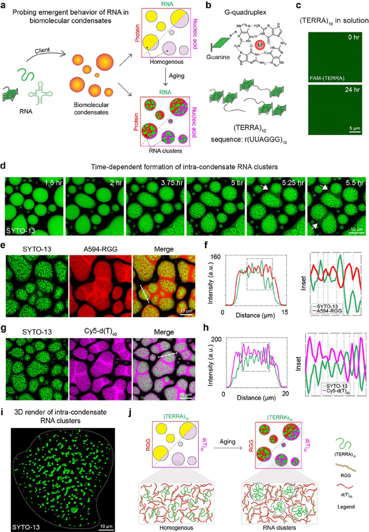
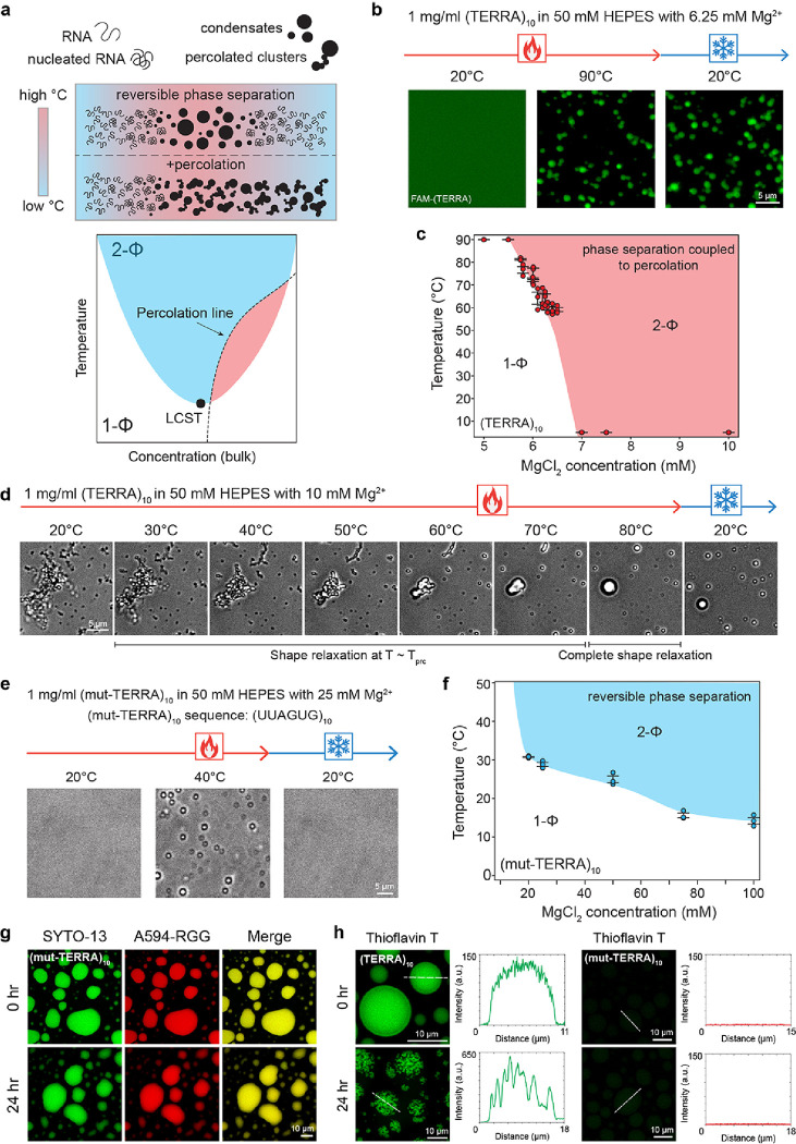
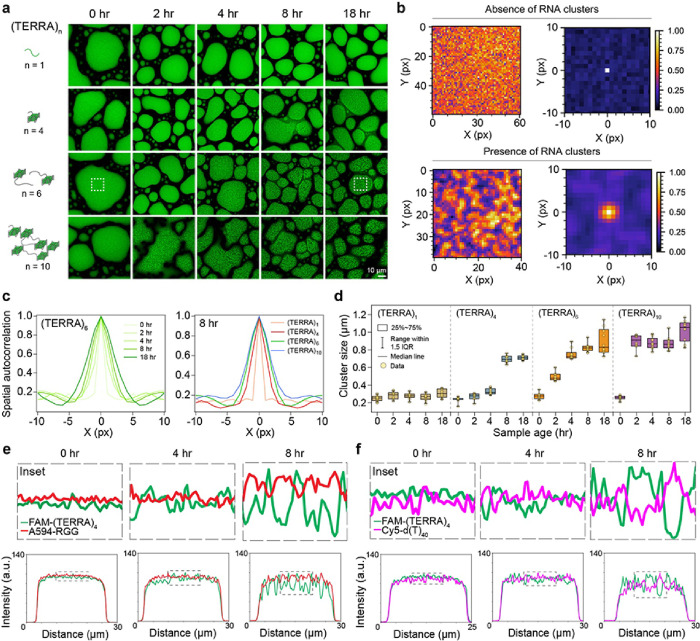
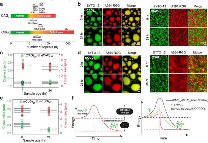
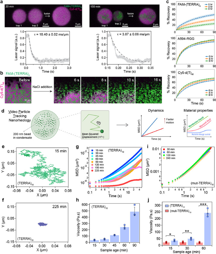
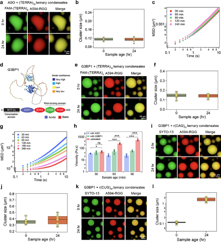
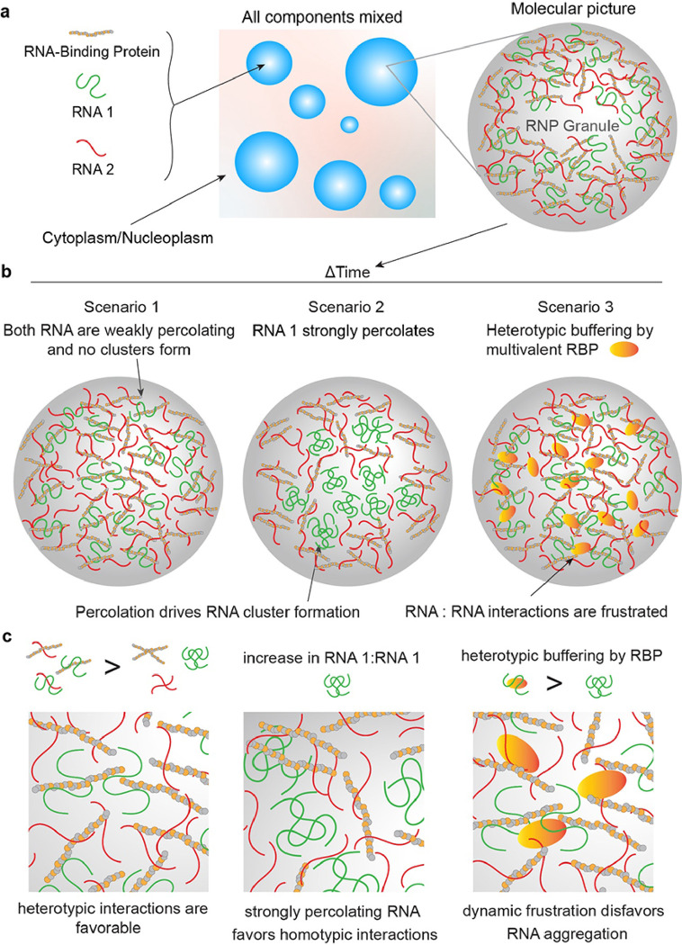
Intracellular aggregation of repeat expanded RNA has been implicated in many neurological disorders. Here, we study the role of biomolecular condensates on irreversible RNA clustering. We find that physiologically relevant and disease-associated repeat RNAs spontaneously undergo an age-dependent percolation transition inside multi-component protein-nucleic acid condensates to form nanoscale clusters. Homotypic RNA clusters drive the emergence of multiphasic condensate structures with an RNA-rich solid core surrounded by an RNA-depleted fluid shell. The timescale of the RNA clustering, which drives a liquid-to-solid transition of biomolecular condensates, is determined by the sequence features, stability of RNA secondary structure, and repeat length. Importantly, G3BP1, the core scaffold of stress granules, introduces heterotypic buffering to homotypic RNA-RNA interactions and impedes intra-condensate RNA clustering in an ATP-independent manner. Our work suggests that biomolecular condensates can act as sites for RNA aggregation. It also highlights the functional role of RNA-binding proteins in suppressing aberrant RNA phase transitions.


Conflict of interest statement

Competing interests P.R.B. is a member of the Biophysics Reviews (AIP Publishing) editorial board. This affiliation did not influence the work reported here. All other authors have no conflicts to report. Additional Declarations: There is NO Competing Interest.

Figures

Figure 1
Figure 1. RNA aggregation is enhanced within multi-component biomolecular condensates.
(a) Depiction of the experimental approach to probing the emergent properties of a client RNA in a heterotypic biomolecular condensate system comprised of RGG and d(T)40. (a) A top-view schematic of Hoogsteen base-pairing in a G-quartet that forms the structural foundation of (TERRA)10. (c) In dilute solution, (TERRA)10 at 10 mg/ml remains soluble and does not show age-dependent aggregation, as probed by FAM-labeled (TERRA)4, over a period of 24 hours at room temperature. (d) In multicomponent condensates containing 1.0 mg/ml (TERRA)10, 5.0 mg/ml RGG, 1.5 mg/ml d(T)40 [buffer = 25 mM Tris-HCl (pH 7.5), 25 mM NaCl, 20 mM DTT], (TERRA)10 undergoes an age-dependent demixing transition into fractal-like clusters, visualized with SYTO-13 (green). RNA demixing results in a core-shell architecture of the condensates. Fusion of the shell phase of neighboring condensates is indicated by white arrows (see SupplementaryVideo 1). Localization of (TERRA)10 in 18 hours aged condensates [same composition as (d)] is negatively correlated with the localizations of RGG (e, f) and d(T)40 (g, h) as visualized by Alexa594-RGG and Cy5-d(T)40. (i) Three-dimensional rendering of super-resolution Z-stack images of (TERRA)10 clusters from an 18-hour-aged sample [same composition as (d)]. (j)Schematic of age-dependent intra-condensate RNA clustering. In experiments utilizing fluorescently labeled components, the concentration is 250 nM to 500 nM. Each experiment was independently repeated at least three times.
Figure 2
Figure 2. TERRA undergoes phase separation coupled to percolation that can be perturbed by mutations.
(a) (top) A schematic representing reversible phase separation to form RNA condensates as well as the phase separation coupled to percolation behavior to form irreversible condensates, in response to heating/cooling ramps. (bottom) Depiction of an RNA state diagram, highlighting regions of reversible LCST-type phase separation (blue) and irreversible percolation (red) demarcated by the percolation line (black dashed line). (b) (TERRA)10 with 6.25 mM Mg2+ remains homogenous in solution at 20°C, as visualized using FAM-(TERRA)4. At an elevated temperature, (TERRA)10 phase separates into condensates and does not revert to solution upon cooling, indicative of percolated network formation (Supplementary Video 2) (c) State diagram of (TERRA)10 for a set of experiments similar to (b) with titrations of Mg2+ concentration. (d)Temperature-controlled microscopy shows that (TERRA)10 with 10 mM Mg2+ forms percolated clusters at 20°C, which upon heating above the percolation temperature (Tprc), undergo shape relaxation into spherical condensates that persist when cooled to 20°C (Supplementary Video 3). (e) Temperature-controlled microscopy of 1 mg/ml (mut-TERRA)10 in 50 mM HEPES with 25 mM Mg2+ shows reversible RNA phase separation (Supplementary Video 4).(f) State diagram of (mut-TERRA)10 for a set of experiments similar to (e) with titrations of Mg2+ concentration. (mut-TERRA)10 in RGG-d(T)40 condensates do not show intra-condensate RNA percolation. (h) Thioflavin T (ThT) staining of (TERRA)10 containing RGG-d(T)40 condensates shows homogenous ThT fluorescence at 0 hours and ThT fluorescence within intra-condensate RNA clusters at 24 hours after sample preparation. ThT staining of (mut-TERRA)10 containing RGG-d(T)40 condensates shows the absence of ThT fluorescence at 0 hours and 24 hours after sample preparation. The concentration of ThT used is 50 μM. The concentration of RNA used for temperature-controlled microscopy measurements is 1 mg/ml in 50 mM HEPES (pH 7.5) with the specified Mg2+ concentrations. The composition of the ternary condensate system is 1 mg/ml RNA, 5 mg/ml RGG, and 1.5 mg/ml d(T)40 in a buffer containing 25 mM Tris-HCl (pH 7.5), 25 mM NaCl, and 20 mM DTT. In experiments utilizing fluorescently labeled components, the concentration range is 250 nM to 500 nM. Each experiment was independently repeated at least three times.
Figure 3
Figure 3. TERRA repeat numbers dictate the timescale of RNA clustering.
(a) The effect of (TERRA)n repeat number (n) on the timescale of RNA clustering in RGG-d(T)40 condensates. TERRA clusters are visualized with SYTO-13 (green). The white box corresponds to analyses done in (b). (b) Color map images of (TERRA)6 containing RGG-d(T)40 condensates as shown in (a) at 15 minutes (above; absence of RNA clustering) and 18 hours (below; presence of RNA clustering) after sample preparation along with corresponding x-y spatial autocorrelation functions from SAC. (c) SAC line profiles of (TERRA)6 at various time points as shown in (b) (left) and SAC line profiles of (TERRA)n (n= 1, 4, 6, 10) at a sample age of 8 hours (right). (d) Cluster sizes derived from SAC analysis of (TERRA)n at increasing sample age. Pairwise line profile analyses of (TERRA)4 images with respect to RGG (e) and d(T)40 (f) as a function of time (for corresponding images, see Supplementary Fig. 9). Each line profile shown here is normalized with respect to the maximum intensity value, wherein all values were first offset by the minimum intensity value. The composition of the ternary RGG-d(T)40 condensate system is 1 mg/ml RNA, 5 mg/ml RGG, and 1.5 mg/ml d(T)40 in a buffer containing 25 mM Tris-HCl (pH 7.5), 25 mM NaCl, and 20 mM DTT. In experiments utilizing fluorescently labeled components, the concentration range is 250 nM to 500 nM. Each experiment was independently repeated at least three times.
Figure 4
Figure 4. Repeat expanded RNAs form intra-condensate clusters in a length-dependent manner.
(a) Schematic of disease-associated triplet RNA repeat expansions highlighting the threshold number of RNA repeats (n) that correspond to healthy states versus pathological states. Diseases are listed in an order according to the minimum RNA repeat length required for disease onset. The disease threshold for each GC-rich repeat RNA is marked according to the lowest repeat length linked to disease onset, , . Disease abbreviations are as follows. SCA6: Spinocerebellar Ataxia (SCA) Type 6, HD: Huntington’s disease, SBMA: Spinal and Bulbar Muscular Atrophy, SCA7: SCA Type 7, SCA2: SCA Type 2, SCA17: SCA Type 17, SCA1: SCA Type 1, SCA12: SCA Type 12, SCA3: SCA Type 3, DRPLA: Dentatorubral-Pallidoluysian Atrophy, HDL2: Huntington’s Disease-Like 2, DM1: Myotonic Dystrophy Type 1, FECD: Fuchs’ Endothelial Corneal Dystrophy, SCA8: SCA Type 8, CDM: Congenital Myotonic Dystrophy. (b)r(CAG)20 containing RGG-d(T)40 condensates remain homogenous and do not form clusters with time (left) whereas r(CAG)31 containing condensates form microscale RNA clusters. The 0-hour image was acquired after 15 minutes of sample preparation (right). (c) Cluster sizes derived from SAC analysis corresponding to (b). (d) r(CUG)31 containing RGG-d(T)40 condensates remain homogenous for 24 hours whereas r(CUG)47 containing condensates form microscale RNA clusters. The 0-hour image was acquired after 15 minutes of sample preparation. (e) Cluster sizes derived from SAC analysis corresponding to (d). (f)A schematic showing the hierarchy of activation energy barrier for an RNA monomer to form percolated RNA clusters. The composition of the ternary RGG-d(T)40 condensate system is 1 mg/ml RNA [0.45 mg/ml in the case of r(CUG)47], 5 mg/ml RGG, and 1.5 mg/ml d(T)40 in a buffer containing 25 mM Tris-HCl (pH 7.5), 25 mM NaCl, and 20 mM DTT. In experiments utilizing fluorescently labeled components, the concentration range is 250 nM to 500 nM. Each experiment was independently repeated at least three times.
Figure 5
Figure 5. Intra-condensate RNA clustering drives a liquid-to-solid transition.
(a) (top) Optical tweezer-mediated fusion of (TERRA)10 containing RGG-d(T)40 condensates at two different time points, as indicated, after sample preparation (see Supplementary Videos 5, 6). (bottom) Corresponding force relaxation profiles and the estimated fusion relaxation times (error represents ±1 standard deviation). (b) The addition of 0.5 μl of 5 M NaCl to (TERRA)10 containing RGG-d(T)40 condensates at 6 hours after sample preparation shows the dissolution of Cy5-d(T)40-rich shell phase (magenta) but not the (TERRA)10 clusters (green) (see Supplementary Fig. 15; Supplementary Video 7). (c) FRAP recovery profiles of FAM-(TERRA)4, Alexa 594-RGG, and Cy5-d(T)40 in (TERRA)4 containing RGG-d(T)40 condensate system with age (Supplementary Fig. 16; Supplementary Videos 10–15). Shaded regions in each plot signify the standard error. (d) Schematic of video particle tracking (VPT) nanorheology using beads passively embedded inside condensates, which is used to estimate their mean squared displacements (MSD) to ascertain condensate material properties. Created with BioRender.com. Individual bead trajectories at 15 minutes (e) and 225 minutes (f) inside (TERRA)10 containing RGG-d(T)40 condensates. (g) MSDs of beads inside (TERRA)10 containing RGG-d(T)40 condensates as a function of condensate age. (h) The corresponding terminal viscosities are reported. (i) MSDs of beads inside (mut-TERRA)10 containing RGG-d(T)40 condensates as a function of condensate age. (j) The corresponding terminal viscosities are reported in comparison to that of (TERRA)10 containing RGG-d(T)40 condensates as a function of condensate age. Statistical significance was determined by performing a two-sided Student’s t-test (* means p<0.05, ** means p<0.01, *** means p<0.001) between viscosities of (TERRA)10 and (mut-TERRA)10 condensates; the p-values determined at sample age of 15 minutes, 30 minutes, and 60 minutes are 0.0423, 0.0100, and 0.0002, respectively. The composition of the ternary condensate system is 1 mg/ml RNA, 5 mg/ml RGG, and 1.5 mg/ml d(T)40 for the optical tweezer and FRAP experiments; 0.5 mg/ml RNA, 2.5 mg/ml RGG, and 0.75 mg/ml d(T)40 for the condensate dissolution experiments; and 2 mg/ml RNA, 10 mg/ml RGG, and 3.0 mg/ml d(T)40 for the VPT measurements. Buffer composition for all experiments is 25 mM Tris-HCl (pH 7.5), 25 mM NaCl, and 20 mM DTT. In experiments utilizing fluorescently labeled components, the concentration range is 250 nM. The left panel in (a) is representative of three samples, and the right panel in (a) is representative of a single sample. All other measurements were independently repeated at least three times.
Figure 6
Figure 6. Heterotypic buffering by ASO and G3BP1 can prevent homotypic RNA clustering in biomolecular condensates.
(a) (TERRA)10 containing RGG-d(T)40 condensates treated with TERRA antisense oligonucleotide [ASO; sequence: r(CCCUAA)] does not form age-dependent RNA clusters. (b) The corresponding cluster sizes derived from SAC analysis are reported. (c) Age-dependent MSDs of 200 nm beads inside (TERRA)10 containing RGG-d(T)40 condensates treated with ASO (also see Supplementary Fig. 21). (d) Alphafold2 predicted structure of G3BP1 (identifier: AF-Q13283-F1) with color coding corresponding to model confidence. Domain architecture of G3BP1 (NTF2L, nuclear transport factor 2-like domain; IDR, intrinsically disordered region; RRM, RNA recognition motif; RGG, Arg/Gly-rich domain). (e) (TERRA)10 containing RGG-d(T)40 condensates with G3BP1 do not show age-dependent morphological changes. (f).The corresponding cluster sizes derived from SAC analysis indicate a lack of microscale RNA clustering (g) Age-dependent MSDs of 200 nm beads inside (TERRA)10 containing RGG-d(T)40 condensates with G3BP1 (also see Supplementary Fig. 24). (h) Comparison of terminal viscosities of (TERRA)10 containing RGG-d(T)40 condensates with or without the ASO or G3BP1 [“untreated” data taken from Fig. 5h]. Statistical significance was determined by performing a two-sided Student’s t-test (* means p<0.05, ** means p<0.01, *** means p<0.001, ns means ‘not significant’) between viscosities of untreated condensates versus condensates containing either the ASO or G3BP1. The p-values, between ‘untreated’ and ‘with ASO’ condition at 30 minutes, 60 minutes, and 90 minutes sample age, are 0.041, 0.0003, and 0.0008, respectively. The p-values between ‘untreated’ and ‘with G3BP1’ conditions at 30 minutes, 60 minutes, and 90 minutes sample age are 0.4766, 0.0009, and 0.0004, respectively. (i) r(CAG)31 containing RGG-d(T)40 condensates with G3BP1 do not show age-dependent morphological (j) changes. The corresponding cluster size analysis indicates an absence of RNA clustering. (k) r(CUG)47 containing RGG-d(T)40 condensates with G3BP1 do not show visible RNA clusters at 15 minutes after sample preparation but show some RNA clusters at an age of 24 hours. (l) The corresponding cluster size analysis is reported. The concentrations of the ASO and G3BP1 are 1 mg/ml and 10 μM, respectively. The composition of the condensate system used for imaging is 1 mg/ml RNA [0.45 mg/ml in the case of r(CUG)47], 5 mg/ml RGG, and 1.5 mg/ml d(T)40. For the nanorheology measurements, the relative proportion of the condensate components was kept the same, but the overall concentration of each component was doubled to achieve a higher volume fraction of the dense phase. Buffer composition for all experiments is 25 mM Tris-HCl (pH 7.5), 25 mM NaCl, and 20 mM DTT. In experiments utilizing fluorescently labeled components, the concentration range is 250 nM to 500 nM. Each experiment was independently repeated at least three times, except for the sample with G3BP1 in (h) that aged for 90 minutes, which was independently repeated two times.
Figure 7
Figure 7. schematic showing the proposed model of intra-condensate RNA percolation and heterotypic buffering.
(a) A model of multi-component condensates formed by two RNAs with strong (as shown in green) and weak (as shown in red) percolation propensity, respectively, and an RBP. (b) Three possible scenarios of RNA percolation-driven condensate aging or a lack thereof in the presence of a multivalent RBP. (c) Zoomed-in views of the panels shown above.

References

    1. Shin Y, Brangwynne CP. Liquid phase condensation in cell physiology and disease. Science 2017, 357(6357): eaaf4382. - PubMed
    1. Banani SF, Lee HO, Hyman AA, Rosen MK. Biomolecular condensates: organizers of cellular biochemistry - Nature Reviews Molecular Cell Biology. Nat Rev Mol Cell Biol 2017, 18(5): 285–298. - PMC - PubMed
    1. Boeynaems S, Alberti S, Fawzi NL, Mittag T, Polymenidou M, Rousseau F, et al. Protein Phase Separation: A New Phase in Cell Biology. Trends Cell Biol 2018, 28(6): 420–435. - PMC - PubMed
    1. Brangwynne CP, Eckmann CR, Courson DS, Rybarska A, Hoege C, Gharakhani J, et al. Germline P Granules Are Liquid Droplets That Localize by Controlled Dissolution/Condensation. Science 2009, 324(5935): 1729–1732. - PubMed
    1. Li P, Banjade S, Cheng HC, Kim S, Chen B, Guo L, et al. Phase transitions in the assembly of multivalent signalling proteins. Nature 2012, 483(7389): 336–340. - PMC - PubMed

Publication types

LinkOut - more resources