[Preprint]. 2024 Jul 16:2024.07.16.603789.
doi: 10.1101/2024.07.16.603789.

Diffusing protein binders to intrinsically disordered proteins

Affiliations

Diffusing protein binders to intrinsically disordered proteins

Caixuan Liu et al. bioRxiv. .

Update in

  • Diffusing protein binders to intrinsically disordered proteins.
    Liu C, Wu K, Choi H, Han HL, Zhang X, Watson JL, Ahn G, Zhang JZ, Shijo S, Good LL, Fischer CM, Bera AK, Kang A, Brackenbrough E, Coventry B, Hick DR, Qamar S, Li X, Decarreau J, Gerben SR, Yang W, Goreshnik I, Vafeados D, Wang X, Lamb M, Murray A, Kenny S, Bauer MS, Hoofnagle AN, Zhu P, Knowles TPJ, Baker D. Liu C, et al. Nature. 2025 Aug;644(8077):809-817. doi: 10.1038/s41586-025-09248-9. Epub 2025 Jul 30. Nature. 2025. PMID: 40739343 Free PMC article.

Abstract

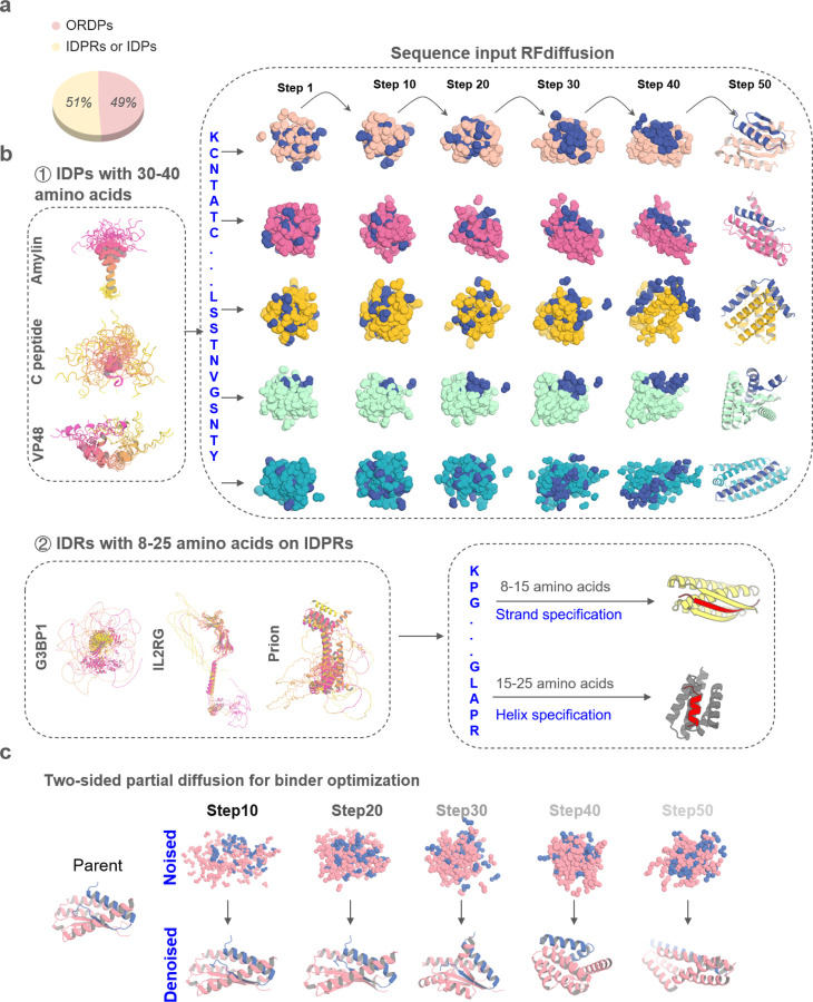
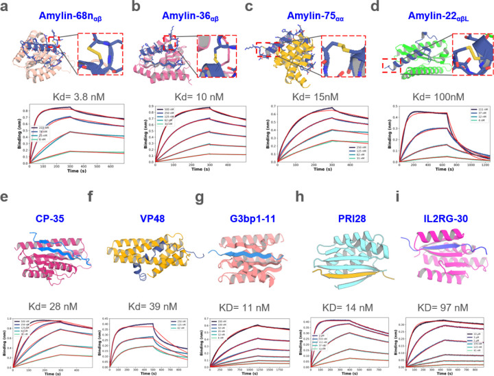
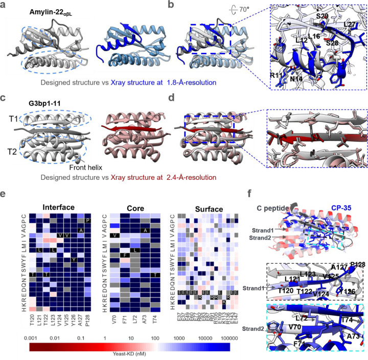
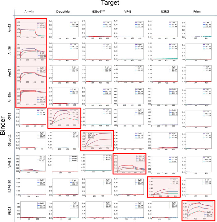
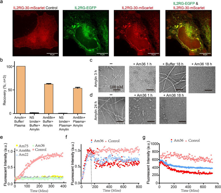
Proteins which bind intrinsically disordered proteins (IDPs) and intrinsically disordered regions (IDRs) with high affinity and specificity could have considerable utility for therapeutic and diagnostic applications. However, a general methodology for targeting IDPs/IDRs has yet to be developed. Here, we show that starting only from the target sequence of the input, and freely sampling both target and binding protein conformation, RFdiffusion can generate binders to IDPs and IDRs in a wide range of conformations. We use this approach to generate binders to the IDPs Amylin, C-peptide and VP48 in a range of conformations with Kds in the 3 -100nM range. The Amylin binder inhibits amyloid fibril formation and dissociates existing fibers, and enables enrichment of amylin for mass spectrometry-based detection. For the IDRs G3bp1, common gamma chain (IL2RG) and prion, we diffused binders to beta strand conformations of the targets, obtaining 10 to 100 nM affinity. The IL2RG binder colocalizes with the receptor in cells, enabling new approaches to modulating IL2 signaling. Our approach should be widely useful for creating binders to flexible IDPs/IDRs spanning a wide range of intrinsic conformational preferences.

Keywords: Amyloid fibril dissociation; Intrinsically disordered protein; RFdiffusion; Rosetta; deep learning; diagnostics; intrinsically disordered region; protein design.


Figures

Figure. 1
Figure. 1. Design strategies for binding conformational flexible peptides
a, Frequency of ORDPs (ordered proteins), IDPRs /IDPs (intrinsically disordered proteins) in the human proteome. b, ① Left, the NMR structure of Amylin (PDBID: 2KB8), C peptide (PDBID: 1T0C), the predicted structures of VP48 by five AlphaFold models. The 5 predicted structures of VP48 are aligned together, revealing the flexibility of the intrinsically disordered protein. Right, Diffusion models for proteins are trained to recover noised protein structures and to generate new structures by reversing the corruption process through iterative denoising of initially random noise into a realistic structure. Here, A modified version of RFdiffusion was trained on two chain systems from the PDB to permit the design of protein binders to targets, for which only the sequence of the target was specified. The fine-tuned was found to generate binders to peptides in finely varying helix conformationsWith solely sequence input. ② Left, the predicted structures of G3BP1, IL2RG and prion by five AlphaFold models. Right, A modified version of RFdiffusion was trained, allowing for specification of the secondary structure of a region, along with its sequence (See Method). When provided with the same target sequence input but different secondary structure specifications (helix or strand), the resulting conformations of the target could vary. c, Top: two sided partial diffusion. RFdiffusion is used to denoise a randomly noised starting parent design for both target and binder ; varying the extent by different noised step of initial noising (top row) enables control over the extent of introduced structural variation (bottom row; colours, new designs; grey, parent design).
Figure. 2
Figure. 2. Design of disordered region binder
a-d, Binder design of Amylin using sequence input diffusion. Top, from left to right, design model of Amylin and its binder Amylin-68nαβ, Amylin-36αβ, Amylin-75αα and Amylin-22αβL, respectively. The secondary structure of Amylin is indicated in the subscript of the binder’s name. For each of the designs, the Amylin disulfide bonds between 2nd Cysteine and 7th Cysteine were retained well. Bottom, from left to right, the BLI measurement indicated that the binding affinity between Amylin-68nαβ, Amylin-36αβ, Amylin-75αα, Amylin-22αβL and Amylin are 3.8, 10, 15, 100 nM respectively. e-f, Binder design of CP and VP48 using sequence input diffusion, the binder affinity of CP and VP48 are 28 and 39 nM, respectively. g-i, Binder design using strand specification. Top, from left to right, design model of G3BP1RBD, prion and IL2RG and their binders G3bp1–11, PRI28 and IL2RG-30. Bottom, the BLI measurement indicated that the binding affinity of G3bp1–11, PRI28 and IL2RG-30 binders are 11, 14 and 97 nM, respectively.
Figure. 3
Figure. 3. Structural characterizations.
a, Left, the designed model of Amylin-22αβL, with target and binder proteins rendered in dim gray and gray, respectively. The helical and strand segments that create the groove in the binder, docking the helical segment of Amylin, are highlighted with blue dashed ellipsoid. Right, the crystal structure of Amylin-22αβL at 1.8 Å-resolution, with target and binder proteins rendered in blue and cornflower blue, respectively. b, Left, the overlay of the design model and the crystal structure of Amylin-22αβL. Right, magnified views of the regions indicated with black dotted frames in the left panel are provided to illustrate the detailed interface view of the design and crystal structure. The binder proteins are rendered with 90% transparency to enhance the visibility of the peptide target. The key residues on the Amylin are labeled to illustrate the good alignment of the key residues between designed protein and crystal structure. c, Left, the designed model of G3bp1–11, with target and binder proteins rendered in dim gray and gray, respectively. The two α/β topologies (T1 and T2) of the binders, forming the cleft where the target strand is positioned, are highlighted with blue dashed ellipses. The front helix of T2 is denoted by a black arrow. Right, the crystal structure of G3bp1–11 at 2.4 Å-resolution, with target and binder proteins rendered in dark red and rosy brown, respectively. d, Left, the overlay of the design model and the crystal structure of G3bp1–11. Right, magnified views of the regions indicated with black dotted frames in the left panel. The front helix of T2 has been surface capped to reveal the strand pairing interface. e, Heat maps representing C peptide-binding Kd (nM) values for single mutations in the designed interface (left), core (middle) and the surface (right). Substitutions that are heavily depleted are shown in blue, and beneficial mutations are shown in red, gray color indicates the lost yeast strains. For the interface region, we highlighted and showcased strand 1 (indicated by the arrow), which serves as the primary interaction secondary structure with the C peptide. For the core region, we showcased the right segment of strand 2 (indicated by the arrow), representing a main core region that does not form interactions with the C peptide. For the surface region, we selected the most exposed surface residues that don’t form any connections with other residues (Supplementary Fig. 6c). Full SSM map over all positions for CP35 is provided in Supplementary Fig. 6b. f, Top, designed binding proteins are colored by positional Shannon entropy from site saturation mutagenesis, with blue indicating positions of low entropy (conserved) and red those of high entropy (not conserved). Bottom, zoomed-in views of central regions of the design interface and core with the C peptide.
Figure. 4
Figure. 4. Specificity profile of designed binders in BLI.
Biotinylated peptides were immobilized onto octet streptavidin biosensors at equal densities and incubated with all binders in separate experiments at three concentrations (2, 0.667 and 0.222 μM except VP48 binder at 0.833, 0.277 and 0.093 μM). Amylin-68nαβ, Amylin-36αβ, Amylin-75αα, Amylin-22αβL are abbreviated as Am68n, Am36, Am75 and Am22, respectively. The designed on-target interactions are indicated with a light red background.
Figure. 5
Figure. 5. Applications of designed binders
a, Colocalization of binder IL2RG-30 and target membrane receptor IL2RG in HeLa Cells. Cells with endogenous IL2RG knocked out express only the red fluorescent mScarlet-tagged binder IL2RG-30, which is uniformly distributed throughout the cell (left). In contrast, cells co-expressing green EGFP-tagged IL2RG and red mScarlet-tagged IL2RG-30 show specific colocalization of both proteins. b, The LC–MS/MS recovery percent of Amylin from PBS-0.1% CHAPS buffer and EDTA-anticoagulated plasma was compared between BSA-blocked tosyl-activated bead, an off-target binder, and amylin-targeted binders (Am68n). Percent recovery was calculated using the peak area of a sample of pure amylin peptide in elution solvent as the denominator (i.e., 100% recovery of the peptide). Error bars represent SD (n=3). c-d, Visualization of fibril dissociation by Amylin-36αβ binder using negative staining electron microscopy. panels (c) and (d) demonstrate the dissociation of existing fibrils at elongation phase (c) and mature phase (d) following the addition of Amylin-36αβ. Scale bars, 100 nM. e, Thioflavin T (ThT) assay revealed that all 4 binders could strongly inhibit fibril formation at molar ratio of binder to Amylin 1:4. f, Amylin-36αβ could dissociate fibrils at elongation phase in concentration-dependent manner. The Tht assay was performed since the Amylin monomer, Amylin-36αβ was added at 3h when Amylin fibrils were at elongation phase, marked with a dotted line. Red dot and blue dot indicate that Amylin-36αβ to Amylin is 1:4 and 1:40, respectively. g, Tht assay was performed after the mature Amylin fibrils were formed for 24 h, at the same time, Amylin-36αβ was added, the data revealed that fibril fluorescence decreased in a concentration-dependent manner. Red dot and blue dot indicate that Amylin-36αβ to Amylin is 1:4 and 1:40, respectively.

References

    1. Zhang G. et al. Islet amyloid polypeptide cross-seeds tau and drives the neurofibrillary pathology in Alzheimer’s disease. (2022). - PMC - PubMed
    1. Dyson H. J. & Wright P. E. Intrinsically unstructured proteins and their functions. Nature Reviews Molecular Cell Biology 6, 197–208, doi: 10.1038/nrm1589 (2005). - DOI - PubMed
    1. Tompa P. The interplay between structure and function in intrinsically unstructured proteins. FEBS Lett 579, 3346–3354, doi: 10.1016/j.febslet.2005.03.072 (2005). - DOI - PubMed
    1. Uversky V. N. Intrinsically disordered proteins in overcrowded milieu: Membrane-less organelles, phase separation, and intrinsic disorder. Curr Opin Struct Biol 44, 18–30, doi: 10.1016/j.sbi.2016.10.015 (2017). - DOI - PubMed
    1. Bradbury A. & Plückthun A. Reproducibility: Standardize antibodies used in research. Nature 518, 27–29, doi: 10.1038/518027a (2015). - DOI - PubMed

Publication types

LinkOut - more resources