[Preprint]. 2024 Jul 16:2024.07.11.603104.
doi: 10.1101/2024.07.11.603104.

Female sex hormones exacerbate retinal neurodegeneration

Affiliations

Female sex hormones exacerbate retinal neurodegeneration

Ashley A Rowe et al. bioRxiv. .

Update in

  • Female sex hormones exacerbate retinal neurodegeneration.
    Rowe AA, Velasquez MJ, Aumeier JW, Reyes S, Yee T, Nettesheim ER, McDonald JG, Wert KJ. Rowe AA, et al. Sci Adv. 2025 Apr 11;11(15):eadr6211. doi: 10.1126/sciadv.adr6211. Epub 2025 Apr 11. Sci Adv. 2025. PMID: 40215317 Free PMC article.

Abstract

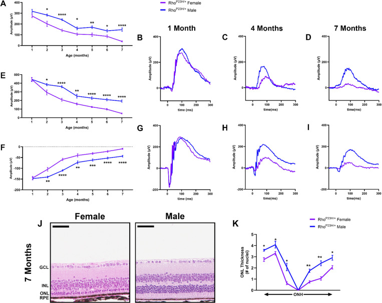
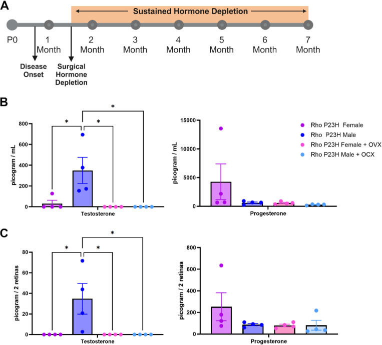
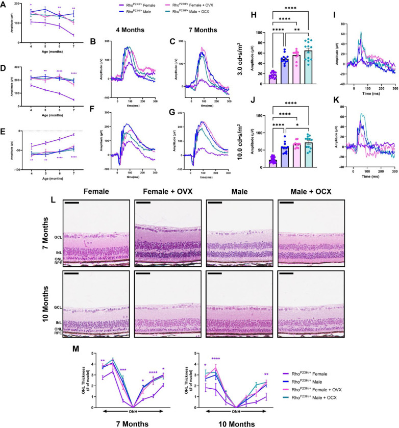
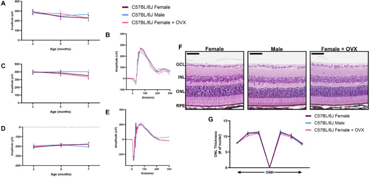
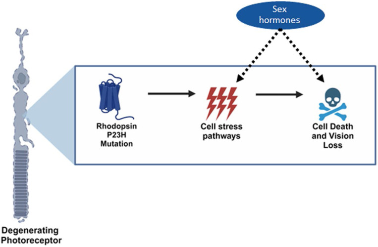
Neurodegenerative disorders such as Alzheimer's disease and macular degeneration represent major sources of human suffering, yet the factors influencing disease severity remain poorly understood. Sex has been implicated as one potential modifying factor. Here, we show that female sex is a risk factor for worsened outcomes in a model of retinal degeneration. Further, we show that this susceptibility is caused by the presence of female-specific circulating sex hormones. The adverse effect of female sex hormones was specific to diseased retinal neurons, and depletion of these hormones ameliorated this phenotypic effect. These findings provide novel insights into the pathogenesis of neurogenerative diseases and how sex hormones can impact the severity of disease. These findings have far-reaching implications for clinical trial design and the use of hormonal therapy in females with certain neurogenerative disorders.


Conflict of interest statement

Competing interests: Authors declare that they have no competing interests.

Figures

Figure 1.
Figure 1.. Rho P23H mice display a sexually dimorphic loss of photoreceptor function and survival.
(A) 0.01 cd•s/m2 scotopic ERG b-wave values in male (blue) and female (purple) Rho P23H mice from one to seven months of age. (B-D) Representative 0.01 cd•s/m2 scotopic ERG traces at one, four, and seven months of age. (E) 1.0 cd•s/m2 scotopic ERG b- and (F) a-wave amplitudes. (G-I) Representative 1.0 cd•s/m2 scotopic ERG traces at one, four, and seven months of age. Error bars = SEM. *, p<0.05. **, p<0.01. ***, p<0.001. ****, p<0.0001. Statistical analysis performed by multiple unpaired t-test with a Holm-Šídák Correction with α=0.05. N=16 eyes per group. (J) Representative H&E-stained histology from seven-month Rho P23H female and male mouse retinas. (K) Quantification of outer nuclear layer (ONL) thickness at six different regions spanning from the optic nerve head (ONH). GCL, ganglion cell layer; INL, inner nuclear layer; RPE, retinal pigmented epithelium. Statistics performed via multiple unpaired t-tests and a Holm-Šídák multiple comparisons test. *, p-value<0.05; **, p-value<0.01. Scale bar = 50 μM. N ≥ 3 mice per group.
Figure 2.
Figure 2.. Surgical gonadectomy depletes steroidal sex hormones in the serum and retina.
(A) Timeline depicting surgical procedure at six weeks of age, after disease onset, followed by sustained hormone depletion through the duration of the study. P0, post-natal day zero. (B) Mass spectrometry analysis of serum and (C) retina testosterone and progesterone in Rho P23H females (purple), males (blue), females with bilateral ovariectomy (OVX; pink), and males with orchiectomy (OCX; light blue), two-weeks post-surgery. *, p < 0.05. Statistics performed via One-way ANOVA with Tukey’s multiple comparison’s test. N = 4 mice per group. Part of figure made with Biorender.com (agreement #QH271R0NYW).
Figure 3.
Figure 3.. Ovariectomy ameliorates female photoreceptor neurodegeneration.
(A) 0.01 cd•s/m2 scotopic b-wave amplitudes from four to seven months of age in Rho P23H females (purple), males (blue), females with OVX (pink), and males with OCX (light blue). (B-C) Representative 0.01 cd•s/m2 scotopic traces. (E) 1.0 cd•s/m2 scotopic b- and (F) a-wave amplitudes from four to seven months of age, and (F-G) representative traces. Statistics performed via Two-way ANOVA with Tukey’s multiple comparison’s test. Statistical results are shown for females compared to females + OVX and males compared to males + OCX. N≥16 eyes. (H) 3.0 cd•s/m2 and (J) 10.0 cd•s/m2 photopic amplitudes at seven months of age, (I, K) with respective representative traces. Statistics analyzed by One-way ANOVA with Tukey’s multiple comparison’s test. N≥10 eyes. Error bars=SEM. (L) H&E-stained histology from seven and ten-month-old Rho P23H female and male mice with and without OVX or OCX, respectively. (M) Quantification of ONL thickness spanning from the ONH at both seven and ten months of age. Statistics performed via Two-way ANOVA with Tukey’s multiple comparison’s test. Statistical results are shown for females compared to females + OVX. N≥3 mice per group. Scale bar=50 μM. *, p<0.05; **, p<0.01; ***, p<0.001; ****, p<0.0001.
Figure 4.
Figure 4.. Sex hormone depletion does not affect healthy retinal neurons.
(A) 0.01 cd•s/m2 scotopic ERG b-wave amplitudes, (C) 1.0 cd•s/m2 scotopic ERG b- and (D) a-wave amplitudes from male (blue), female (purple), and females with bilateral ovariectomy (OVX; pink) C57BL/6J mice at three, five and seven months of age. Representative ERG traces for the (B) 0.01 cd•s/m2 and (E) 1.0 cd•s/m2 scotopic ERGs settings for all three groups at seven months of age. Statistics analyzed via Two-way ANOVA (α=0.05) with Tukey’s multiple comparison’s test. N = 10 eyes per group. (F) H&E-stained histology of retinas from seven-month-old C57BL/6J females, males, and females + OVX. (G) Quantification of the outer nuclear layer (ONL) thickness at six different regions in the retina spanning out from the optic nerve head (ONH) for all groups. GCL, ganglion cell layer; INL, inner nuclear layer; RPE, retinal pigmented epithelium. Statistics performed via Two-way ANOVA with Tukey’s multiple comparison’s test. Scale bar = 50 μM. N = 3 eyes per group.
Figure 5.
Figure 5.. Sex hormones impact cellular pathways underlying the rhodopsin P23H mutation in photoreceptor neurons.
Graphical schematic depicting our findings that female systemic sex hormones interact within pathways found in diseased, but not healthy, retinal neurons to exacerbate neurodegeneration in a sexually dimorphic manner. Part of figure made with BioRender.com (agreement #XT271R0LO4).

References

    1. Lopez-Lee C., Torres E. R. S., Carling G., Gan L., Mechanisms of sex differences in Alzheimer’s disease. Neuron 112, 1208–1221 (2024). - PMC - PubMed
    1. Marin A. I., Poppelaars F., Wagner B. D., Palestine A. G., Patnaik J. L., Holers V. M., Frazer-Abel A. A., Mathias M. T., Manoharan N., Fonteh C. N., Mandava N., Lynch A. M., Sex and Age-Related Differences in Complement Factors Among Patients With Intermediate Age-Related Macular Degeneration. Trans. Vis. Sci. Technol. 11, 22–22 (2022). - PMC - PubMed
    1. Mielke M. M., Sex and Gender Differences in Alzheimer’s Disease Dementia. Psychiatr. Times 35, 14–17 (2018). - PMC - PubMed
    1. Sasaki M., Harada S., Kawasaki Y., Watanabe M., Ito H., Tanaka H., Takeuchi A., Tsubota K., Takebayashi T., Nishiwaki Y., Kawasaki R., Gender-specific association of early age-related macular degeneration with systemic and genetic factors in a Japanese population. Sci. Rep. 8, 785 (2018). - PMC - PubMed
    1. Nuzzi R., Scalabrin S., Becco A., Panzica G., Gonadal hormones and retinal disorders: a review. Front. Endocrinol. 9, 66 (2018). - PMC - PubMed

Publication types

LinkOut - more resources