[Preprint]. 2024 Jul 19:2024.07.17.603936.
doi: 10.1101/2024.07.17.603936.

Immune disease dialogue of chemokine-based cell communications as revealed by single-cell RNA sequencing meta-analysis

Affiliations

Immune disease dialogue of chemokine-based cell communications as revealed by single-cell RNA sequencing meta-analysis

Mouly F Rahman et al. bioRxiv. .

Abstract

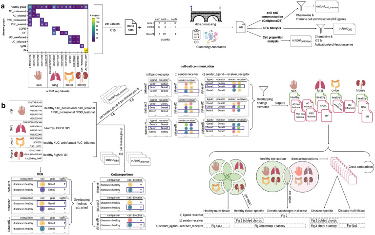
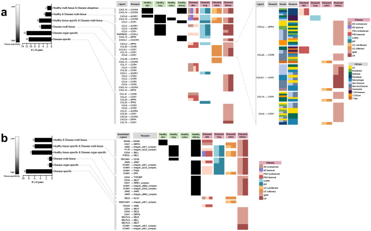
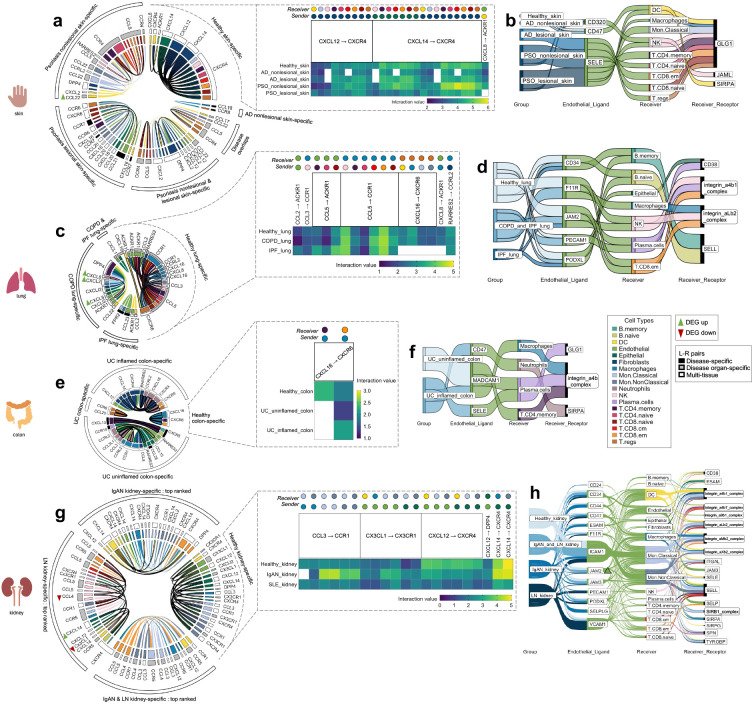
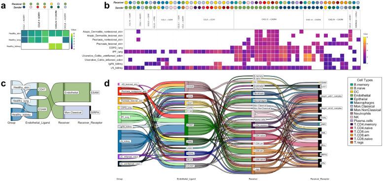
Immune-mediated diseases are characterized by aberrant immune responses, posing significant challenges to global health. In both inflammatory and autoimmune diseases, dysregulated immune reactions mediated by tissue-residing immune and non-immune cells precipitate chronic inflammation and tissue damage that is amplified by peripheral immune cell extravasation into the tissue. Chemokine receptors are pivotal in orchestrating immune cell migration, yet deciphering the signaling code across cell types, diseases and tissues remains an open challenge. To delineate disease-specific cell-cell communications involved in immune cell migration, we conducted a meta-analysis of publicly available single-cell RNA sequencing (scRNA-seq) data across diverse immune diseases and tissues. Our comprehensive analysis spanned multiple immune disorders affecting major organs: atopic dermatitis and psoriasis (skin), chronic obstructive pulmonary disease and idiopathic pulmonary fibrosis (lung), ulcerative colitis (colon), IgA nephropathy and lupus nephritis (kidney). By interrogating ligand-receptor (L-R) interactions, alterations in cell proportions, and differential gene expression, we unveiled intricate disease-specific and common immune cell chemoattraction and extravasation patterns. Our findings delineate disease-specific L-R networks and shed light on shared immune responses across tissues and diseases. Insights gleaned from this analysis hold promise for the development of targeted therapeutics aimed at modulating immune cell migration to mitigate inflammation and tissue damage. This nuanced understanding of immune cell dynamics at the single-cell resolution opens avenues for precision medicine in immune disease management.

Keywords: cell communication; chemokine; extravasation; immune disease; meta-analysis; scRNA-seq.


Conflict of interest statement

CONFLICT OF INTEREST The authors are or were employees of Sanofi US at the time of this work.

Figures

Figure 1.
Figure 1.. Overview of the single cell (sc)-RNA-seq datasets selected and how they were processed and analyzed for the meta-analysis of chemokine and extravasation-based cell-cell communication.
a) 15 scRNA-seq datasets were selected, encompassing the following 14 groups: healthy skin, atopic dermatitis (AD) nonlesional skin, AD lesional skin, psoriasis (PSO) nonlesional skin, PSO lesional skin, healthy lung, chronic obstructive pulmonary disease (COPD) lung, idiopathic pulmonary fibrosis (IPF) lung, healthy colon, ulcerative colitis (UC) uninflamed colon, UC inflamed colon, healthy kidney, IgA nephropathy (IgAN) kidney, lupus nephritis (LN) kidney. The counts and metadata files for each dataset were processed using the CellBridge pipeline. The Seurat RDS output was used for the down-stream analyses: cell-cell communication using CellphoneDB, differentially expressed gene (DEG) analysis (disease vs healthy), and cell proportion (disease vs healthy) analysis. Cell-cell communication results were filtered for interactions involving chemokine and immune cell extravasation genes, and DEG results were filtered for chemokine, immune cell extravasation, immune cell activation and proliferation genes. b) Per a set of datasets belonging to a particular group, common findings were extracted for each of the three types of analyses (cell-cell communication, DEG, cell proportions). For the cell-cell communication results: 1) healthy-tissue specific findings were extracted by comparing each tissue to each other (e.g. to extract interactions only occurring in healthy kidney), 2) all healthy interactions were compared to all disease interactions to extract those only occurring in disease, and then 3) those disease interactions were compared against each other to extract interactions that were specific to a particular disease group (e.g. only in COPD). These comparisons resulted in findings that were: healthy multi-tissue, healthy tissue specific, directional changes in disease (i.e. healthy interactions that occurred in disease, but with an increased/decreased interaction value, hence the ‘directional’ change), disease-specific and disease multi-tissue.
Figure 2.
Figure 2.. All of the unique chemokine and immune cell extravasation-based ligand-receptor pairs extracted from the meta-analysis of cell-cell communication predictions based on CellphoneDB.
a) The chemokine-based ligand-receptor pairs that took place in: multiple healthy tissues and all diseases (“Healthy multi-tissue & Disease ubiquitous”), multiple healthy and disease tissues (“Healthy & Disease multi-tissue”), a specific healthy tissue along with multiple disease tissues (“Healthy tissue-specific & Disease multi-tissue”), only disease tissue and not in any healthy tissue, but not specific to a particular diseased tissue (“Disease multi-tissue”), a specific diseased organ (“Disease organ-specific”), a particular disease (“Disease-specific”). For “Disease multi-tissue”, sender-receiver cell type-pairs were completely unshared across disease organ types (shown in the blow-out). b) The immune cell extravasation-based Endothelial_ligand – receptor pairs that took place in the various sets mentioned above. AD atopic dermatitis, COPD chronic obstructive pulmonary disease, IgAN IgA nephropathy, IPF idiopathic pulmonary fibrosis, LN lupus nephritis, PSO psoriasis, UC ulcerative colitis.
Figure 3.
Figure 3.. The tissue-specific sender-receiver ligand-receptor cell-cell communications identified with CellphoneDB; segmented by the four different organs (skin, lung, colon, kidney). Heatmaps display healthy tissue chemokine communications (which typically co-occurred in disease with either increased/decreased interaction values, i.e. with directional changes). Chord diagrams display healthy-tissue specific and disease-specific chemokine communications. Sankey diagrams display immune cell extravasation communications.
a) Healthy skin, atopic dermatitis and psoriasis-specific chemokine-based communication & b) extravasation-based communication. c) Healthy lung, chronic obstructive pulmonary disease and idiopathic pulmonary fibrosis-specific chemokine-based communication & d) extravasation-based communication. e) Healthy colon and ulcerative colitis-specific chemokine-based communication & f) extravasation-based communication. g) Top-ranked healthy kidney, IgA nephropathy and lupus nephritis-specific chemokine-based communication & h) extravasation-based communication. Bolded chords indicate sender_cell-receiver_cell communication that are unique to a particular disease. AD atopic dermatitis, COPD chronic obstructive pulmonary disease, DEG differently expressed gene, IgAN IgA nephropathy, IPF idiopathic pulmonary fibrosis, les lesion, LN lupus nephritis, nl nonlesion, PSO psoriasis, UC ulcerative colitis.
Figure 4.
Figure 4.. The tissue-nonspecific sender-receiver ligand-receptor cell-cell communications identified with CellphoneDB, with heatmaps for chemokine-based communications and Sankey diagrams for immune cell extravasation-based communications.
a) Healthy chemokine-based communication that were common across tissues, specifically, healthy skin, healthy lung and healthy kidney, at either increased/decreased interaction strength values. b) Disease chemokine-based communication that were common across all 10 disease groups examined. c) Healthy tissue extravasation-based communication that were common across three of the tissues examined. d) Disease extravasation-based communication that were common across nine of the disease groups examined. AD atopic dermatitis, COPD chronic obstructive pulmonary disease, IgAN IgA nephropathy, IPF idiopathic pulmonary fibrosis, LN lupus nephritis, PSO psoriasis, UC ulcerative colitis.
Figure 5.
Figure 5.. Experimental investigation of the CXCL2 --> CD8 T effector memory cell_DPP4 interaction found in the IgAN kidney cell-cell communication analysis.
a) Flow cytometry analysis of CD8+ memory T cells. b) Identification of CD8+ memory T cells as predominantly effector memory (EM) subtype (CD45RO+ and CD62L−), with a minor fraction of central memory (CM) cells (CD45RO+ and CD62L+). c) Cartoon depiction of the chemotaxis assay using the transwell; overtime, cells inputted to the top chamber of the transwell may be chemoattracted to the bottom chamber, similar to how within tissues, a chemotactic gradient chemoattracts cells to a site of inflammation. d) CXCL2 transwell migration assay conducted on day 0 revealed chemotaxis of CD8+ memory T cells towards CXCL2 (at all dilutions examined for both donors, with the exception of 10 ng/ml for donor 2), consistent with the meta-analysis findings. e-f) In CD8+ memory T cells, reduced DPP4 expression was observed only in the DPP4 KO group, while the wild-type (WT), electroporation only, and non-target control (NTC) KO groups showed no significant alterations on day 11. g) Subsequent CXCL2 transwell migration assay conducted with CD8+ memory T cells to assess the impact of DPP4 KO on CXCL2-induced chemotaxis. DPP4 KO cell’s CXCL2-induced migration was not significantly different from the medium-only condition (for both donors), contrasting with wild-type (WT) and NTC cells in donor 1 and 2 respectively, that showed significantly stronger migration towards CXCL2 compared to the medium-only control (p < 0.05). CXCL12 positive control showed similar levels of significantly higher migration compared to the medium-only control (p < 0.05 for both donors) for all groups (KO, WT, NTC). * p < 0.05. ** p < 0.01, *** p < 0.0001 compared to the medium control condition per donor.

Similar articles

References

    1. Adams T. S., Schupp J. C., Poli S., Ayaub E. A., Neumark N., Ahangari F., et al. (2020). Single-cell RNA-seq reveals ectopic and aberrant lung-resident cell populations in idiopathic pulmonary fibrosis. Sci. Adv. 6, eaba1983. doi: 10.1126/sciadv.aba1983 - DOI - PMC - PubMed
    1. Amersfoort J., Eelen G., and Carmeliet P. (2022). Immunomodulation by endothelial cells — partnering up with the immune system? Nat. Rev. Immunol. 22, 576–588. doi: 10.1038/s41577-022-00694-4 - DOI - PMC - PubMed
    1. Arazi A., Rao D. A., Berthier C. C., Davidson A., Liu Y., Hoover P. J., et al. (2019). The immune cell landscape in kidneys of patients with lupus nephritis. Nat. Immunol. 20, 902–914. doi: 10.1038/s41590-019-0398-x - DOI - PMC - PubMed
    1. Ardain A., Marakalala M. J., and Leslie A. (2020). Tissue-resident innate immunity in the lung. Immunology 159, 245–256. doi: 10.1111/imm.13143 - DOI - PMC - PubMed
    1. Arif N., Zinnhardt M., Nyamay’Antu A., Teber D., Brückner R., Schaefer K., et al. (2021). PECAM-1 supports leukocyte diapedesis by tension-dependent dephosphorylation of VE-cadherin. EMBO J. 40, e106113. doi: 10.15252/embj.2020106113 - DOI - PMC - PubMed

Publication types

LinkOut - more resources