[Preprint]. 2024 Jul 18:2023.12.07.570729.
doi: 10.1101/2023.12.07.570729.

"Off-pore" nucleoporins relocalize heterochromatic breaks through phase separation

Affiliations

"Off-pore" nucleoporins relocalize heterochromatic breaks through phase separation

Chiara Merigliano et al. bioRxiv. .

Abstract

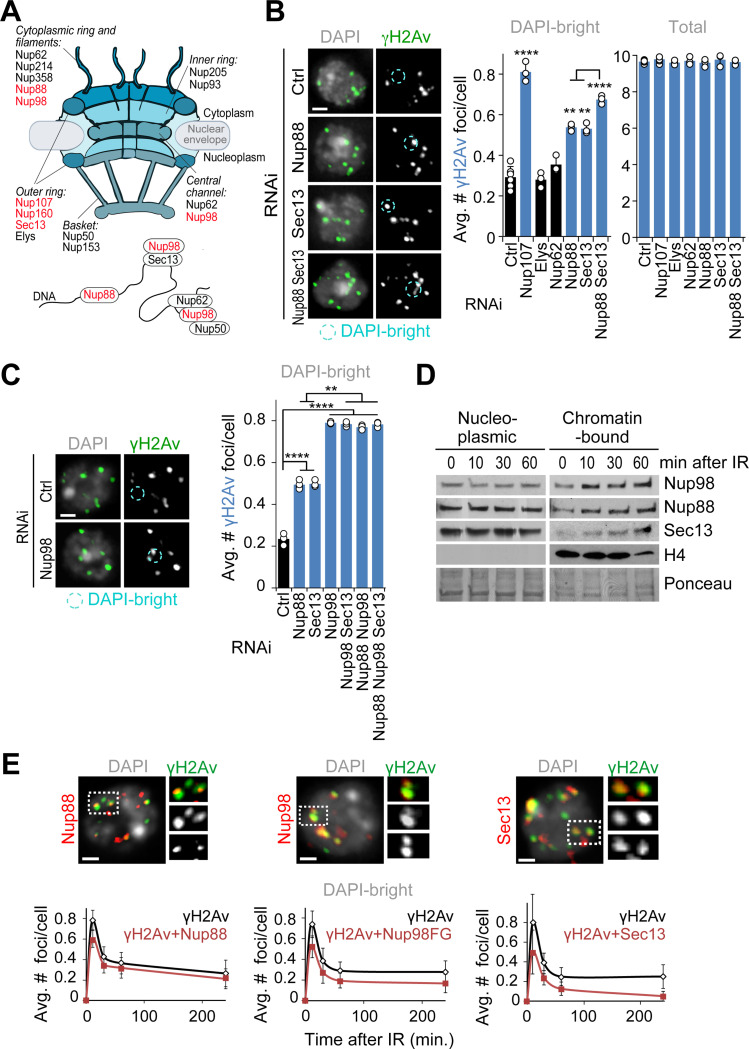
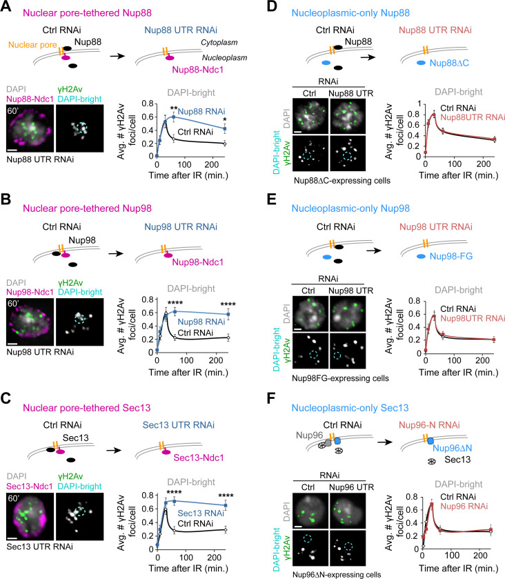
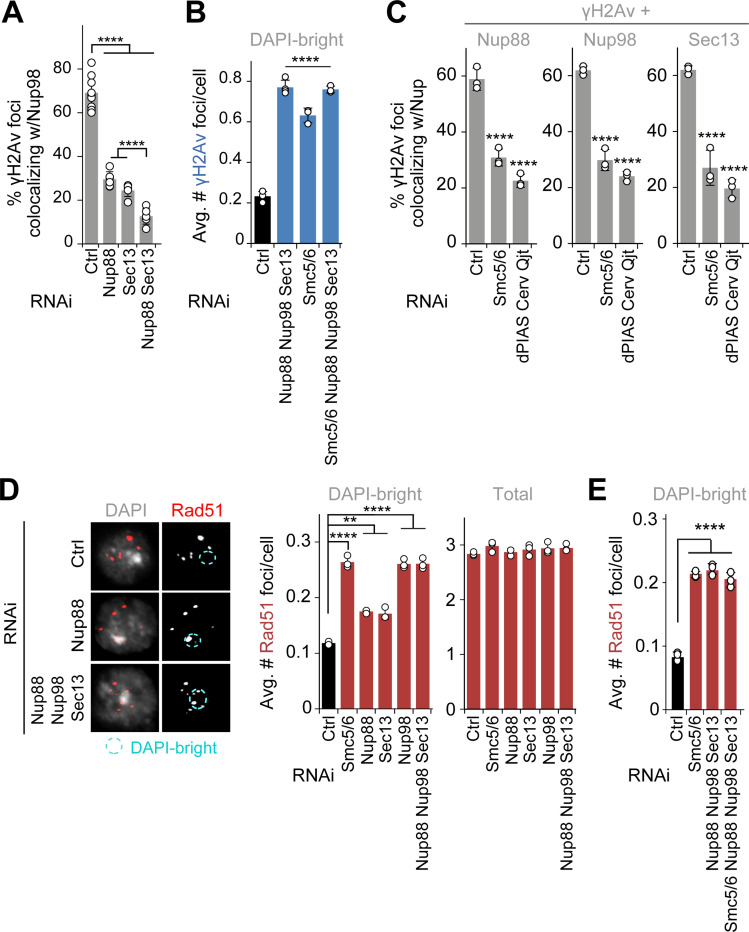
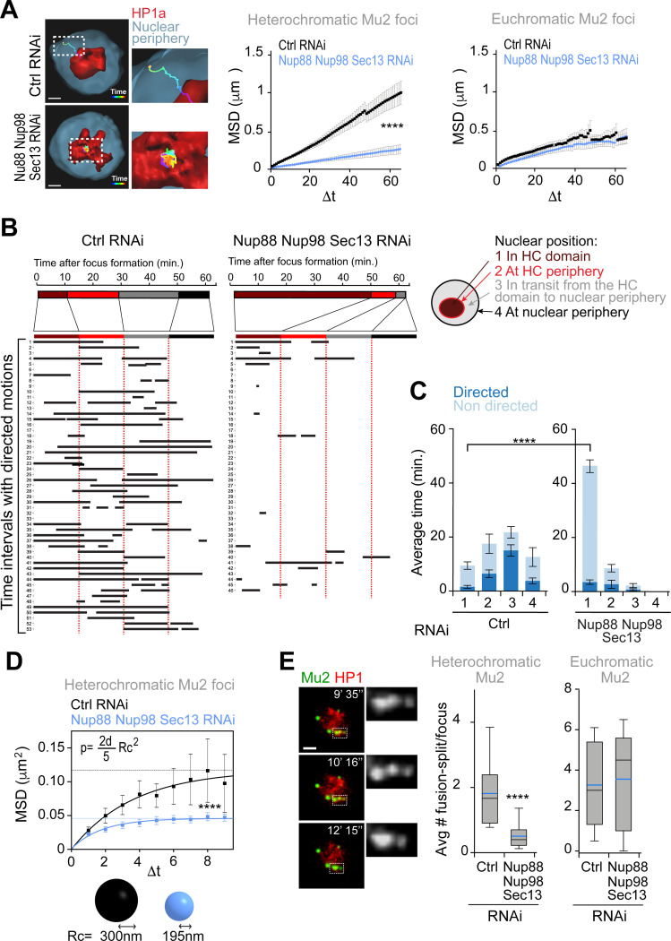
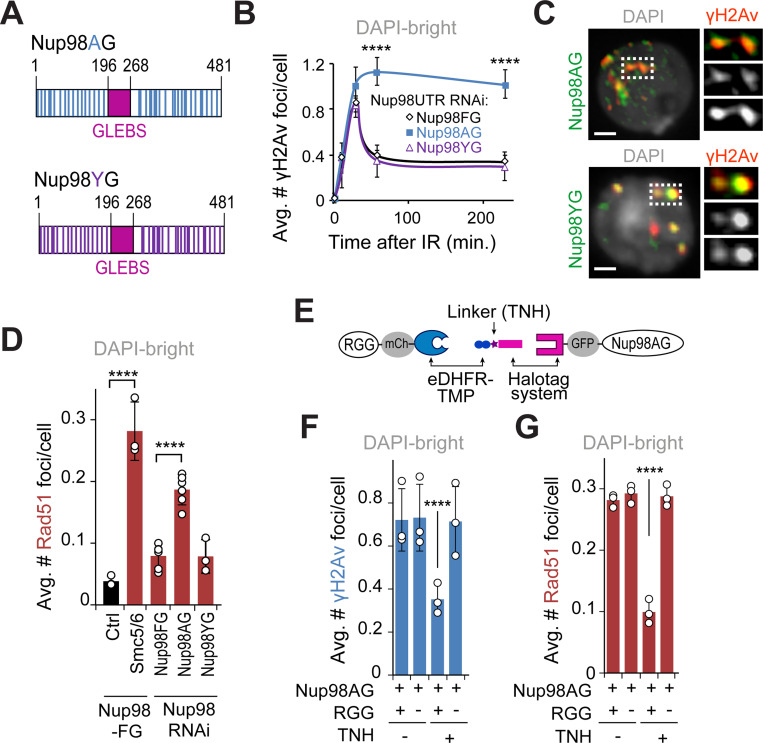
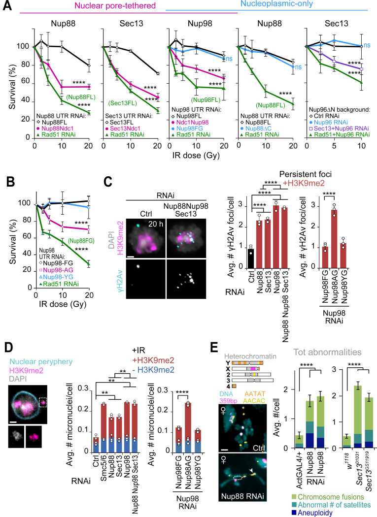
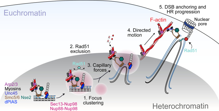
Phase separation forms membraneless compartments in the nuclei, including by establishing heterochromatin "domains" and repair foci. Pericentromeric heterochromatin mostly comprises repeated sequences prone to aberrant recombination, and "safe" homologous recombination (HR) repair of these sequences requires the movement of repair sites to the nuclear periphery before Rad51 recruitment and strand invasion. How this mobilization initiates is unknown, and the contribution of phase separation to these dynamics is unclear. Here, we show that Nup98 nucleoporin is recruited to heterochromatic repair sites before relocalization through Sec13 or Nup88 nucleoporins, and downstream from the Smc5/6 complex and SUMOylation. Remarkably, the phase separation properties of Nup98 are required and sufficient to mobilize repair sites and exclude Rad51, thus preventing aberrant recombination while promoting HR repair. Disrupting this pathway results in heterochromatin repair defects and widespread chromosome rearrangements, revealing a novel "off-pore" role for nucleoporins and phase separation in nuclear dynamics and genome integrity in a multicellular eukaryote.

Keywords: Heterochromatin repair; Nup88; Nup98; Sec13; condensates; double-strand break repair; homologous recombination; nuclear architecture; phase separation; repair dynamics.


Conflict of interest statement

Declaration of Interests The authors declare no competing interests.

Figures

Figure 1.
Figure 1.. Nup88, Nup98 and Sec13 are required for relocalization of heterochromatic DSBs.
(A) Schematic representation of nuclear pore proteins including chromatin-associated nucleoporins. Red indicates nucleoporins whose depletion affects relocalization/anchoring of heterochromatic DSBs from this study and12. (B) Immunofluorescence (IF) analysis and quantification of Kc cells fixed 60 min after IR show the number of γH2Av foci in DAPI-bright and total foci following indicated RNAi depletions. *P<0.05, **P<0.01, ****P<0.0001, n>195 cells for each RNAi condition. (C) As described in b, except cells expressing Nup96-MycFLAG were used. **P<0.01, ****P<0.0001, n>328 cells for each RNAi condition. (D) Chromatin fractionation and Western Blot (Wb) analysis of Kc cells shows the chromatin-bound fractions of Nup88, Nup98, and Sec13 at indicated time points after IR. Histone H4 and Ponceau are loading controls. (E) IF analysis shows γH2Av foci colocalizing with the indicated nucleoporins in DAPI-bright, at different timepoints after IR. Dashed boxes and zoomed details highlight colocalizations 10 min after IR. Kc cells were used for Nup88 and Sec13 staining. GFP-Nup98FG-expressing cells were used in place of Nup98 WT due to a stronger nucleoplasmic signal associated with this mutant. n>118 foci for each time point. Error bars: SEM in E, and SD in B,C from three or more independent experiments. P values calculated with two-tailed Mann–Whitney test. Images are max intensity projections of Z-stacks spanning the DAPI-bright region. Ctrl = Control. Scale bar = 1μm.
Figure 2.
Figure 2.. The nucleoplasmic fractions of Nup88, Nup98, and Sec13 are required for relocalization.
Schematic representations (top of each figure panel) show the experimental setup. (A) Cells expressing Nup88-MycFLAG-Ndc1 are treated with dsRNAs for Nup88 UTR to deplete endogenous Nup88, or a control (Ctrl). IF (left) and quantification (right) show the number of γH2Av foci in DAPI-bright at different timepoints after IR, following indicated RNAi depletions. *P<0.05, **P<0.01, n>60 cells/time point. (B) As in (A), except cells express MycFLAG-Ndc1-Nup98 (plus Nup96-MycFLAG), and RNAi depletion is done against Nup98 UTR. ****P<0.0001, n>100 cells for each time point. (C) As in (A), except cells express Sec13-MycFLAG-Ndc1 and RNAi depletion targets Sec13 UTR. ****P<0.0001, n>100 cells for each time point. (D) Cells expressing FHA-Nup88ΔC are treated with dsRNAi for Nup88 UTR to deplete endogenous Nup88, or a control (Ctrl). n>77 cells for each time point. (E) As in (D), except cells express FHA-Nup98FG and dsRNAs target Nup98 UTR. n>100 cells for each time point. (F) As in (D), except cells express Nup96ΔN-MycFLAG and dsRNAs target only endogenous Nup96 (see also Figures S4A and S4D-F). n>44 cells for each time point. P values calculated with two-tailed Mann–Whitney test. Error bars are SEM. Images are max intensity projections of Z-stacks spanning the DAPI-bright region. Scale bar = 1μm.
Figure 3.
Figure 3.. Nup88, Nup98 and Sec13 act downstream from Smc5/6 to promote relocalization and to prevent abnormal Rad51 recruitment.
(A) Quantification of cells fixed 10 min after IR shows the number of γH2Av foci colocalizing with GFP-Nup98FG foci in DAPI-bright following indicated RNAi depletions. ****P<0.0001, n>74 foci for RNAi condition. (B) Quantification of cells expressing Nup96-MycFLAG fixed 60 min after IR, shows the number of γH2Av foci in DAPI-bright after indicated RNAi depletions. ****P<0.0001 relative to Ctrl, n>226 cells for each condition. (C) Quantification of cells expressing Nup88-MycFLAG, GFP-Nup98FG or Sec13-MycFLAG shows colocalizations with γH2Av foci 10 min after IR, following indicated RNAi depletions. ****P<0.0001 relative to Ctrl, n>495 foci for each condition. (D) IF and quantification of cells fixed 60 min after IR show the number of Rad51 foci in DAPI-bright and total foci following indicated RNAi depletions. Smc5/6 RNAi results in abnormal Rad51 focus formation in heterochromatin,, and is used as positive control. n>246 cells for each condition. **P<0.01, ****P<0.0001 relative to Ctrl. (E) As in (D). ****P<0.0001 for comparisons to Ctrl, n>581 cells for each RNAi condition. Error bars are SD from three independent experiments. P values calculated with two-tailed Mann–Whitney test. Scale bar = 1μm.
Figure 4.
Figure 4.. Nup88, Nup98, and Sec13 nucleoporins are required for mobilizing heterochromatic DSBs.
(A) 3D reconstructions in Imaris (left) and MSD analysis (right) of GFP-Mu2 foci that form inside (heterochromatic) or outside (euchromatic) the mCh-HP1a domain after IR, following indicated RNAi depletions. n≥46 tracks for each condition from three or more experiments. Dashed boxes and zoomed details highlights the tracks. Δt, time intervals (40s each). ****P>0.0001. (B) LDM analysis of heterochromatic foci from the experiment described in (A). Duration and distribution of LDMs (black lines) along each track relative to different nuclear positions, adjusted relative to a ‘pseudo-trajectory’ defined by the average time that foci spend in each nuclear space, and displayed for equalized time intervals. Numbers on the left side indicate individual foci. (C) Average duration of directed and non-directed motions for heterochromatic foci in (A,B), in the indicated nuclear locations. ****P>0.0001. (d) MSD analysis of time points from (A) characterized by non-directed motion inside the heterochromatin domain, following indicated RNAi depletions. ****P>0.0001, n=29 foci for Ctrl and n=38 foci for Nup88+Nup98+Sec13 RNAi from (A). The confinement radius (below, Rc) was calculated using the plateau of the curve (p, dashed line), as indicated in54. d: dimensions (d=3 in this analysis) (E) Images from a movie (left) and quantification (right) show fusion and spitting events for heterochromatic Mu2/Mdc1 foci in cells expressing GFP-Mu2 and mCh-HP1a, following indicated RNAi depletions. Dashed boxes and zoomed details highlight fusion-splitting events. In the box plot blue line is the mean; black line is the median n>97 foci per each condition. ***P<0.001 for the indicated comparison. Error bars are SEM in A, C, D. The whiskers are drawn down to the 10th percentile and up to the 90th in e. P values calculated with extra sum-of-squares F-test, nonlinear regression for curve fitting for A, D and two-tailed Mann–Whitney test in C, E. Scale bars = 1 μm.
Figure 5.
Figure 5.. Nup98 mediates relocalization of heterochromatic DSBs via phase separation.
(A) Schematic representation of Nup98AG and YG mutations. Each Phenylalanine of the FG-domain of Nup98FG is substituted with Ala in Nup98AG or Tyr in Nup98YG. (B) Quantification of γH2Av foci in DAPI-bright at different time points after IR, in cells expressing FHA-Nup98-FG, -AG or -YG following RNAi depletion of endogenous of Nup98 (Nup98 UTR RNAi). ****P<0.0001 for Nup98AG relative to Nup98FG, n>50 cells for each time point. (C) IF shows Nup98AG and Nup98YG foci colocalizing with γH2Av foci. Dashed boxes and zoomed details highlight colocalizations. (D) Quantification of cells expressing FHA-Nup98-FG, -AG or -YG following depletion of endogenous Nup98, fixed 60 min after IR, shows the number of Rad51 foci in DAPI-bright. ****P<0.0001 for indicated comparisons. n>363 cells for each RNAi condition. Smc5/6 RNAi in cells expressing Nup98FG is shown as a positive control. (E) Schematic representation of the dimerizer system (adapted from). The RGG domains are tagged with mCherry (mCh) and fused to eDHFR, whereas Nup98AG is tagged with GFP and fused to 3xHalo. The dimerizer is TNH. (F) Quantification of cells expressing (+) or non-expressing (−) RGG-mCherry-RGG-eDHFR, plus Halo-GFP-Nup98AG, treated for 120 min with (+) or without (−) TNH as indicated, and fixed 60 min after IR, shows the number of γH2Av foci in DAPI-bright. ****P<0.0001. n>346 cells for each condition. (G) As in F, except Rad51 foci were quantified. ****P<0.0001. n>342 cells for each condition. Error bars are SEM in B, and SD in D,F,G from three independent experiments. P values calculated with two-tailed Mann–Whitney test. Scale bars = 1 μm.
Figure 6.
Figure 6.. Nup88, Nup98, and Sec13 nucleoporins are required for heterochromatin repair and stability.
(A) IR sensitivity assays show cell survival at different IR doses and after indicated RNAi depletions and protein expression. Rad51 RNAi results in cell sensitivity to IR12,13 and is used as positive control in cells expressing each FL (full-length) protein or Nup96ΔN, as indicated. (Left to right) First graph: Survival of cells expressing Nup88-MycFLAG-Ndc1 or Nup88FL-MycFLAG in the absence of endogenous Nup88 (Nup88 UTR RNAi). ****P<0.0001 for all comparisons vs Nup88FL. Second graph: Survival of cells expressing Sec13-MycFLAG-Ndc1 or Sec13FL-MycFLAG, after Sec13 UTR RNAi. ****P<0.0001 for comparisons vs Sec13FL. Third graph: Survival of cells expressing FHA-Nup98FL, MycFLAG-Ndc1-Nup98, or FHA-Nup98FG, after Nup98 UTR RNAi. ****P<0.0001 for Ndc1-Nup98 or Rad51 RNAi vs Nup98FL, P=ns for Nup98FG vs Nup98FL. Fourth graph: Survival of cells expressing FHA-Nup88ΔC or FHA-Nup88FL, after Nup88 UTR RNAi. P=ns for Nup88ΔC vs Nup88FL. ****P<0.0001 for Rad51 vs FHA-Nup88FL. Fifth graph: Survival of cells expressing Nup96ΔN-MycFLAG after RNAi depletion of: Ctrl; endogenous Nup96 (Nup96 UTR RNAi); Sec13+Nup96 (positive control); or Rad51+Nup96 (positive control). ****P<0.0001 for comparisons vs Ctrl RNAi, P=ns for Nup96 vs Ctrl RNAi. n>150 cells per RNAi per dose. (B) As in (A), except cells expressing FHA-Nup98-FG, -AG or -YG were used, following Nup98 UTR RNAi depletion. ****P<0.0001 for Nup98AG or Rad51 vs Nup98FG. n>100 cells per RNAi per dose. Rad51 RNAi is used as positive control in cells expressing Nup98FG. (C) IF and quantifications of cells fixed 20 hrs after IR and stained for γH2Av and H3K9me2 following indicated RNAi depletions, or in cells expressing FHA-Nup98-FG, -AG or -YG following Nup98 UTR RNAi. ****P<0.0001, n>273 cells for each condition. +H3K9me2: γH2Av foci associated with H3K9me2 signals. (D) IF and quantification of micronuclei in cells stained for DNA (DAPI), heterochromatin (H3K9me2), and nuclear periphery (Nup62), after indicated RNAi depletions or in cells expressing FHA-Nup98-FG, -AG or -YG, after Nup98 UTR RNAi depletion, 2 days post IR. **P<0.01, ****P=0.0001, n>447 cells for each condition. +/− H3K9me2: micronuclei with/without H3K9me2. Dashed box and the zoomed detail highlights a micronucleus. (E) Images of chromosome preparations from larval brains and quantifications from Ctrl and indicated RNAi depleted or mutant larval brains stained by FISH for indicated satellites. Examples of fusion (arrowhead) and chromosome arm losses (dashed circle) are highlighted. n>46 karyotypes/genotype from at least two independent crosses and three brains. Nup98 RNAi lines also express Nup96-Myc. ****P<0.0001 for indicated comparisons. The diagram of Drosophila chromosomes indicates satellite positions. Error bars are SEM in E and SD from three or more experiments in A-D. P values calculated with extra sum-of-squares F-test, nonlinear regression for curve fitting for A and B, two-tailed Mann–Whitney test in C-D, and unpaired T-test in E. Images are max intensity projections of a few Z-stacks. Scale bar = 1μm.
Figure 7.
Figure 7.. Model for the role of Nup88, Nup98 and Sec13 in heterochromatic DSBs repair in Drosophila.
DSBs are resected inside the heterochromatin domain, where Smc5/6 promotes the loading of Nup88-Nup98 and Nup98-Sec13. Nup98-FG repeats create a phase separated environment that facilitates focus clustering inside the heterochromatin domain while excluding Rad51. We hypothesize that a multiphase condensate established by Nup98 inside the HP1a domain generates capillary forces that promote the relocalization of Nup98 condensates at repair sites to the surface of the heterochromatin domain. Here, Nup98-driven wetting behavior might also promote the association of repair foci with actin filaments generated by Arp2/3. Unc45 recruitment by Smc5/6 activates nuclear myosins to “walk” repair sites along filaments, thus relocalizing DSBs to nuclear pores where HR continues with Rad51 recruitment. Relocalization prevents ectopic recombination by isolating damaged sites and their homologous templates (black lines) from undamaged repeats before strand invasion.

References

    1. Ho J. W. et al. Comparative analysis of metazoan chromatin organization. Nature 512, 449–452 (2014). 10.1038/nature13415 - DOI - PMC - PubMed
    1. Smith C. D., Shu S., Mungall C. J. & Karpen G. H. in Science Vol. 316 1586–1591 (2007). - PMC - PubMed
    1. Hoskins R. A. et al. The Release 6 reference sequence of the Drosophila melanogaster genome. Genome Res 25, 445–458 (2015). 10.1101/gr.185579.114 - DOI - PMC - PubMed
    1. Riddle N. C. et al. Plasticity in patterns of histone modifications and chromosomal proteins in Drosophila heterochromatin. Genome Res 21, 147–163 (2011). 10.1101/gr.110098.110 - DOI - PMC - PubMed
    1. Peng J. C. & Karpen G. H. Epigenetic regulation of heterochromatic DNA stability. Curr Opin Genet Dev 18, 204–211 (2008). https://doi.org:S0959-437X(08)00020-8 [pii] 10.1016/j.gde.2008.01.021 - DOI - PMC - PubMed

Publication types