[Preprint]. 2023 Oct 6:2023.10.05.561076.
doi: 10.1101/2023.10.05.561076.

Variants in the DDX6-CXCR5 autoimmune disease risk locus influence the regulatory network in immune cells and salivary gland

Mandi M Wiley  1 Bhuwan Khatri  1 Michelle L Joachims  1   2 Kandice L Tessneer  1 Anna M Stolarczyk  1 Astrid Rasmussen  1 Juan-Manuel Anaya  3 Lara A Aqrawi  4   5 Sang-Cheol Bae  6 Eva Baecklund  7 Albin Björk  8 Johan G Brun  9   10 Sara Magnusson Bucher  11 Nick Dand  12 Maija-Leena Eloranta  7 Fiona Engelke  13 Helena Forsblad-d'Elia  14 Cecilia Fugmann  7 Stuart B Glenn  1 Chen Gong  12 Jacques-Eric Gottenberg  15 Daniel Hammenfors  10 Juliana Imgenberg-Kreuz  7 Janicke Liaaen Jensen  5 Svein Joar Auglænd Johnsen  16 Malin V Jonsson  9 Jennifer A Kelly  1 Sharmily Khanam  2 Kwangwoo Kim  17 Marika Kvarnström  8 Thomas Mandl  18 Javier Martín  19 David L Morris  12 Gaetane Nocturne  20   21 Katrine Brække Norheim  16 Peter Olsson  18 Øyvind Palm  22 Jacques-Olivier Pers  23 Nelson L Rhodus  24 Christopher Sjöwall  25 Kathrine Skarstein  9 Kimberly E Taylor  26 Phil Tombleson  12 Gudny Ella Thorlacius  8 Swamy Venuturupalli  27 Edward M Vital  28 Daniel J Wallace  27 Kiely M Grundahl  1   2 Lida Radfar  29 Michael T Brennan  30 Judith A James  2   31 R Hal Scofield  2   31   32 Patrick M Gaffney  1   31 Lindsey A Criswell  26   33 Roland Jonsson  9 Silke Appel  9 Per Eriksson  25 Simon J Bowman  34 Roald Omdal  9   16 Lars Rönnblom  7 Blake M Warner  35 Maureen Rischmueller  36 Torsten Witte  13 A Darise Farris  2   31 Xavier Mariette  20   21 Caroline H Shiboski  26 Sjögren’s International Collaborative Clinical Alliance (SICCA)Marie Wahren-Herlenius  8   9 Marta E Alarcón-Riquelme  8   37 PRECISESADS Clinical ConsortiumWan-Fai Ng  38   39 UK Primary Sjögren’s Syndrome RegistryKathy L Sivils  2 Joel M Guthridge  2   31 Indra Adrianto  40   41 Timothy J Vyse  12 Betty P Tsao  42 Gunnel Nordmark  7 Christopher J Lessard  1   31
Affiliations

Variants in the DDX6-CXCR5 autoimmune disease risk locus influence the regulatory network in immune cells and salivary gland

Mandi M Wiley et al. bioRxiv. .

Update in

  • Variants in the DDX6-CXCR5 autoimmune disease risk locus influence the regulatory network in immune cells and salivary gland.
    Wiley MM, Radziszewski M, Khatri B, Joachims ML, Tessneer KL, Stolarczyk AM, Yao S, Li J, Pritchett-Frazee C, Johnston AA, Rasmussen A, Anaya JM, Aqrawi LA, Bae SC, Baecklund E, Björk A, Brun JG, Bucher SM, Dand N, Eloranta ML, Engelke F, Forsblad-d'Elia H, Fugmann C, Glenn SB, Gong C, Gottenberg JE, Hammenfors D, Imgenberg-Kreuz J, Jensen JL, Johnsen SJA, Jonsson MV, Kelly JA, Khanam S, Kim K, Kvarnström M, Mandl T, Martín J, Morris DL, Nocturne G, Norheim KB, Olsson P, Palm Ø, Pers JO, Rhodus NL, Sjöwall C, Skarstein K, Taylor KE, Tombleson P, Thorlacius GE, Venuturupalli SR, Vital EM, Wallace DJ, Radfar L, Brennan MT, James JA, Scofield RH, Gaffney PM, Criswell LA, Jonsson R, Appel S, Eriksson P, Bowman SJ, Omdal R, Rönnblom L, Warner BM, Rischmueller M, Witte T, Farris AD, Mariette X, Shiboski CH; Sjögren’s International Collaborative Clinical Alliance (SICCA); Wahren-Herlenius M, Alarcón-Riquelme ME; PRECISESADS Clinical Consortium; Ng WF; UK Primary Sjögren’s Syndrome Registry; Sivils KL, Guthridge JM, Adrianto I, Vyse TJ, Tsao BP, Nordmark G, Lessard CJ. Wiley MM, et al. Ann Rheum Dis. 2025 Sep;84(9):1512-1527. doi: 10.1016/j.ard.2025.04.023. Epub 2025 May 30. Ann Rheum Dis. 2025. PMID: 40447495

Abstract

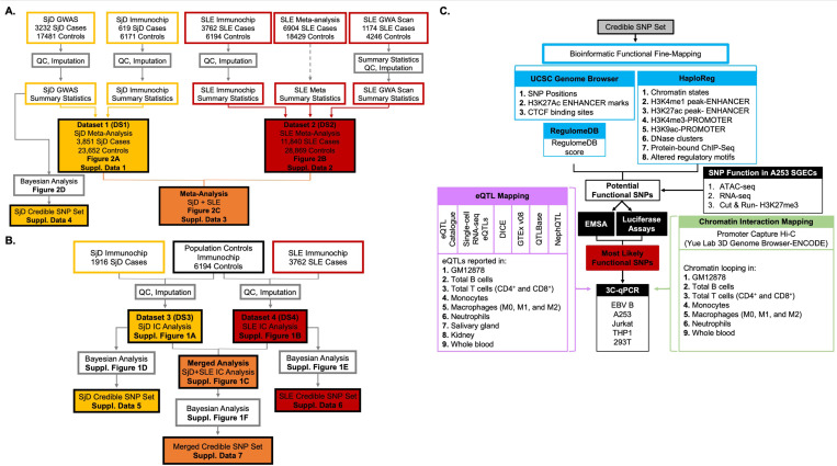
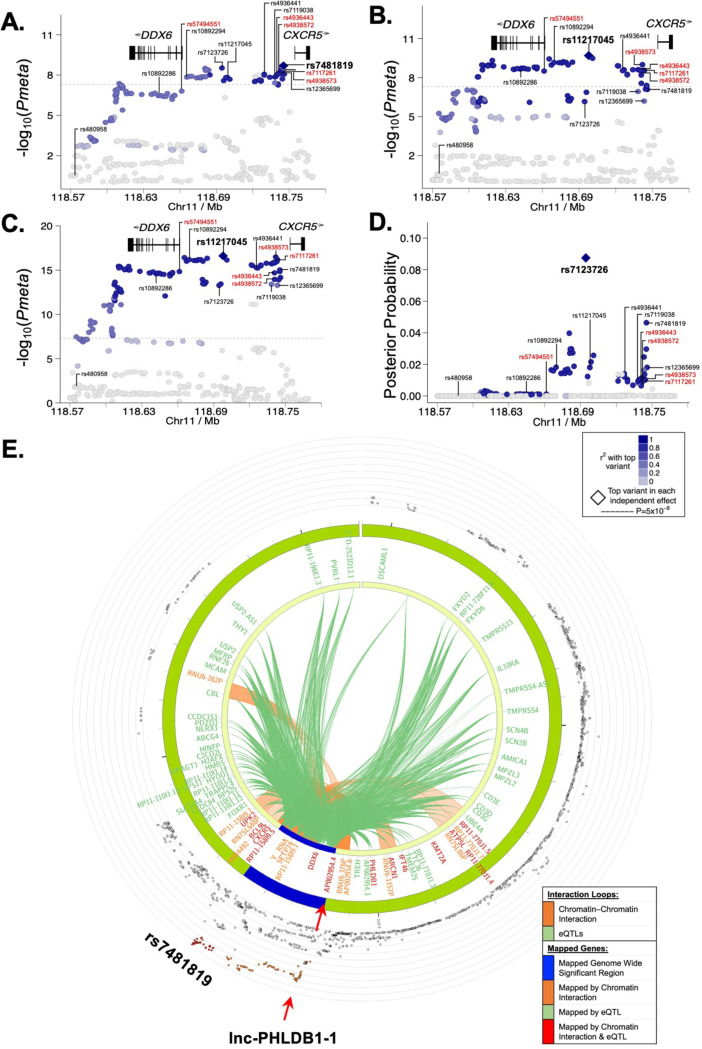
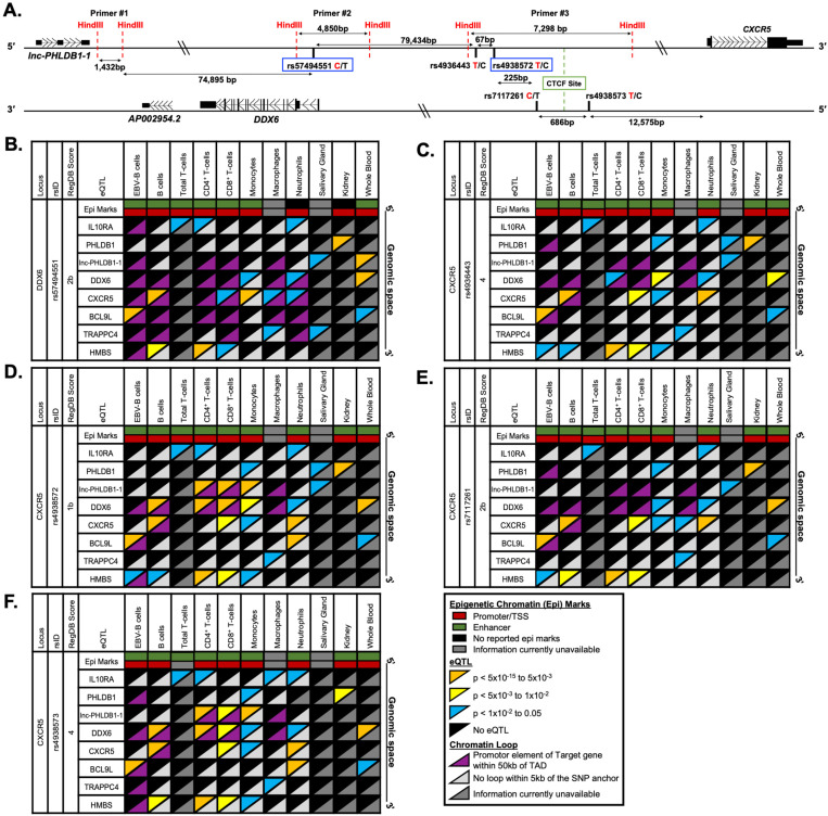
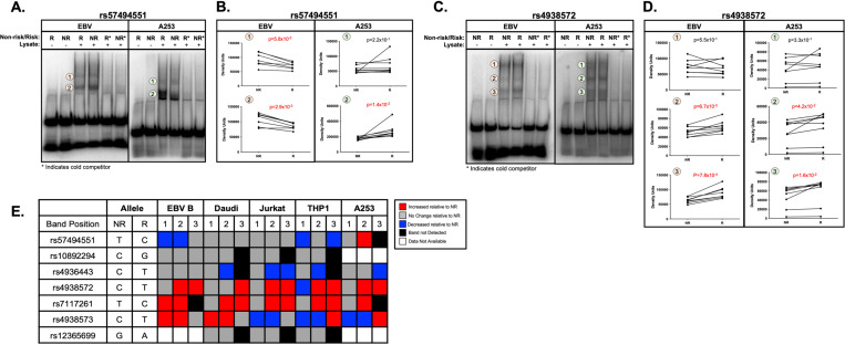
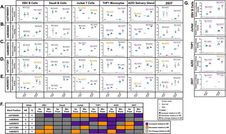
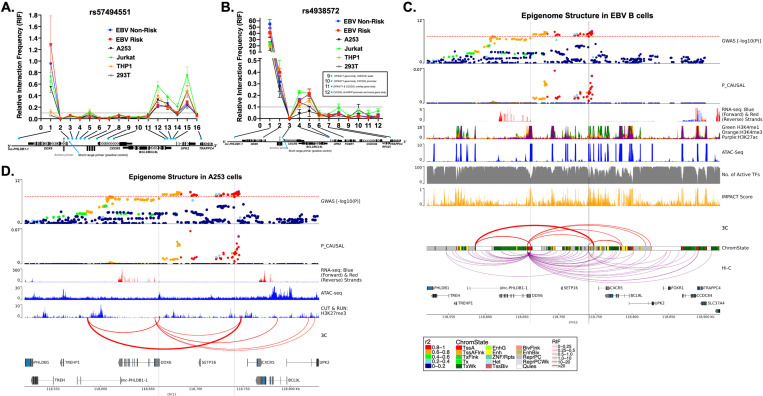
Fine mapping and bioinformatic analysis of the DDX6-CXCR5 genetic risk association in Sjögren's Disease (SjD) and Systemic Lupus Erythematosus (SLE) identified five common SNPs with functional evidence in immune cell types: rs4938573, rs57494551, rs4938572, rs4936443, rs7117261. Functional interrogation of nuclear protein binding affinity, enhancer/promoter regulatory activity, and chromatin-chromatin interactions in immune, salivary gland epithelial, and kidney epithelial cells revealed cell type-specific allelic effects for all five SNPs that expanded regulation beyond effects on DDX6 and CXCR5 expression. Mapping the local chromatin regulatory network revealed several additional genes of interest, including lnc-PHLDB1-1. Collectively, functional characterization implicated the risk alleles of these SNPs as modulators of promoter and/or enhancer activities that regulate cell type-specific expression of DDX6, CXCR5, and lnc-PHLDB1-1, among others. Further, these findings emphasize the importance of exploring the functional significance of SNPs in the context of complex chromatin architecture in disease-relevant cell types and tissues.

Keywords: C-X-C motif chemokine receptor 5; CXCR5; DDX6; DEAD-Box helicase 6; SLE; Sjögren’s Disease; chromatin regulatory network; lnc-PHLDB1-1; systemic lupus erythematosus.


Conflict of interest statement

Competing Interests C.J.L.* and A.D.F. have an active collaborative research agreement with Janssen. E.B. has an active research collaboration with Pfizer. T.M. is employed as medical solutions lead in rheumatology at UCB. R.H.S. is a consultant for Jansen Pharmaceuticals. S.J.B. provided consultancy services for Abbvie, BMS, Galapagos, Iqvia, J&J, Kiniksa, and Novartis in 2020–2021. L.R. provided consultancy services for AstraZeneca. B.M.W. has active collaborative research agreements with Astellas Bio and Pfizer, Inc. M.R. received grants from Amgen, AstraZeneca, Bristol Myers-Squibb, Novartis, and Servier for clinical trials in Sjögren’s Syndrome and SLE. All other authors have reported that they have no competing interests to report.

Figures

Figure 1.
Figure 1.. Schematic of the meta-analysis, fine mapping, and bioinformatic workflow of the shared DDX6-CXCR5 interval associated with Sjögren’s disease (SjD) and systemic lupus erythematosus (SLE).
(A) Composition and workflow of three datasets (DS) used in the meta-analysis of SjD (DS1; yellow), SLE (DS2; red), and SjD and SLE merged (DS1+DS2; orange). DS1 was also used to perform SNP-SjD single marker trait analysis and identify a SjD credible SNP set. (B) Composition and workflow of the SjD (DS3; yellow) and SLE (DS4; Red) Immunochip datasets used for SNP-single marker trait analysis and identification of credible SNP sets for SjD, SLE, and SjD and SLE merged. (C) Workflow of the bioinformatic fine mapping applied to the 95% credible SNP sets from the DDX6-CXCR5 risk locus to identify, prioritize, and functionally characterize SNPs common between the SjD and SLE risk associations.
Figure 2.
Figure 2.. Fine mapping of the DDX6-CXCR5 region in Sjögren’s disease (SjD) and systemic lupus erythematosus (SLE) after imputation and meta-analysis.
(A-C) Logistic regression analysis was performed on (A) Dataset 1 (DS1)-SjD (3851 SjD cases; 23652 controls), (B) DS2-SLE (11840 SLE cases; 28869), and (C) DS1+DS2 (merged SjD and SLE) after quality control and imputation, identifying the top SNPs (e.g., index SNPs indicated in bold) of the DDX6-CXCR5 region. (D) Posterior probability distribution of SNPs in the DDX6-CXCR5 region of DS1-SjD, identifying rs7123726 as having the highest posterior probability. For A-D, SNPs prioritized for bioinformatic screening are indicated in black; five SNPs prioritized for functional characterization are labeled in red. (E) Circos plot showing chromatin-chromatin interactions in GM12878 EBV B cell line (orange) and reported eQTLs from different cells/tissues (green). The outer ring shows the logistic regression analysis of DS1-SjD with index SNP rs7481819 indicated.
Figure 3.
Figure 3.. Reported expression quantitative trait loci (eQTLs) and chromatin-chromatin interactions in immune and disease-relevant tissues for the prioritized SNPs on the DDX6-CXCR5 risk interval.
(A) Schematic of the five prioritized SNPs arranged in genomic space. SNP rs57494551 is in the first intron of DDX6. The other four SNPs span 978 bp in a shared promoter/enhancer region between DDX6 and CXCR5. Risk alleles are indicated by red font; non-risk alleles by black. SNPs rs57494551 and rs4938572 (blue boxes) are representative SNPs. HindIII restriction sites (red dotted line), CTCF site (green box), and positions of 3C-qPCR primers #1–3 are also indicated. (B-F) Publicly reported cell type-specific functional annotations (horizontal rectangles), select eQTLs (top triangles), and chromatin-chromatin interactions (bottom triangles) are shown for (B) rs57494551, (C) rs4936443, (D) rs4938572, (E) rs7117261, and (F) rs4938573 across 10 different immune cell types or disease-specific tissues (GM12878 EBV B cells and primary human B cells, CD4+ T cells, CD8+ T cells, monocytes, macrophages, neutrophils, salivary gland tissue, kidney tissue, and whole blood).
Figure 4.
Figure 4.. Allele- and cell-type specific differential nuclear protein affinities of the prioritized SNPs rs57494551 and rs4938572 on the shared DDX6-CXCR5 risk region.
(A-D) Radiolabeled electromobility shift assays (EMSA) were performed to assess the binding affinity of ribonucleoproteins isolated from EBV B or A253 cells to oligonucleotides containing the non-risk (NR) or risk (R) allele of (A-B) rs57494551 or (C-D) rs4938572. Probes incubated in the absence of nuclear lysate were used as negative control (Lanes 1, 2). Cold competitors were used to assess non-specific binding (Lanes 5, 6). Images shown in (A) and (C) are representative of n>6 biological replicates. (B, D) Bands indicated in (A, C) by the orange or green circles were quantified by densitometry and analyzed using paired t-test (n>6); p-values indicated. (E) Summary analysis of the allele-specific nuclear protein affinities of the five prioritized SNPs in EBV B, Daudi, Jurkat, THP1, and A253 cells shown in A-D and Supplemental Figures 4-9. Increases in binding relative to NR are shown in red; decreases relative to NR in blue; no change relative to NR in grey; no detected band in black; data not available in white.
Figure 5.
Figure 5.. Allele- and cell-type specific promoter and enhancer activity of the prioritized SNPs on the shared DDX6-CXCR5 risk region.
(A-E) gBlocks carrying the non-risk or risk alleles of (A) rs57494551, (B) rs4936443, (C) rs4938572, (D) rs7117261, or (E) rs4938573 were cloned into a promoter-less (pGL4.14; noP) or minimal promoter (pGL4.26; minP) luciferase vector. Plasmids were transfected into EBV B, Daudi, Jurkat, THP1, A253, or 293T cells. Luciferase activity was measured after 24 hours and normalized to the Renilla transfection control and then the vector-only control; reported as Relative Luciferase Activity. Statistical comparisons were performed using a paired t-test (n>3); p-values are indicated. (F) Summary analysis of the allele-specific luciferase activity of the five prioritized SNPs in EBV B, Daudi, Jurkat, THP1, A253 and 293Tcells. Increases in luciferase activity relative to non-risk are shown in purple; decreases relative to non-risk in orange; no change relative to non-risk in grey. (G) gBlocks carrying all non-risk or all risk alleles of rs4936443, rs4938572, and rs7117261 were cloned into the promoter-less or minimal promoter above, transfected into EBV B, Jurkat, THP1, A253, or 293T cells, and luciferase activity tested as described above. Statistical comparisons were performed using a paired t-test (n>3); p-values are indicated.
Figure 6.
Figure 6.. Complex chromatin architecture revealed across the DDX6-CXCR5 region in immune cells, salivary gland, and kidney by 3C-qPCR.
(A, B) Chromatin conformation capture with quantitative PCR (3C-qPCR) across the DDX6-CXCR5 region where (A) rs57494551 or (B) rs4938572 is the anchor SNP (grey dot). Relative interaction frequency (RIF) is plotted relative to the primer number in 5’−3’ genomic orientation (blue dots; see Figure 3A for additional detail). Primers 9–12 are shown in a text box for simplicity in (B). (C) SjD GWAS association (top panel) and publicly available epigenomic enrichment across the DDX6-CXCR5 region in GM12878 EBV B cells. Vertical grey lines indicate the locations of rs57494551 or rs4938572, respectively. Promoter-capture Hi-C looping (purple lines) contrasts the summary 3C-qPCR results (red lines); line thickness indicates relative interaction frequency (RIF) of the 3C data. (D) SjD GWAS association (top panel) across the DDX6-CXCR5 region in A253 cells. In house ATAC-seq, CUT & RUN: H3K27me3 (Epicypher), and RNA-seq data from A253 cells are shown because of limited publicly available epigenetic data on salivary gland. Vertical grey lines indicate the locations of rs57494551 or rs4938572, respectively. Summary 3C-qPCR results are shown (red lines); line thickness indicates relative interaction frequency (RIF).

References

    1. Khatri B. et al. Genome-wide association study identifies Sjogren’s risk loci with functional implications in immune and glandular cells. Nat Commun 13, 4287 (2022). 10.1038/s41467-022-30773-y - DOI - PMC - PubMed
    1. Thorlacius G. E., Bjork A. & Wahren-Herlenius M. Genetics and epigenetics of primary Sjogren syndrome: implications for future therapies. Nat Rev Rheumatol 19, 288–306 (2023). 10.1038/s41584-023-00932-6 - DOI - PMC - PubMed
    1. Baer A. N. & Hammitt K. M. Sjogren’s Disease, Not Syndrome. Arthritis Rheumatol 73, 1347–1348 (2021). 10.1002/art.41676 - DOI - PubMed
    1. Mariette X. & Criswell L. A. Primary Sjogren’s Syndrome. N Engl J Med 379, 97 (2018). 10.1056/NEJMc1804598 - DOI - PubMed
    1. Nocturne G. & Mariette X. Advances in understanding the pathogenesis of primary Sjogren’s syndrome. Nat Rev Rheumatol 9, 544–556 (2013). 10.1038/nrrheum.2013.110 - DOI - PubMed

Publication types

LinkOut - more resources