. 2024 Jul 24:19:7509-7527.
doi: 10.2147/IJN.S471623. eCollection 2024.

A New Nanoplatform Under NIR Released ROS Enhanced Photodynamic Therapy and Low Temperature Photothermal Therapy for Antibacterial and Wound Repair

Affiliations

A New Nanoplatform Under NIR Released ROS Enhanced Photodynamic Therapy and Low Temperature Photothermal Therapy for Antibacterial and Wound Repair

Licai Miao et al. Int J Nanomedicine. .

Abstract

Purpose: Skin injury, often caused by physical or medical mishaps, presents a significant challenge as wound healing is critical to restore skin integrity and tissue function. However, external factors such as infection and inflammation can hinder wound healing, highlighting the importance of developing biomaterials with antibiotic and wound healing properties to treat infections and inflammation. In this study, a novel photothermal nanomaterial (MMPI) was synthesized for infected wound healing by loading indocyanine green (ICG) on magnesium-incorporated mesoporous bioactive glass (Mg-MBG) and coating its surface with polydopamine (PDA).

Results: In this study, Mg-MBG and MMPI was synthesized via the sol-gel method and characterized it using various techniques such as scanning electron microscopy (SEM), the energy dispersive X-ray spectrometry (EDS) system and X-ray diffraction (XRD). The cytocompatibility of MMPI was evaluated by confocal laser scanning microscopy (CLSM), CCK8 assay, live/dead staining and F-actin staining of the cytoskeleton. The antibacterial efficiency was assessed using bacterial dead-acting staining, spread plate method (SPM) and TEM. The impact of MMPI on macrophage polarization was initially evaluated through flow cytometry, qPCR and ELISA. Additionally, an in vivo experiment was performed on a mouse model with skin excision infected. Histological analysis and RNA-seq analysis were utilized to analyze the in vivo wound healing and immunomodulation effect.

Conclusion: Collectively, the new photothermal and photodynamic nanomaterial (MMPI) can achieve low-temperature antibacterial activity while accelerating wound healing, holds broad application prospects.

Keywords: immunomodulation; infection-related wounds; magnesium-doped bioactive glass; photodynamic therapy; photothermal therapy.


Conflict of interest statement

The authors declare that they have no known competing financial interests or personal relationships that could have appeared to influence the work reported in this paper.

Figures

Scheme 1
Scheme 1
The synthesis process of MMPI and the mechanism for antibacterial and immunomodulation.
Figure 1
Figure 1
Characterization of the Mg-MBG and MMPI. (A) SEM and TEM-EDS images of Mg-MBG and MMPI. Scale bar: 100 nm. (B) FTIR spectra,(C) XRD patterns,(D) Pore-size distribution, (E) XPS spectrum, and (F and G) the concentrations of Mg2+ as determined by ICP-OES of Mg-MBG and MMPI.
Figure 2
Figure 2
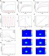
Photothermal performance of various nanoparticles. (A) Heating curves of various nanoparticles with NIR laser (1.5 W/cm2). (B) Heating curves of MMPI with different concentration. (C) Heating curves of MMPI with different laser power densities. (D) Temperature changes for MMPI over five cycles of irradiation/cooling processes (100 μg/mL). (E) Irradiation/cooling temperature change of MMPI under laser irradiation. (F) Liner time data versus -Ln (θ). (G) Time-dependent absorbance spectra of MMPI co-incubated with DPBF after NIR irradiation. (H) The released ICG from MMPI at different temperatures. (I) Representative real-time infrared thermal images of MMPI in PBS under NIR laser irradiation (1.5 W/cm2).
Figure 3
Figure 3
Cytocompatibility assessment in vitro. (A) Viability of NIH-3T3 treated with different dosages of MMPI for 24 h and 48 h. (B) Cell morphology and (C) The fluorescence images of living cells (green) and dead cells (red) of NIH-3T3 treated with PBS, Mg-MBG, MMPI. Scale bar: 100 μm. Data are expressed as average ± SD (n = 3). **P < 0.01.
Figure 4
Figure 4
Antibacterial properties in vitro of various samples. (A) The fluorescence images of living bacterial (green) and dead bacterial (red). Scale bar: 100 μm. (B) Representative plates of MRSA and E. coli colonies formed by the bacteria with various samples after 24 h of culture. (C), (D) The bacterial inhibition rate of various samples. (E), (F) The fluorescence intensity analysis of various samples. (G) ROS release from various samples which co-cultured with bacteria with or without NIR irradiation. Scale bar: 100 μm. (H)Representative TEM images of MRSA (Scale bar: 10 μm) and E. coli (Scale bar: 20 μm) from biofilms after different treatments. ****P < 0.0001.
Figure 5
Figure 5
Immunomodulatory effects on macrophages in vitro. (A) Typical scatter plots of RAW264.7 macrophages surface markers CD11C (M1 macrophages marker) and CD206 (M2 macrophages marker) detected using flow cytometer. (B) RT-PCR detection of macrophage polarization and inflammation related gene expression. *P < 0.05, **P < 0.01.
Figure 6
Figure 6
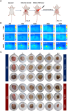
Antibacterial and photothermal performance in vivo. (A) Schematic diagram of infected wound formation and the following treatments. (B) Representative real-time thermographic images under NIR irradiation during 600 s. (C) Representative pictures of infected wounds during the wound healing process.
Figure 7
Figure 7
Histopathological examination of infected wounds after different treatments. (A) Representative HE staining images of skin tissues. (B) Representative Masson’s trichrome (MT) staining images. (C) Immunofluorescent staining of skin tissues: green (iNOS), red (CD206), and blue (DAPI). (D) The inflammation related gene expression of wound tissue. **P < 0.01, ***P < 0.001, ****P < 0.0001.
Figure 8
Figure 8
Exploration of anti-inflammatory mechanisms of MMPI by RNA-seq analysis in vivo. (A) FPKM distribution for all samples. (B) Volcano plots of DEGs. (C) Microarray heat map of all samples. (D) Biological process downregulated with MMPI samples were analyzed using the GO enrichment terms. (E) KEGG enrichment for the downregulated pathways of MMPI samples.

References

    1. Li R, Liu K, Huang X. et al. Bioactive materials promote wound healing through modulation of cell behaviors. Adv Sci. 2022;9(10):e2105152. doi:10.1002/advs.202105152 - DOI - PMC - PubMed
    1. Xu M, Chen Z, Chen K, Ma D, Chen L, DiPietro LA. Phagocytosis of apoptotic endothelial cells reprograms macrophages in skin wounds. J Immunol Regen Med. 2021;1:12. - PMC - PubMed
    1. Tottoli EM, Dorati R, Genta I, Chiesa E, Pisani S, Conti B. Skin wound healing process and new emerging technologies for skin wound care and regeneration. Pharmaceutics. 2020;12(8):735. doi:10.3390/pharmaceutics12080735 - DOI - PMC - PubMed
    1. Ocsoy I, Yusufbeyoglu S, Yılmaz V, McLamore ES, Ildız N, Ülgen A. DNA aptamer functionalized gold nanostructures for molecular recognition and photothermal inactivation of methicillin-Resistant Staphylococcus aureus. Colloids Surf B. 2017;159:16–22. doi:10.1016/j.colsurfb.2017.07.056 - DOI - PubMed
    1. Wang S, Wang Y, Peng Y, Yang X. Exploring the antibacteria performance of multicolor Ag, Au, and Cu nanoclusters. ACS Appl Mater Interfaces. 2019;11(8):8461–8469. doi:10.1021/acsami.8b22143 - DOI - PubMed

MeSH terms