. 2024 Jul 12:11:1419792.
doi: 10.3389/fvets.2024.1419792. eCollection 2024.

Comparative subcutaneous and submuscular implantation of an electroencephalography device for long term electroencephalographic monitoring in dogs

Affiliations

Comparative subcutaneous and submuscular implantation of an electroencephalography device for long term electroencephalographic monitoring in dogs

Casey B Rogers et al. Front Vet Sci. .

Abstract

Background: Implantable electroencephalography (EEG) recording devices have been used for ultra-long-term epilepsy monitoring both in clinical and home settings in people. Objective and accurate seizure detection and recording at home could be of great benefit in diagnosis, management and research in canine idiopathic epilepsy (IE). Continuous EEG monitoring would allow accurate detection of seizure patterns, seizure cycles, and seizure frequency. An EEG acquisition system usable in an "out of clinic" setting could improve owner and veterinary compliance for EEG diagnostics and seizure management.

Objectives: Whether a subcutaneous ultra-long term EEG monitoring device designed for humans could be implanted in dogs.

Animals: Cadaver study with 8 medium to large breed dogs.

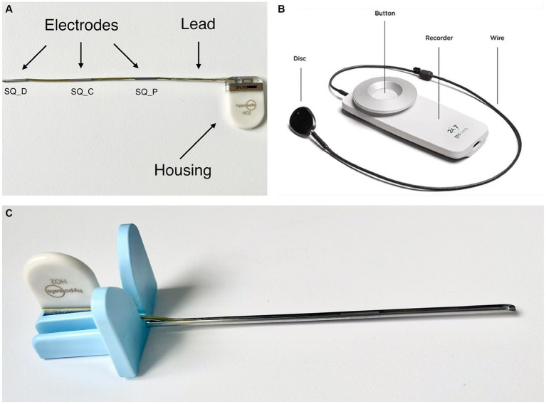
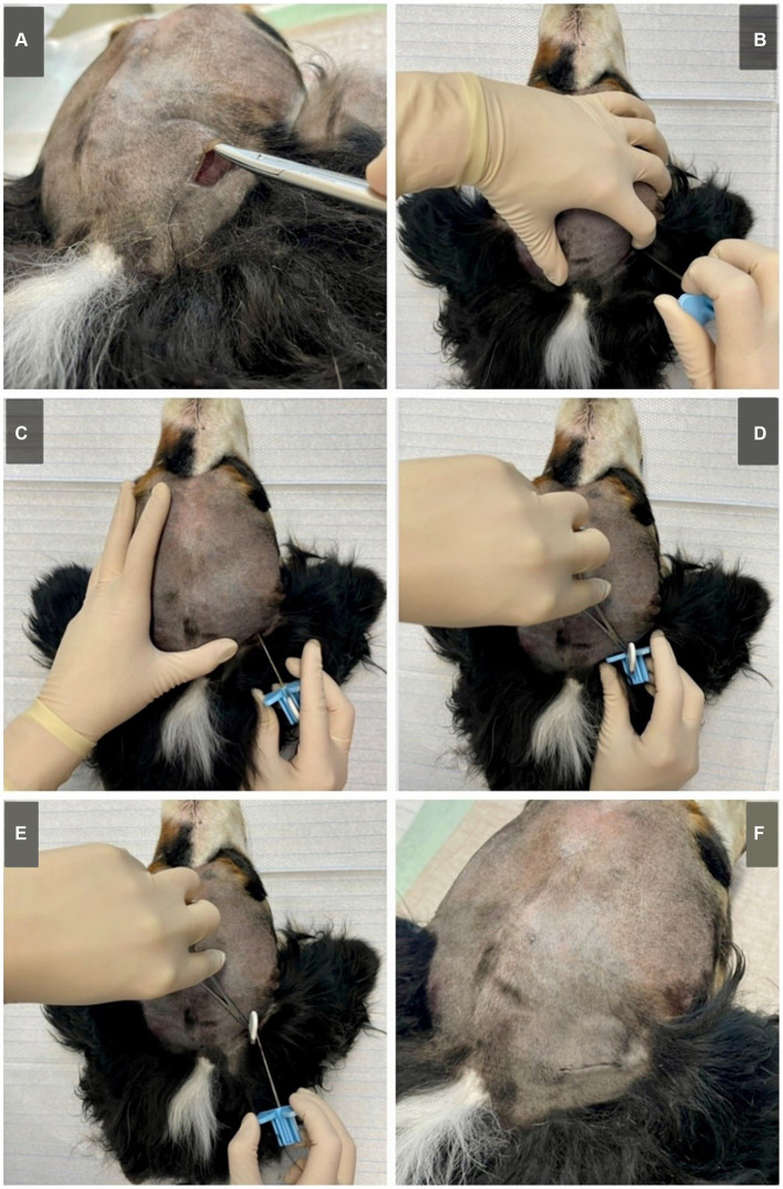
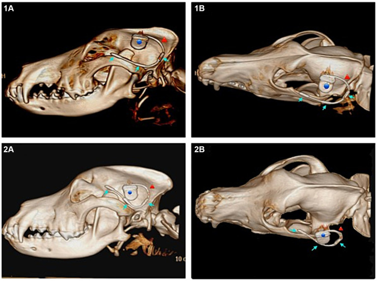
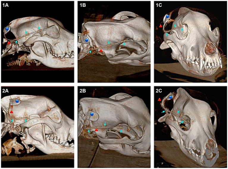
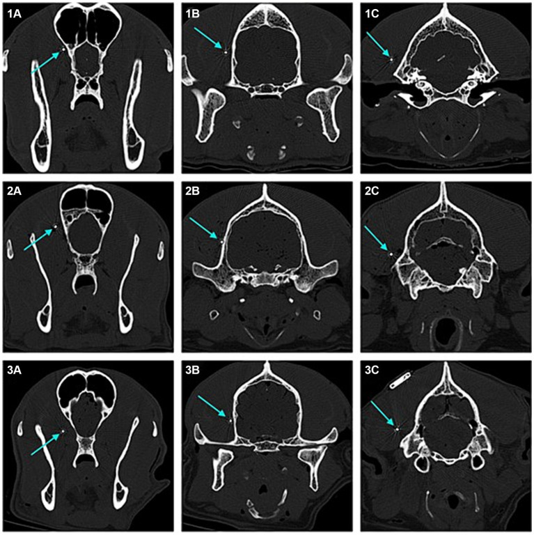
Methods: Comparatively using a subcutaneous and submuscular approach to implant the UNEEG SubQ-Implant in each dog. Positioning was controlled via CT post implantation and cranial measurements were taken.

Results: In four of the eight dogs a submuscular implantation without any complications was possible. Complications were close contact to the optic nerve in the first approaches, before the implantation angle was changed and in the smallest dog contact of the implant with the orbital fat body. Cranial measurements of less than 95 mm length proved to be too small for reliable implantation via this approach. The subcutaneous approach showed severe limitations and the implant was prone to dislocation.

Conclusion: The UNEEQ SubQ-Implant can be implanted in dogs, via submuscular approach. CT imaging and cranial measurements should be taken prior to implantation.

Keywords: EEG; EEG implant; continuous EEG; epilepsy; long-term epilepsy monitoring.


Conflict of interest statement

The authors declare that the research was conducted in the absence of any commercial or financial relationships that could be construed as a potential conflict of interest. The author(s) declared that they were an editorial board member of Frontiers, at the time of submission. This had no impact on the peer review process and the final decision.

Figures

Figure 1
Figure 1
(A) UNEEG SubQ-implant. The electrodes are named SQ_P (subcutaneous contact point proximal), SQ_C (subcutaneous contact point central) and SQ_D (subcutaneous contact point distal). (B) 24/7 EEG SubQ recorder, the wearable, external part of the electroencephalography monitoring solution. The disk attaches to the implanted housing. In people it can be attached to clothing via a magnet. Picture source: UNEEG Medical, https://www.uneeg.com/recorder. (C) UNEEG Sub-Q introduction needle with inserted implant.
Figure 2
Figure 2
Submuscular approach on a Bernese Mountain Dog (cadaver, dog 6). (A) A subcutaneous pocket is created to fit the housing of the implant. (B) The implant is vertically inserted through the masseter, at about 1/3 of the distance between the caudal part of the zygomatic arch and the occiput using the introduction needle. When reaching the skull, (C) the introduction needle is orientated horizontally and angled towards the zygomatic process of the frontal bone. It is then inserted, following the skull. (D) When fully inserted (or in smaller dogs inserted to the desired length) the lead can be fixated using forceps. (E) The introduction needle is carefully removed, keeping the implant fixated in place. (F) The implant is fitted in the subcutaneous pocket and the skin is closed using surgical sutures (pictured here, intracutaneous stiches). Although not pictured, when applying to live animals, a sterile approach is needed. Standard surgical preparation including shaving, washing, disinfection of the skin and sterile covering of the surgical area are required.
Figure 3
Figure 3
Subcutaneous approach. (1A) Lateral view of the subcutaneous implant in a 3D reconstruction of the computed tomography (CT) images of dog 4, ideal positioning. (1B) Dorsolateral view of the same dog (dog 4). (2A) Lateral view of the dislocated subcutaneous implant in a 3D reconstruction of the CT images of dog 6. (2B) Dorsolateral view of the same (dog 6). Blue dot marking the housing, red arrow marking the lead, blue arrows marking the three electrodes of the implant.
Figure 4
Figure 4
Showing 3D reconstructions of the skull from computed tomography (CT) images after submuscular implantation. Dog 6 (images numbered 1) with ideal implantation and dog 1 (images numbered 2) implanted with probable contact to the optic nerve. (1A) Lateral view of dog 6. (1B) Dorsolateral view of dog 6. (1C) Rostral view of dog 6. (2A) Lateral view of dog 1. (2B) Dorsolateral view of dog 1. (2C) Rostral view of dog 1. Blue dot marking the housing, red arrow marking the lead, blue arrow marking the three electrodes of the implant.
Figure 5
Figure 5
Transverse 2D computed tomography (CT) images (bone window, metal artefact reduction) of the dogs’ heads taken at the position of each of the three electrodes of the implant (submuscular implantation). The electrodes are pointed at with blue arrows. Images numbered 1 show the positions for dog 5, with good implant position. Images numbered 2 show the positions for dog 6, with good implant position. Images numbered 3 show the positions for dog 1, with the rostral laying electrode close to the optic nerve. (1A) Rostral laying electrode of the implant located at the level of the frontal lobe in dog 5. (1B) Middle electrode located at the level of the temporal lobe in dog 5. (1C) Caudal laying electrode located at the level of the occipital lobe in dog 5. (2A) Rostral laying electrode of the implant located at the level of the frontal lobe in dog 6. (2B) Middle electrode located at the level of the temporal lobe in dog 6. (2C) Caudal laying electrode located at the level of the occipital lobe in dog 6. (3A) Rostral laying electrode of the implant located at the level of the frontal lobe, in the area of the optic nerve, in dog 1. (3B) Middle electrode located at the level of the temporal lobe in dog 1. (3C) Caudal laying electrode located at the level of the occipital lobe in dog 1.

Similar articles

References

    1. Kearsley-Fleet L, O’Neill DG, Volk HA, Church DB, Brodbelt DC. Prevalence and risk factors for canine epilepsy of unknown origin in the UK. Vet Rec. (2013) 172:338. doi: 10.1136/vr.101133 - DOI - PubMed
    1. Heske L, Nødtvedt A, Jäderlund KH, Berendt M, Egenvall A. A cohort study of epilepsy among 665,000 insured dogs: incidence, mortality and survival after diagnosis. Vet J. (2014) 202:471–6. doi: 10.1016/j.tvjl.2014.09.023, PMID: - DOI - PubMed
    1. De Risio L, Bhatti S, Muñana K, Penderis J, Stein V, Tipold A, et al. . International veterinary epilepsy task force consensus proposal: diagnostic approach to epilepsy in dogs. BMC Vet Res. (2015) 11:148. doi: 10.1186/s12917-015-0462-1, PMID: - DOI - PMC - PubMed
    1. Ukai M, Parmentier T, Cortez MA, Fischer A, Gaitero L, Lohi H, et al. . Seizure frequency discrepancy between subjective and objective ictal electroencephalography data in dogs. J Vet Intern Med. (2021) 35:1819–25. doi: 10.1111/jvim.16158 - DOI - PMC - PubMed
    1. Mondino A, Catanzariti M, Mateos DM, Khan M, Ludwig C, Kis A, et al. . Sleep and cognition in aging dogs. A polysomnographic study. Front Vet Sci. (2023) 10:1151266. doi: 10.3389/fvets.2023.1151266 - DOI - PMC - PubMed

LinkOut - more resources