SNO-EANO-EURACAN consensus on management of pineal parenchymal tumors
- PMID: 39073785
- PMCID: PMC11630543
- DOI: 10.1093/neuonc/noae128
SNO-EANO-EURACAN consensus on management of pineal parenchymal tumors
Abstract
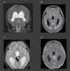
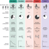
Pineal parenchymal tumors are rare neoplasms for which evidence-based treatment recommendations are lacking. These tumors vary in biology, clinical characteristics, and prognosis, requiring treatment that ranges from surgical resection alone to intensive multimodal antineoplastic therapy. Recently, international collaborative studies have shed light on the genomic landscape of these tumors, leading to refinement in molecular-based disease classification in the 5th edition of the World Health Organization (WHO) classification of tumors of the central nervous system. In this review, we summarize the literature on diagnostic and therapeutic approaches, and suggest pragmatic recommendations for the clinical management of patients presenting with intrinsic pineal region masses including parenchymal tumors (pineocytoma, pineal parenchymal tumor of intermediate differentiation, and pineoblastoma), pineal cyst, and papillary tumors of the pineal region.
Keywords: clinical treatment guidelines; papillary tumor of pineal region; pineal cyst; pineal parenchymal tumors; risk-stratification.
© The Author(s) 2024. Published by Oxford University Press on behalf of the Society for Neuro-Oncology. All rights reserved. For commercial re-use, please contact reprints@oup.com for reprints and translation rights for reprints. All other permissions can be obtained through our RightsLink service via the Permissions link on the article page on our site—for further information please contact journals.permissions@oup.com.
Conflict of interest statement
JRH declares honoraria from Bayer and Alexion Pharmaceuticals and Institutional funding from Servier International for work on their Scientific Advisory Board unrelated to this work. GL declares consulting or advisory role funding from AbbVie, Bayer, Novartis, Orbus Therapeutics, BrainFarm, Celgene, Cureteq, Health4U, Braun, Janssen, BioRegio STERN, Servier, and Novocure; and travel funding from Roche and Bayer. All other authors declare no conflict of interest.
Figures


References
-
- Jussila M-P, Olsén P, Salokorpi N, Suo-Palosaari M.. Follow-up of pineal cysts in children: Is it necessary? Neuroradiology. 2017;59(12):1265–1273. - PubMed
-
- Jouvet A, Vasiljevic A, Champier J, Fèvre Montange M.. Pineal parenchymal tumours and pineal cysts. Neurochirurgie. 2015;61(2-3):123–129. - PubMed
Publication types
MeSH terms
Grants and funding
LinkOut - more resources
Full Text Sources
Medical

