. 2024 Nov;31(11):755-768.
doi: 10.1002/jhbp.12056. Epub 2024 Jul 29.

Japanese classification of pancreatic carcinoma by the Japan Pancreas Society: Eighth edition

Affiliations

Japanese classification of pancreatic carcinoma by the Japan Pancreas Society: Eighth edition

Masaharu Ishida et al. J Hepatobiliary Pancreat Sci. 2024 Nov.

Abstract

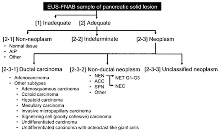
In 2023, the Japan Pancreas Society (JPS) published the new eighth edition of the Japanese classification of pancreatic carcinoma. We present here an excerpted version in English, based on the latest edition. The major changes in this revision are as follows: In the eighth edition of the Union for International Cancer Control (UICC), the T category was changed to be based on tumor size; however, the eighth edition of the Japanese classification retains the previous T category based on local invasion factors. Lymph nodes have been renamed, and regional lymph nodes have been defined by location. Peritoneal cytology, which was not previously included in distant metastasis (M), has now been included in the M category. Moreover, significant additions have been made regarding the pathological diagnosis of endoscopic ultrasound-guided fine-needle aspiration biopsy (EUS-FNAB) and criteria for histological assessment of the effects after chemotherapy and radiation therapy. Although this classification is aimed at carcinoma originating in the pancreas, not in the bile duct or duodenum, if the differentiation of the primary organ is difficult, this classification should be applied. It is also desirable to describe tumors other than carcinoma and metastatic tumors to the pancreas in accordance with this classification.

Keywords: Japanese; classification; neoplasm staging; pancreatic cancer; pancreatic neoplasms.


Conflict of interest statement

The authors declare there are no conflicts of interest.

Figures

FIGURE 1
FIGURE 1
Anatomical portions of the pancreas. Reprinted with permission from the Japan Pancreas Society and Kanehara & Co., Ltd.: Japanese Classification of Pancreatic Carcinoma, eighth edition, 2023. The border between the pancreatic head and body is defined as the left side of the SMV and PV. The neck of the pancreas (the part anterior to the SMV and PV) and the uncinate process are included in the pancreatic head. The border between the pancreatic body and tail is defined as the left border of the abdominal aorta. Pb, pancreatic body; Ph, pancreatic head; Pt, pancreatic tail; PV, portal vein, SMA, superior mesenteric artery; SMV, superior mesenteric vein; UP, uncinate process.
FIGURE 2
FIGURE 2
Extrapancreatic nerve plexus. Reproduced with permission from the Japan Pancreas Society and Kanehara & Co., Ltd.: Japanese Classification of Pancreatic Carcinoma, eighth edition, 2023. (a) The extra‐pancreatic nerve plexus is differentiated into seven categories (PLph I, pancreatic head nerve plexus I; PLph II, pancreatic head nerve plexus II; PLsma, superior mesenteric artery nerve plexus; PLcha, common hepatic artery nerve plexus; PLhdl, hepatoduodenal ligament nerve plexus; PLspa; splenic artery nerve plexus; PLce, celiac nerve plexus). (b) PLphI and PLphII are blue and green, respectively. (c) PLphII and PLsma are histologically examined in the anatomical samples. CHA, common hepatic artery; GDA, gastroduodenal artery; IPDA, inferior pancreatoduodenal artery; J1A, first jejunal artery; MCA, middle colic artery; SMA, superior mesenteric artery.
FIGURE 3
FIGURE 3
Station numbers and locations of lymph nodes. Reproduced with permission from the Japan Pancreas Society and Kanehara & Co., Ltd.: Japanese Classification of Pancreatic Carcinoma, eighth edition, 2023. Station numbers of lymph nodes in relation to the pancreas. (a) The location and boundaries of the lymph nodes within the hepatoduodenal ligament. The diagram on the right shows the transverse view at line A in the diagram on the left (b), with details provided along the superior mesenteric artery (c) and around the aorta (d). Ao, aorta; CA, celiac artery; IMA, inferior mesenteric artery; IVC, inferior vena cava; LRA, left renal artery; LRV, left renal vein; SMA, superior mesenteric artery.
FIGURE 4
FIGURE 4
Inking example. Reprinted with permission from the Japan Pancreas Society and Kanehara & Co., Ltd.: Japanese Classification of Pancreatic Carcinoma, eighth edition, 2023. Pancreatoduodenectomy specimen after fixation. (a) View from the posterior surface. (b) View from the transection margin (blue, portal vein margin; red, superior mesenteric artery margin; green, posterior surface; yellow, transection margin).
FIGURE 5
FIGURE 5
Diagnostic categories of EUS‐FNAB samples of pancreatic solid lesions. Adapted from reference. [1] Inadequate: This category should be applied to specimens that are not suitable for diagnosis due to insufficient quantity or quality. The reasons for unsuitability, such as degeneration or artifacts, should be noted. [2] Adequate: This category should be applied to specimens suitable for diagnosis. The diagnostic procedures should be evaluated according to the following subcategories: Non‐neoplasm, indeterminate, and neoplasm. Whether the sample size is large or small should be assessed. [2‐1] Non‐neoplasm: This subcategory should be applied to specimens with non‐neoplastic lesions. Specimens suggestive of autoimmune pancreatitis can be further evaluated by elastica staining and immunohistochemistry for IgG4. [2‐2] Indeterminate: This subcategory should be applied to specimens that have sufficient quantity and quality but present challenges in evaluation. This subcategory may include specimens that are suggestive of malignancy but pose challenges in definitive determination. The term “suggestive of malignancy” should be employed in consultation with local clinicians to guide management decisions. Samples in this subcategory can be further evaluated using stepwise sections and/or immunohistochemistry. [2‐3] Neoplasm: This subcategory should be applied to specimens evaluated as neoplastic lesions, which should be further categorized into the following diagnostic categories, including ductal carcinoma, nonductal neoplasm, and unclassified neoplasm. [2‐3‐1] Ductal carcinoma: This diagnostic category should be applied to specimens sufficiently evaluated for ductal carcinoma according to the histological features listed in Table 7. Acinar ductal metaplasia can be differentiated using immunohistochemical staining for p53 and maspin. [2‐3‐2] Non‐ductal neoplasm: This diagnostic category should be applied to specimens sufficiently evaluated as nonductal neoplasms, such as neuroendocrine neoplasms, acinar cell carcinomas, solid pseudopapillary neoplasms, metastatic neoplasms, and nonepithelial neoplasms. Immunohistochemical analysis may be useful for this evaluation. [2‐3‐3] Unclassified neoplasm: This diagnostic category should be applied to specimens evaluated as neoplasms with an indeterminate ductal nature according to further immunohistochemical examinations. ACC, acinar cell carcinoma; AIP, autoimmune pancreatitis, EUS‐FNAB, endoscopic ultrasound‐guided fine needle aspiration biopsy; NEC, neuroendocrine carcinoma; NEN, neuroendocrine neoplasm; NET, neuroendocrine tumor; SPN, solid pseudopapillary neoplasm.

References

    1. Japan Pancreas Society . Japanese classification of pancreatic carcinoma. 1st ed. Tokyo: Kanehara; 1980. (in Japanese).
    1. Japan Pancreas Society . Japanese classification of pancreatic carcinoma. 8th ed. Tokyo: Kanehara; 2023. (in Japanese).
    1. Japan Pancreas Society . Japanese classification of pancreatic carcinoma. 7th ed. Tokyo: Kanehara; 2016. (in Japanese).
    1. Tsuchida H, Fujii T, Mizuma M, Satoi S, Igarashi H, Eguchi H, et al. Prognostic importance of peritoneal washing cytology in patients with otherwise resectable pancreatic ductal adenocarcinoma who underwent pancreatectomy: a nationwide, cancer registry‐based study from the Japan pancreas society. Surgery. 2019;166:997–1003. - PubMed
    1. Yoshioka H, Wakabayashi T. Procedure of neurotomy on the head of the pancreas (in Japanese). Shujutsu Oper. 1957;11:849–857.