. 2024 Jul 29;22(1):158.
doi: 10.1186/s12915-024-01958-2.

Plancitoxin-1 mediates extracellular trap evasion by the parasitic helminth Trichinella spiralis

Affiliations

Plancitoxin-1 mediates extracellular trap evasion by the parasitic helminth Trichinella spiralis

Jing Ding et al. BMC Biol. .

Erratum in

Abstract

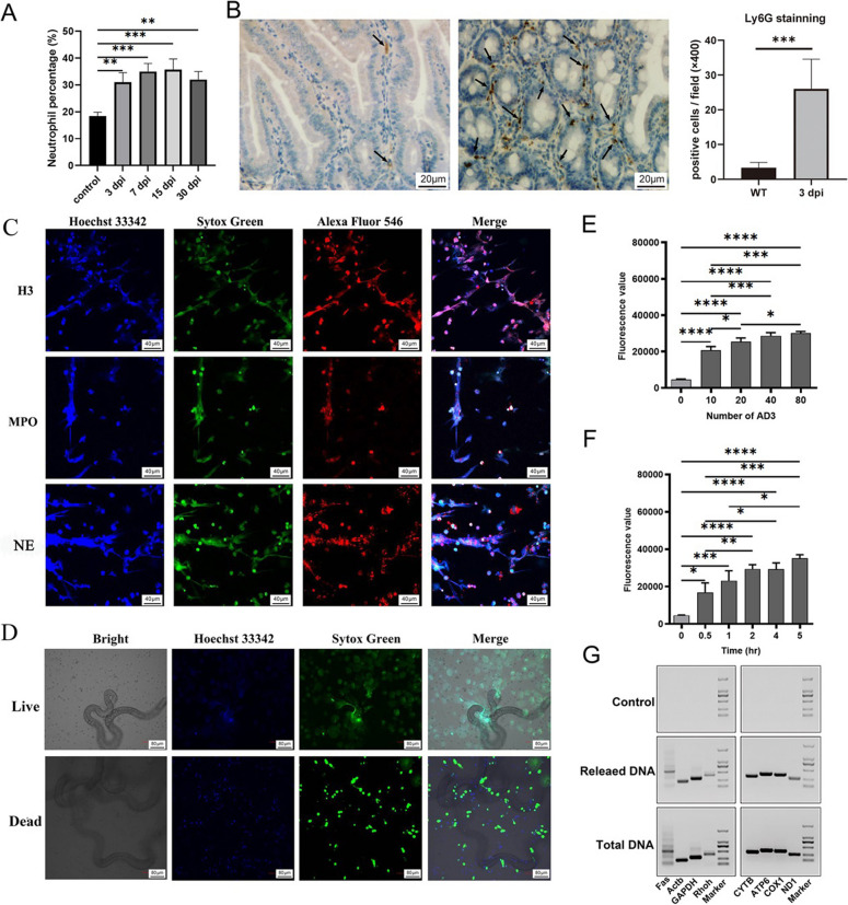
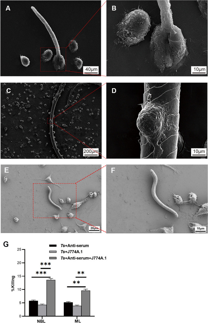
Background: Trichinella spiralis (T. spiralis) is a parasitic helminth that causes a globally prevalent neglected zoonotic disease, and worms at different developmental stages (muscle larvae, adult worms, newborn larvae) induce immune attack at different infection sites, causing serious harm to host health. Several innate immune cells release extracellular traps (ETs) to entrap and kill most pathogens that invade the body. In response, some unicellular pathogens have evolved a strategy to escape capture by ETs through the secretion of nucleases, but few related studies have investigated multicellular helminths.

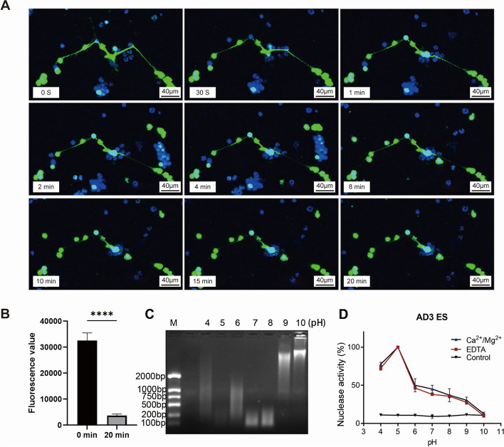
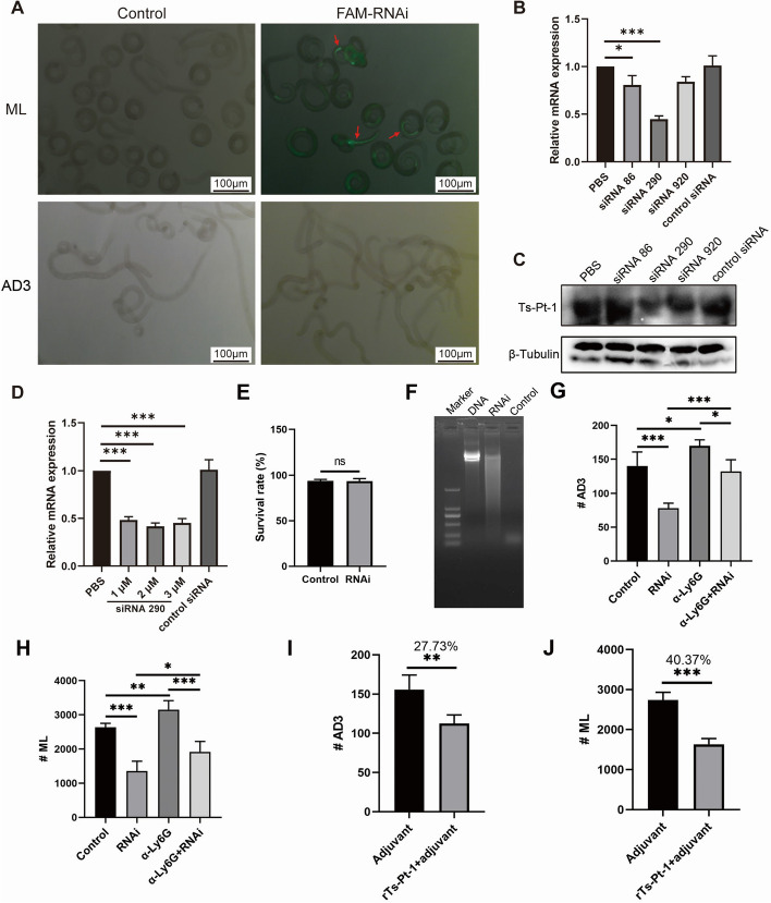
Results: In the present study, we observed that ETs from neutrophils capture adult worms of T. spiralis, while ETs from macrophages trap muscle larvae and newborn larvae, and ETs had a killing effect on parasites in vitro. To defend against this immune attack, T. spiralis secretes plancitoxin-1, a DNase II-like protein, to degrade ETs and escape capture, which is essential for the survival of T. spiralis in the host.

Conclusions: In summary, these findings demonstrate that T. spiralis escapes ET-mediated capture by secreting deoxyribonuclease as a potential conserved immune evasion mechanism, and plancitoxin-1 could be used as a potential vaccine candidate.

Keywords: Trichinella spiralis; Extracellular traps; Immune evasion; Nuclease; Vaccine candidate.


Conflict of interest statement

The authors declare that they have no competing interests.

Figures

Fig. 1
Fig. 1
Neutrophils release NETs in response to T. spiralis adults. A Neutrophil counts of mice at 0, 3, 7, 15, and 30 days after infection with 300 muscle larvae. Data (mean ± SD) were collected from two independent experiments (n = 3 mice/time point), *** (P < 0.001) in one way ANOVA test. B Representative microphotographs showing Ly6G-immunopositive cells (corresponding to neutrophils, black arrows) in the small intestine of control and infected mice at 3 dpi; scale bar: 20 µm. Data (mean ± SD) are representative of two independent experiments performed (n = 3 mice/group), *** (P < 0.001) in Student’s t test. C The components of NETs induced by adults of T. spiralis include histone 3 (H3), myeloperoxidase (MPO), and neutrophil elastase (NE). Fifty adult worms were cocultured with 2 × 105 PMNs in medium containing ATA for 3 h at 37 °C in 5% CO2. H3, MPO, and elastase were detected by immunofluorescence and are shown in red. The DNA scaffold of NETs and the nuclei of dead cells were dyed green with SYTOX Green, and the nuclei of all cells were dyed blue with Hoechst 33,342. Scale bar, 40 μm. D Live adults but not dead worms induced NETs. Fifty live adults and 50 dead adults were cocultured with PMNs separately. The culture method and staining method were the same as in C. NETs were dyed green, as shown in the white square. Scale bar, 80 μm. Data in C and D were collected from two independent experiments (n = 3), and representative images are shown. E and F The NETs quantity increased with the number of adults (0, 10, 20, 40, 80 adults) and the coculture time (0, 0.5, 1, 2, 4, 5 h) (with ATA in the medium). The results are the averages of three independent experiments (mean ± SD, *** P < 0.001 in one-way ANOVA). G The DNA scaffold of NETs derived from both the nucleus and mitochondria. Fifty adult worms were cocultured with 2 × 105 PMNs in medium containing ATA for 3 h at 37 °C in 5% CO2. The culture supernatant and the total DNA of cells of the control group and experimental group were used as templates. Marker genes of mouse nuclear genome genes (Rhoh, GAPDH, Actb, Fas) and mitochondrial genome genes (Nd1, Cox1, Atp6, Cyb) were detected by PCR. Data were collected from two independent experiments (n = 3), and representative images are shown
Fig. 2
Fig. 2
Macrophages can release METs, and ETs kill T. spiralis in vitro. A and B Scanning electron micrographs obtained after a total of 400 newborn larvae (NBL) cocultured with 1 × 104 J774A.1 cells in medium containing ATA for 3 h at 37 °C in 5% CO2. Scale bar, 30 μm (A) and 5 μm (B). C and D SEM micrographs obtained after 100 muscle larvae (ML) cocultured with 1 × 104 J774A.1 cells in medium containing ATA for 3 h at 37 °C in 5% CO2. Scale bar, 100 μm (C) and 30 μm (D). Data in A–D were collected from two independent experiments (n = 3), and representative images are shown. E Approximately 50 adults were cocultured with 2 × 105 PMNs in medium containing ATA, and the surviving worms were counted after 6 and 12 h of cocultivation. Data were collected from two independent experiments (n = 3) and analyzed by ANOVA. F and G Approximately 100 ML (F) or 400 NBL (G) were cocultured with 1 × 104 J774A.1 cells in medium containing ATA, and the surviving worms were counted after 24 and 48 h of cocultivation. Data in E–G were collected from two independent experiments (n = 3) and analyzed by one-way ANOVA. * (P < 0.05), ** (P < 0.01), and *** (P < 0.001) indicate a statistically significant difference
Fig. 3
Fig. 3
The antibody-dependent cell-mediated phagocytosis effect of macrophages kills ML and NBL. A and B Scanning electron micrographs of NBL-J774A.1 cocultivations. A total of 400 newborn larvae (NBL) were cocultured with 1 × 104 J774A.1 cells with anti-T. spiralis serum in culture medium for 3 h at 37 °C in 5% CO2. Scale bar, 40 μm (A) and 10 μm (B). C and D Scanning electron micrographs of ML-J774A.1 cocultivations. One hundred muscle larvae (ML) were cocultured with 1 × 104 J774A.1 cells with anti-T. spiralis serum for 3 h at 37 °C in 5% CO2. Scale bar, 200 μm (C) and 10 μm (D). E and F Scanning electron micrographs of NBL-J774A.1 cocultivations. A total of 400 NBL were cocultured with 1 × 10.4 J774A.1 cells without anti-T. spiralis serum in culture medium for 3 h at 37 °C in 5% CO2. Scale bar, 20 μm (E) and 10 μm (F). Data in A–F were collected from two independent experiments (n = 3), and the representative images are shown. G Killing efficiency statistics (mean ± SD, n = 3, **P < 0.01 in two-tailed Student’s t test)
Fig. 4
Fig. 4
Excretory/secretory products of adult worms obtained at 3 days post infection have nuclease activity to degrade NETs in vitro. A Images of NETs induced by PMA after the addition of excretory/secretory products of adult worms obtained at 3 days post infection (AD3 ES) in the cultivation at various time points. The DNA scaffold of NETs and the nuclei of dead cells were dyed green with SYTOX Green, and the nuclei of all cells were dyed blue with Hoechst 33,342. AD3 ES showed DNase activity in a time-dependent manner. Scale bar, 40 μm. Data were collected from two independent experiments (n = 3), and representative images are shown. B Quantification of NETs detected by PicoGreen in the samples at 0 and 20 min in A. Data were collected from two independent experiments (n = 3) and analyzed by Student’s t test. **** (P < 0.0001) indicates a statistically significant difference. C λDNA was detected by agarose gel electrophoresis after reacting with AD3 ES under different pH conditions, and λDNA was most significantly degraded at pH 5. Data were collected from two independent experiments (n = 3), and a representative image is shown. D λDNA was detected by spectrophotometry after reacting with AD3 ES under different pH conditions with or without Ca2+/Mg2+. The absorbance of λDNA was highest at pH 5, and there was no significant difference with or without Ca2+/Mg.2+ at each pH analyzed by Student’s t test. Data were collected from two independent experiments (n = 3)
Fig. 5
Fig. 5
Identification of nuclease-active proteins in excretory/secretory products of adult worms obtained at 3 days post infection. A The protein with nuclease activity in excretory/secretory products of adult worms obtained at 3 days post infection (AD3 ES) was between 35 and 45 kDa, as detected by 1D zymography. B All proteins obtained by mass spectrometry were blast searched, and only Ts-Pt-1 had typical enzymatic active sites. The protein DNase II-8, which does not have a typical enzymatic active site, had no enzymatic activity detected by agarose gel electrophoresis (C) or the agar diffusion method (D). E Ts-Pt-1 with typical enzymatic active sites had nuclease activity detected by agarose gel electrophoresis, and Ca2+, Mg2+, Co2+, and Zn.2+ had an inhibitory effect on its enzymatic activity in a concentration-dependent manner. F rTs-Pt-1 has the strongest enzymatic activity at pH 5, as detected by agarose gel electrophoresis. G The degradation products of λDNA cleaved by rTs-Pt-1 can be further cleaved by phosphatidase II. Data were collected from two independent experiments (n = 3). * (P < 0.05) and *** (P < 0.001) indicate a statistically significant difference analyzed by one-way ANOVA test. H The mRNA levels of plancitoxin-1 were highest in the adult stage, as assessed by qPCR. The ML group was regarded as 100%. The results are the averages of three independent experiments (mean ± SD)
Fig. 6
Fig. 6
Ts-Pt-1 exerts nuclease activity in excretory/secretory products and can be a potential vaccine candidate. A FAM-siRNA was successfully transferred into muscle larvae (ML), and green fluorescence was observed in the intestinal tube of the worms. Scale bar, 100 µm. qPCR (B) and Western blot (C) analysis of relative Ts-Pt-1 expression levels after T. spiralis ML were transfected with two µM siRNA86, siRNA290, and siRNA920. Data in B were collected from two independent experiments (n = 3). * (P < 0.05) and *** (P < 0.001) indicate a statistically significant difference analyzed by one-way ANOVA. D qPCR analysis of relative Ts-Pt-1 expression levels after T. spiralis ML were transfected with 1, 2, or 3 µM siRNA290. Data were collected from two independent experiments (n = 3). *** (P < 0.001) indicates a statistically significant difference analyzed by one-way ANOVA. E Survival rate analysis of normal ML and Ts-Pt-1-RNAi-treated ML at 24 h after interference. Data were collected from two independent experiments (n = 6 wells) and analyzed by Student’s t test; ns indicates no significant difference. F The nuclease activity of ES derived from Ts-Pt-1-RNAi-treated ML was weaker than that of the control larvae, as detected by agarose gel electrophoresis. G and H Neutrophil-depleted mice were treated with anti-Ly6G mAb by intraperitoneal (i.p.) injection once a day − 1, 0, 1, 2, and 3. On day 0, neutrophil-depleted mice and wild-type mice were infected with 200 normal ML or Ts-Pt-1-RNAi-treated ML. The number of adult worms collected from the intestine was counted in each group (G), and the number of ML collected from muscle was counted in each group (H). Data were collected from two independent experiments (n = 6–8 mice/group) and analyzed by ANOVA; * (P < 0.05), ** (P < 0.01), and *** (P < 0.001) indicate a statistically significant difference. I and J The number of adult worms was reduced by 27.73% in mice immunized with rTs-pt-1 compared with the control mice (I). The number of ML was reduced by 40.37% in mice immunized with rTs-pt-1 compared with control mice (J). Data were collected from two independent experiments (n = 6–8 mice/group) and analyzed by students’ t test; ** (P < 0.01) and *** (P < 0.001) indicate a statistically significant difference compared with the control groups

Similar articles

Cited by

References

    1. Bintsis T. Foodborne pathogens. AIMS Microbiol. 2017;3(3):529–63. - PMC - PubMed
    1. Li C, Bai X, Liu X, Zhang Y, Liu L, Zhang L, Xu F, Yang Y, Liu M. Disruption of epithelial barrier of Caco-2 Cell monolayers by excretory secretory products of Trichinella spiralis might be related to serine protease. Front Microbiol. 2021;12:634185. - PMC - PubMed
    1. Xu J, Liu RD, Bai SJ, Hao HN, Yue WW, Xu YXY, Long SR, Cui J, Wang ZQ. Molecular characterization of a Trichinella spiralis aspartic protease and its facilitation role in larval invasion of host intestinal epithelial cells. Plos Negl Trop Dis. 2020;14(4):e0008269. - PMC - PubMed
    1. Bai Y, Ma KN, Sun XY, Dan Liu R, Long SR, Jiang P, Wang ZQ, Cui J. Molecular characterization of a novel cathepsin L from Trichinella spiralis and its participation in invasion, development and reproduction. Acta Trop. 2021;224:106112. - PubMed
    1. Yi N, Yu P, Wu L, Liu Z, Guan J, Liu C, Liu M, Lu Y. RNAi-mediated silencing of Trichinella spiralis serpin-type serine protease inhibitors results in a reduction in larval infectivity. Vet Res. 2020;51(1):139. - PMC - PubMed

Substances

LinkOut - more resources