. 2024 Oct;17(10):e202400126.
doi: 10.1002/jbio.202400126. Epub 2024 Jul 29.

Cardiac-gated spectroscopic photoacoustic imaging for ablation-induced necrotic lesion visualization

Affiliations

Cardiac-gated spectroscopic photoacoustic imaging for ablation-induced necrotic lesion visualization

Shang Gao et al. J Biophotonics. 2024 Oct.

Abstract

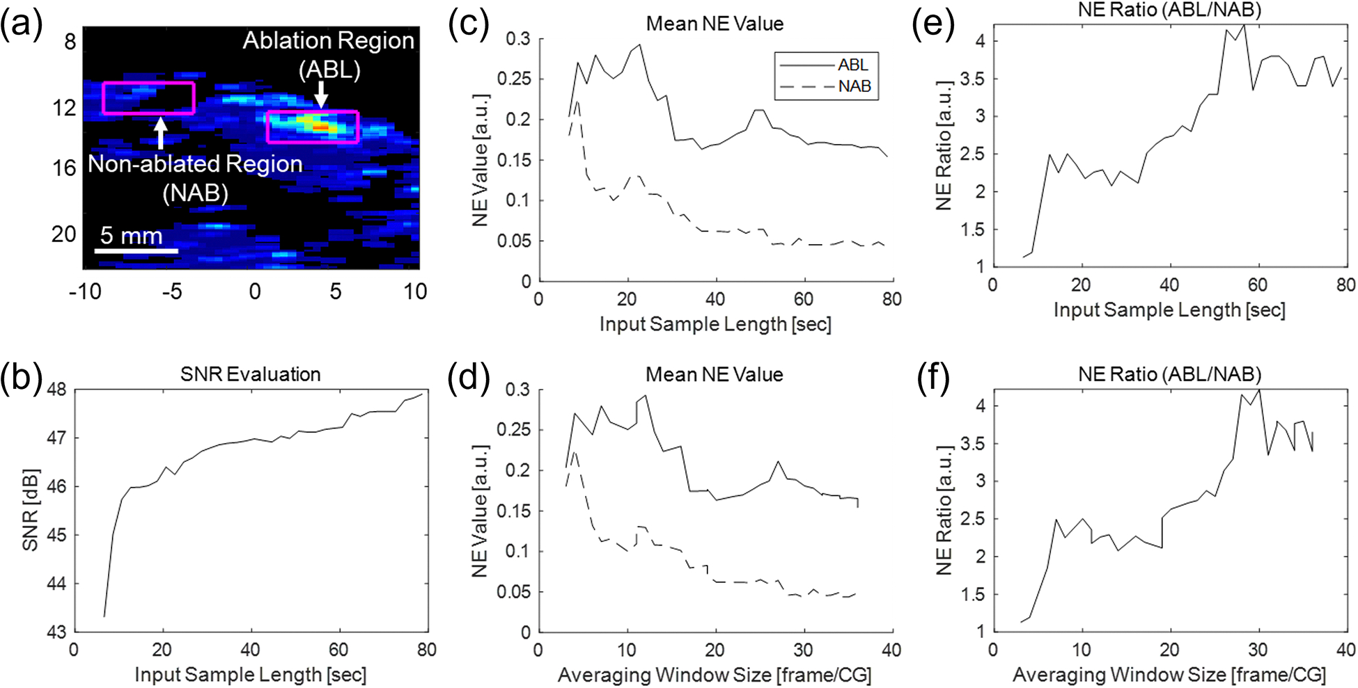
Radiofrequency (RF) ablation is a minimally invasive therapy for atrial fibrillation. Conventional RF procedures lack intraoperative monitoring of ablation-induced necrosis, complicating assessment of completeness. While spectroscopic photoacoustic (sPA) imaging shows promise in distinguishing ablated tissue, multi-spectral imaging is challenging in vivo due to low imaging quality caused by motion. Here, we introduce a cardiac-gated sPA imaging (CG-sPA) framework to enhance image quality using a motion-gated averaging filter, relying on image similarity. Necrotic extent was calculated based on the ratio between spectral unmixed ablated tissue contrast and total tissue contrast, visualizing as a continuous color map to highlight necrotic area. The validation of the concept was conducted in both ex vivo and in vivo swine models. The ablation-induced necrotic lesion was successfully detected throughout the cardiac cycle through CG-sPA imaging. The results suggest the CG-sPA imaging framework has great potential to be incorporated into clinical workflow to guide ablation procedures intraoperatively.

Keywords: cardiac ablation; cardiac gating; image‐guided intervention; in vivo demonstration; spectroscopic photoacoustic imaging.


Conflict of interest statement

Conflict of Interest Statement

Tommaso Mansi was a scientist and employee of Siemens Healthineers USA during this work and currently is a scientist and employee of Janssen: Pharmaceutical Companies of Johnson & Johnson. Young-Ho Kim and Florin-Cristian Ghesu are scientists and employees of Siemens Healthineers USA. The other authors have stated explicitly that there are no conflicts of interest in connection with this article.

Figures

Fig.1.
Fig.1.
The photoacoustic (PA) spectra from ablated and non-ablated tissues used for spectroscopic decomposition [38].
Fig.2.
Fig.2.
Cardiac-gated spectroscopic photoacoustic (CG-sPA) image-processing flowchart
Fig.3.
Fig.3.
(a) Two laser illumination modes: Three-wavelength repetitive illumination (3WL-Rep) is suitable to achieve a high frame sPA imaging, while 16-wavelength individual illumination (16WL-Indiv) offers a robust spectroscopic decomposition. (b) The sketch of the imaging setup and system architecture.
Fig.4.
Fig.4.
(a) The sketch of the imaging setup for in vivo imaging. (b) In vivo cardiac photoacoustic (PA) imaging setup with swine model. (c) The post-procedure heart surface with the ablated region highlighted. The yellow dot-line indicates the imaging slice for necrotic detection.
Fig.5.
Fig.5.
(a) Ex vivo spectroscopic photoacoustic (sPA) mapping of necrotic extent (NE) before and after ablation without motion. (b) Temporal correlation coefficient values at varying axial displacements. (c) Signal-to-noise (SNR) changes with different Temporal correlation coefficient. (d) NE map of Sample 2 within a simulated cardiac cycle. [unit: mm]
Fig.6
Fig.6
Cardiac-gated spectroscopic photoacoustic (CG-sPA) imaging results of the in vivo beating heart based on repetitive illumination. The ablated and non-ablated tissue intensity distribution maps show the outcome of the spectroscopic decomposition algorithm. The sham procedure was performed in a location not containing ablated tissue. The displayed photoacoustic (PA) images were acquired with a 740 nm wavelength. [unit: mm]
Fig.7.
Fig.7.
Necrotic extent (NE) mapping with repetitive illumination mode in one complete cardiac cycle. [unit: mm]
Fig.8.
Fig.8.
Cardiac-gated spectroscopic photoacoustic (CG-sPA) imaging results of the in vivo beating heart based on individual wavelength illumination. The ablated and non-ablated tissue intensity distribution maps show the outcome of the spectroscopic decomposition algorithm. The sham procedure was performed in a location not containing ablated tissue. The displayed photoacoustic (PA) images were acquired with a 740 nm wavelength. [unit: mm]
Fig.9.
Fig.9.
Necrotic extent (NE) mapping calculated with a varied input data length in terms of the time duration of the collected data and the mean number of frames used for averaging across each wavelength. [unit: mm]
Fig.10.
Fig.10.
Quantitative evaluation of necrotic extent (NE) mapping based on the input data size. (a) A NE map based on 80 seconds of input data under repetitive illumination mode. Ablation (ABL) and non-ablation (NAB) regions are marked. (b) The SNR improvement over varied input data duration. The mean NE value in the ABL and NAB regions changes over varied input data duration (c) and averaging window sizes (d). The ratio of NE values between two regions changes over varied input data duration (e) and averaging window sizes (f).
Fig.11.
Fig.11.
Gross pathological analysis of the ablated tissue region. (a) Ablated region (white-dash rectangle) identified by the repetitive illumination scanning mode. (b) Ablated region (white-dash rectangle) identified by the individual wavelength illumination scanning mode. (c) Top view of the post-procedure processed ablated tissue for width measurement. (d) Side-view of the ablated tissue for depth measurement. [unit: mm]

References

    1. “Atrial Fibrillation | cdc.gov,” https://www.cdc.gov/heartdisease/atrial_fibrillation.htm.
    1. Morillo CA, Banerjee A, Perel P, Wood D, and Jouven X, “Atrial fibrillation: The current epidemic,” Journal of Geriatric Cardiology 14(3), 195–203 (2017). - PMC - PubMed
    1. Peters DC, Wylie JV, Hauser TH, Nezafat R, Han Y, Woo JJ, Taclas J, Kissinger KV, Goddu B, Josephson ME, and Manning WJ, “Recurrence of Atrial Fibrillation Correlates With the Extent of Post-Procedural Late Gadolinium Enhancement. A Pilot Study,” JACC Cardiovasc Imaging 2(3), 308–316 (2009). - PMC - PubMed
    1. Nakagawa H and Jackman WM, “The role of contact force in atrial fibrillation ablation,” J Atr Fibrillation 7(1), 78–84 (2014). - PMC - PubMed
    1. Santoro A, Romano A, and Lamberti F, “Steam pop during cavo-tricuspid isthmus ablation shown by intracardiac echocardiography,” J Cardiol Cases 23(1), 13–15 (2021). - PMC - PubMed

Publication types

LinkOut - more resources