. 2024 Jan-Dec:33:9636897241264979.
doi: 10.1177/09636897241264979.

Effects of FGF2 Priming and Nrf2 Activation on the Antioxidant Activity of Several Human Dental Pulp Cell Clones Derived From Distinct Donors, and Therapeutic Effects of Transplantation on Rodents With Spinal Cord Injury

Affiliations

Effects of FGF2 Priming and Nrf2 Activation on the Antioxidant Activity of Several Human Dental Pulp Cell Clones Derived From Distinct Donors, and Therapeutic Effects of Transplantation on Rodents With Spinal Cord Injury

Hidefumi Fukumitsu et al. Cell Transplant. 2024 Jan-Dec.

Abstract

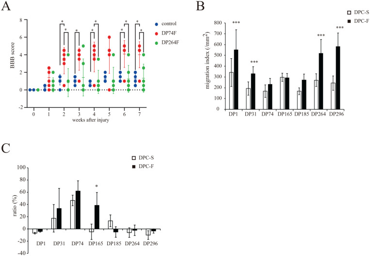
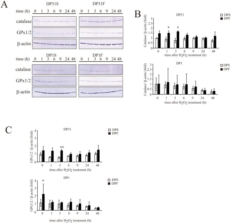
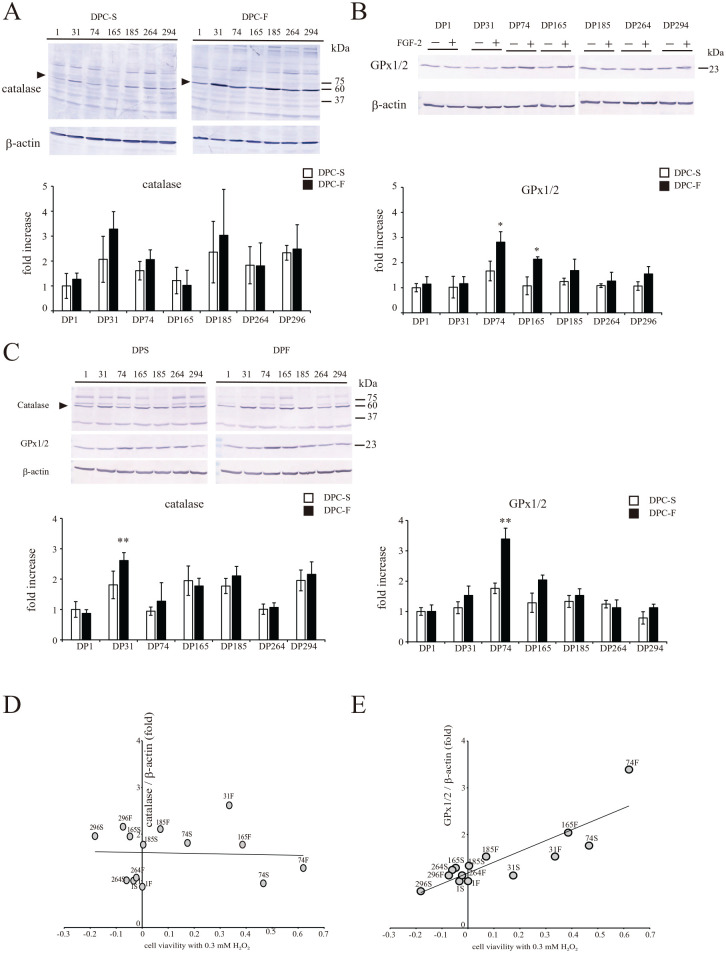
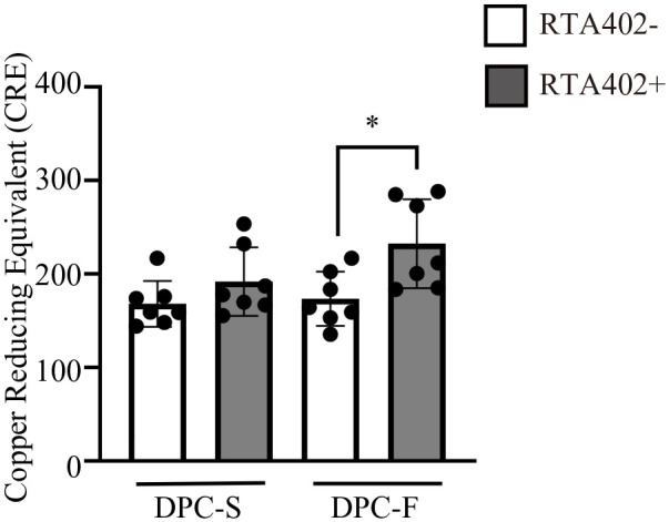
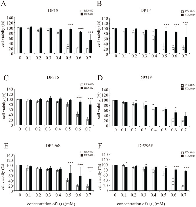
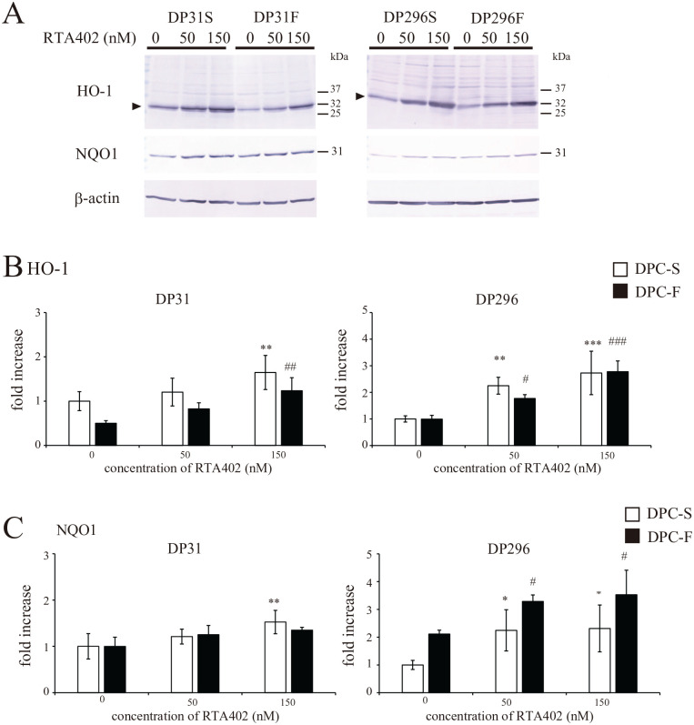
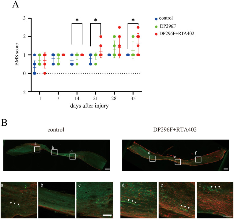
In recent years, the interest in cell transplantation therapy using human dental pulp cells (DPCs) has been increasing. However, significant differences exist in the individual cellular characteristics of human DPC clones and in their therapeutic efficacy in rodent models of spinal cord injury (SCI); moreover, the cellular properties associated with their therapeutic efficacy for SCI remain unclear. Here, using DPC clones from seven different donors, we found that most of the clones were highly resistant to H2O2 cytotoxicity if, after transplantation, they significantly improved the locomotor function of rats with complete SCI. Therefore, we examined the effects of the basic fibroblast growth factor 2 (FGF2) and bardoxolone methyl (RTA402), which is a nuclear factor erythroid 2-related factor 2 (Nrf2) chemical activator, on the total antioxidant capacity (TAC) and the resistance to H2O2 cytotoxicity. FGF2 treatment enhanced the resistance of a subset of clones to H2O2 cytotoxicity. Regardless of FGF2 priming, RTA402 markedly enhanced the resistance of many DPC clones to H2O2 cytotoxicity, concomitant with the upregulation of heme oxygenase-1 (HO-1) and NAD(P)H-quinone dehydrogenase 1 (NQO1). With the exception of a subset of clones, the TAC was not increased by either FGF2 priming or RTA402 treatment alone, whereas it was significantly upregulated by both treatments in each clone, or among all seven DPC clones together. Thus, the TAC and resistance to H2O2 cytotoxicity were, to some extent, independently regulated and were strongly enhanced by both FGF2 priming and RTA402 treatment. Moreover, even a DPC clone that originally exhibited no therapeutic effect on SCI improved the locomotor function of mice with SCI after transplantation under both treatment regimens. Thus, combined with FGF2, RTA402 may increase the number of transplanted DPCs that migrate into and secrete neurotrophic factors at the lesion epicenter, where reactive oxygen species are produced at a high level.

Keywords: antioxidant factors; clonal difference; human dental pulp cells; nuclear factor erythroid 2-related factor 2 (Nrf2) chemical activator (RTA402); spinal cord injury.


Conflict of interest statement

Declaration of Conflicting InterestsThe author(s) declared no potential conflicts of interest with respect to the research, authorship, and/or publication of this article.

Figures

Figure 1.
Figure 1.
Effect of basic fibroblast growth factor (FGF2) priming on the properties of the seven individual donor-derived dental pulp cell (DPC) clones. (A) Effect of FGF2-primed DPC clone (DP74F or DP264F) transplantation on the locomotor function of rats with a completely transected spinal cord at the injury site. The locomotor function of the hind limbs was evaluated weekly for 7 weeks after injury. The values are expressed as individual scores with mean lines. Significant differences from the control group (rat with spinal cord injury with PBS injection) were determined using two-way repeated-measures ANOVA with post-hoc Tukey’s multiple comparison test. *P < 0.05; n = 6 for each group. (B) Effect of FGF2 priming on the cell migration indices of the individual donor-derived DPC clones (DPC-S and DPC-F; DPC clones primed without and with FGF2, respectively). The values are expressed as the mean ± standard deviation (SD). Significant differences from the FGF2 non-treated groups were determined using two-way ANOVA with post-hoc Tukey’s multiple comparison test. ***P < 0.0001; n = 12 for each group. (C) Effect of FGF2 priming on the cell viability of 0.3 mM H2O2-treated individual donor-derived DPC clones. The values are expressed as the mean ± SD Significant differences from the FGF2 non-treated groups were determined using the two-way ANOVA with post-hoc Tukey’s multiple comparison test. *P < 0.05; N = 4 (n = 6) for each group.
Figure 2.
Figure 2.
Effect of basic fibroblast growth factor (FGF2) priming on the time-dependent alterations in the expression levels of catalase and glutathione peroxidase 1/2 (GPx1/2) in DP1 and DP31 after treatment with 0.3 mM H2O2. A. DP1 and DP31 were treated with 0.3 mM H2O2. After the indicated periods, the expression levels of the catalase and GPx1/2 proteins were determined by Western blotting, together with the evaluation of β-actin as a loading control. The results obtained for catalase (B) or GPx1/2 (C) are reported as the mean ± standard deviation. Significant differences from the FGF2 non-treated groups were determined using two-way ANOVA with post-hoc Tukey’s multiple comparison test. *P < 0.05; **P < 0.01, n = 4–5 for each group.
Figure 3.
Figure 3.
Effect of FGF2 priming on the expression levels of catalase and glutathione peroxidase 1/2 (GPx1/2) in individual donor-derived dental pulp cells (DPCs) after treatment with H2O2. Each DPC clone was treated with 0.3 mM H2O2. At 0 h (A, B) or 3 h (C) after the treatment, the expression levels of the catalase (A, C) and GPx1/2 (B, C) proteins were determined by Western blotting, together with the evaluation of β-actin as a loading control. The results obtained for catalase, or GPx1/2, are shown as the mean ± standard deviation. Significant differences from the FGF2 non-treated groups were determined using two-way ANOVA with post-hoc Tukey’s multiple comparison test. *P < 0.05; **P < 0.01, n = 4–5 for each group. Correlation analyses of the expression levels of catalase (D) or GPx1/2 (E) with cell viabilities at 24 h after treatment with 0.3 mM H2O2.
Figure 4.
Figure 4.
Effect of RTA402 and FGF2 on the total antioxidant activities of dental pulp cells (DPCs) [DPC-S and DPC-F; DPC clones primed without and with basic fibroblast growth factor (FGF2), respectively]. The values are expressed as the mean ± standard deviation (SD). Significant differences between groups were determined using two-way ANOVA with post-hoc Tukey’s multiple comparison test. *P < 0.05, n = 7 average copper reducing equivalent of individual DPC clones for each group.
Figure 5.
Figure 5.
Effect of RTA402 on the resistance to H2O2-induced cell death in individual donor-derived dental pulp cells (DPCs) [DPC-S and DPC-F; DPC clones primed without and with basic fibroblast growth factor (FGF2), respectively]. After priming with/without FGF2 and treatment with/without 150 nM RTA402, each DPC clone (A, B: DP1; C, D: DP31; and E, F: DP296) was exposed to H2O2 at various concentrations (0, 0.1, 0.2, 0.3, 0.4, 0.5, 0.6, and 0.7 mM) for 24 h, followed by the MTT assay. The ratios of the absorbance values recorded for the DPCs exposed to H2O2 were calculated relative to the control (neither H2O2- nor RTA402-treated DPCs). The values are expressed as the mean ± standard deviation. Significant differences between the two groups were determined by two-way ANOVA with post-hoc Tukey’s multiple comparison test. *P < 0.05; **P < 0.01; and ***P < 0.001 vs DPC without RTA402 treatment with H2O2 at the same concentration; N = 3 (n = 6) for each group.
Figure 6.
Figure 6.
Effect of RTA402 on the expression levels of the heme oxygenase-1 (HO-1) and NAD(P)H-quinone dehydrogenase 1 (NQO1) proteins in DP31 and DP296 primed with basic fibroblast growth factor (FGF2). After priming with/without FGF2, each dental pulp cell (DPC) clone was treated with various concentrations of RTA402 (0, 50, and 150 nM) for 24 h. (A) The expression of the HO-1 and NQO1 proteins was determined by Western blotting, together with the evaluation of β-actin as a loading control. The results obtained for HO-1 (B) or NQO1 (C) are shown as the mean ± standard deviation. Significant differences between the two groups were determined by two-way ANOVA with post-hoc Tukey’s multiple comparison test. *P < 0.05; **P < 0.01, and ***P < 0.001 vs DPC-S untreated with H2O2 and #P < 0.05; ##P < 0.01, and ###P < 0.001 vs DPC-F untreated with H2O2, n = 4 for each group.
Figure 7.
Figure 7.
Effect of the i.v. transplantation of DP296F treated with/without RTA402 (DP296F+RTA402 or DP296F) on the locomotor function of mice with a compression spinal cord injury (SCI). (A) The locomotor function of the hind limbs was evaluated weekly for 5 weeks after injury. The values are expressed as individual scores with mean lines. Significant differences from the control group (SCI mice with PBS injection) were determined using two-way repeated-measures ANOVA with post-hoc Tukey’s multiple comparison test. n = 8–10 for each group. (B) At 5 weeks after cell transplantation, an immunohistochemical analysis was performed using the central sagittal section of the spinal cord tissues. The lower panels depict a higher magnification of the boxed regions in the upper panels. Note that a measurable number of 5-HT+-nerve fibers were detected in the DP296F+RTA402 groups (three out of four), but not in the remaining groups (0 out of four) below the caudal edge of the injury sites. Single scale bar, 500 μm; double scale bars, 100 μm; respectively.

Similar articles

References

    1. Shingu H, Ohama M, Ikata T, Katoh S, Akatsu T. A nationwide epidemiological survey of spinal cord injuries in Japan from January 1990 to December 1992. Paraplegia. 1995;33(4):183–88. - PubMed
    1. Miyakoshi N, Suda K, Kudo D, Sakai H, Nakagawa Y, Mikami Y, Suzuki S, Tokioka T, Tokuhiro A, Takei H, Katoh S, et al.. A nationwide survey on the incidence and characteristics of traumatic spinal cord injury in Japan in 2018. Spinal Cord. 2021;59(6):626–34. - PubMed
    1. GBD 2016 Traumatic Brain Injury and Spinal Cord Injury Collaborators. Global, regional, and national burden of traumatic brain injury and spinal cord injury, 1990-2016: a systematic analysis for the global burden of disease study 2016. Lancet Neurol. 2019;18(1):56–87. - PMC - PubMed
    1. Tsuji O, Sugai K, Yamaguchi R, Tashiro S, Nagoshi N, Kohyama J, Iida T, Ohkubo T, Itakura G, Isoda M, Shinozaki M, et al.. Concise review: laying the groundwork for a first-in-human study of an induced pluripotent stem cell-based intervention for spinal cord injury. Stem Cells. 2019;37(1):6–13. - PMC - PubMed
    1. Kong H, Liu P, Li H, Zeng X, Xu P, Yao X, Liu S, Cheng CK, Xu J. Mesenchymal stem cell-derived extracellular vesicles: the novel therapeutic option for regenerative dentistry. Stem Cell Rev Rep. 2023;19(1):46–58. - PubMed

LinkOut - more resources