Review
. 2022 Oct 28;23(11):369.
doi: 10.31083/j.rcm2311369. eCollection 2022 Nov.

Atrioventricular Ring Tachycardias: Atypical Fast-Slow Atrioventricular Nodal Reentrant Tachycardia and Atrial Tachycardia Share a Common Arrhythmogenic Substrate-A Unifying Proposal

Affiliations
Review

Atrioventricular Ring Tachycardias: Atypical Fast-Slow Atrioventricular Nodal Reentrant Tachycardia and Atrial Tachycardia Share a Common Arrhythmogenic Substrate-A Unifying Proposal

Yoshiaki Kaneko et al. Rev Cardiovasc Med. .

Abstract

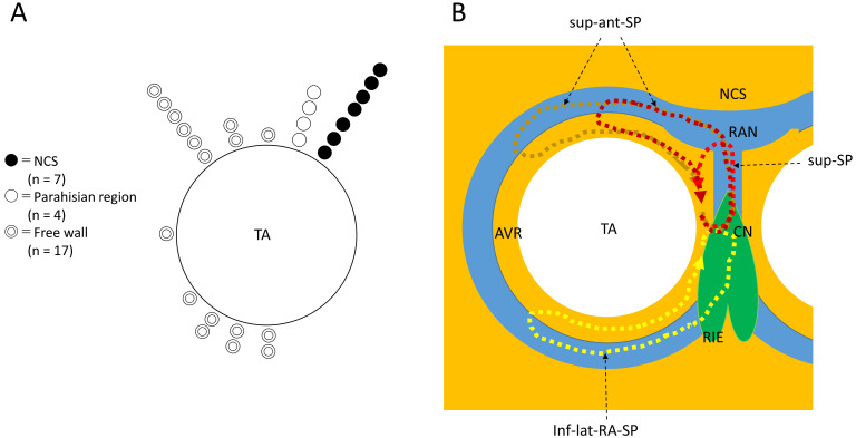
Our understanding of the variants of slow pathway (SP) and associated atypical atrioventricular (AV) nodal reentrant tachycardia (NRT) is still growing. We have identified variants extending outside Koch's triangle along the tricuspid annulus, including superior, superoanterior and inferolateral right atrial SP and associated atypical, fast-slow AVNRT. We review the history of each variant, their electrophysiological characteristics and related atypical AVNRT, and their treatment by catheter ablation. We focused our efforts on organizing the published information, as well as some unpublished, reliable data, and show the pitfalls of electrophysiological observations, along with keys to the diagnosis of atypical AVNRT. The superior-type of fast-slow AVNRT mimics adenosine-sensitive atrial tachycardia originating near the AV node and can be successfully treated by ablation of a superior SP form the right side of the perihisian region or from the non-coronary sinus of Valsalva. Fast-slow AVNRT using a superoanterior or inferolateral right atrial SP also mimics atrial tachycardia originating from the tricuspid annulus. We summarize the similarities among these variants of SP, and the origin of the atrial tachycardias, including their anatomical distributions and electrophysiological and pharmacological characteristics. Moreover, based on recent basic research reporting the presence of node-like AV ring tissue encircling the annuli in adult hearts, we propose the term "AV ring tachycardia" to designate the tachycardias that share the AV ring tissue as a common arrhythmogenic substrate. This review should help the readers recognize rare types of SP variants and associated AVNRT, and diagnose and cure these complex tachycardias. We hope, with this proposal of a unified tachycardia designation, to open a new chapter in clinical electrophysiology.

Keywords: adenosine sensitivity; atrial tachycardia; atrioventricular nodal reentrant tachycardia; atrioventricular ring; slow pathway; tricuspid annulus.


Conflict of interest statement

The authors declare no conflict of interest. Yoshiaki Kaneko is serving as one of the Guest editors of this journal. We declare that Yoshiaki Kaneko had no involvement in the peer review of this article and has no access to information regarding its peer review. Full responsibility for the editorial process for this article was delegated to Giacomo Mugnai.

Figures

Fig. 1.
Fig. 1.
Distribution of the site of earliest atrial activation during fast-slow AVNRT using a superior, superoanterior or inferolateral right atrial SP (A) and schematic illustration of the reentry circuits of an atypical fast-slow AVNRT using variants of SP, the AV ring (AVR) and the retroaortic node (RAN) (B). NCS, non-coronary sinus of Valsalva, TA, tricuspid annulus. (B) The AVR encircling the TA is continuous with the right inferior extension (RIE) of the AV node and with the RAN, just behind to NCS. The inferolateral right atrial slow pathway (inf-lat-RA-SP) is formed by the continuous RIE and AVR, and is used as the retrograde limb of the reentrant inferolateral-type of fast-slow AVNRT. The superior slow pathway (sup-SP), formed by nodal-like tissue connecting the compact node (CN) and RAN, is used as the retrograde limb for the reentrant superior-type of fast-slow AVNRT. The superoanterior slow pathway (sup-ant-SP) is formed by the AVR in continuity with the RAN and the components of sup-SP, and is used as a retrograde limb of the reentry circuit of the superoanterior-type of fast-slow AVNRT.
Fig. 2.
Fig. 2.
Termination of fast-slow AVNRT using a superoanterior SP during ventricular overdrive pacing at an S-S cycle length of 425 ms from the right ventricular apex (RVA1-2). The site of earliest atrial activation during tachycardia is recorded in the HRA (HRA1-2). The atrial cycle length immediately after the 4th and 5th stimuli lengthens slightly without change in the atrial activation sequence, consistent with an orthodromic capture of the atria over the superoanterior SP, with a decremental delay in response to the 4th and 5th stimuli (dotted arrows). The 6th stimulus is blocked (straight arrow), evidenced by the absence of retrograde activation over the SP in its wake. In response to the 6th and 7th ventricular stimuli, the site of earliest retrograde atrial activation was observed in the distal electrogram of the His bundle region (HBE1-2) along with a short ventriculoatrial interval, consistent with retrograde conduction over a FP. The numbers above the HRA1-2 channel are the interatrial intervals. I, II and V1, surface electrocardiogram leads; CS 19-20 to 1-2, proximal to distal CS.
Fig. 3.
Fig. 3.
(A) pseudo-V-A-V and (B) atypical V-A-A-V responses observed upon initiation of the tachycardia after ventricular extrastimuli (S2) at S1-S2 coupling intervals of 280 (A) and 380 ms (B), following trains of simultaneous atrial and ventricular pacing at an S1-S1 cycle length of 400 ms, in a patient presenting with a superior-type of fast-slow AVNRT. Ventriculoatrial conduction was absent at baseline. In both (A) and (B), the interval between the last atrial S1 and the first atrial electrogram of the tachycardia is similar to the interval between the last atrial S and the first atrial electrogram of the tachycardia induced by regular atrial pacing at an S-S cycle length of 400 ms (C). Therefore, the tachycardia is presumed to be induced by the atrial pacing train and follow the ventricular S2, instead of being induced by the ventricular S2. This resulted in an apparent V-A-V, or pseudo-V-A-V (A), and atypical V-A-A-V responses due to anterograde block in the lower common pathway caused by its refractoriness in the wake of S2 (B). II, surface electrocardiogram lead II; HRA1-2, high right atrium; HBE1-2, His bundle electrogram; CS13-14 to 1-2, proximal to distal coronary sinus; RVA1-2, right ventricular apex.
Fig. 4.
Fig. 4.
Representative examples. (A,B) Superior-type of fast-slow AVNRT successfully ablated in the non-coronary sinus of Valsalva (A) and the right perihisian region (B). (C) Superoanterior type fast-slow AVNRT. (D) Inferolateral type fast-slow AVNRT: E: ATP-sensitive atrial tachycardia originating from the inferolateral right atrial wall, along the tricuspid annulus. Left panels (A) to (E): intracardiac electrograms during tachycardia at the site of successful ablation. The asterisks in C, D and E mark the prepotentials preceding the local atrial electrograms. II, surface electrocardiogram lead II; HBE1-2, His bundle electrogram; ABL1-2, distal pole of ablation catheter. Middle panels: right (B) and left (A,C,D,E) anterior oblique fluoroscopic views of the catheter positions at the site of successful ablation. The white arrow points to the tip of the ablation catheter. Right panels: left anterior oblique views of three-dimensional activation maps of the right atrium during tachycardia, using CARTOTM (A to D) and RhythmiaTM (E) systems. The yellow tags indicate the sites of recording of the His bundle electrogram. The middle panel of Fig. 4B is reproduced from Kaneko et al. [19] (Copyright © 2016, the American Heart Association), and the left, middle and right panels of Fig. 4D are reproduced from Kaneko et al. [47] (Copyright © 2019, the Japanese Circulation Society), with permission from the publishers.
Fig. 5.
Fig. 5.
Differential atrial entrainment pacing at an S-S cycle length of 365 ms from the high right atrium (HRA1-2) (A) and the proximal coronary sinus (CS9-10) (B), in a patient presenting with fast-slow AVNRT using an inferolateral, right atrial SP. This patient is the same as in Fig. 4D. Note: (1) the atypical atrial activation sequence during tachycardia, characterized by nearly simultaneous atrial electrograms in the HRA and in the His bundle region (HBE1-2); and (2) shorter His-atrial intervals immediately after entrainment pacing (indicated by horizontal bidirectional arrows and numbers above) in the HRA than in the proximal coronary sinus (CS9-10). II, surface electrocardiogram lead II; CS13-14 to 1-2, proximal to distal coronary sinus; RVA, right ventricular apex.
Fig. 6.
Fig. 6.
Termination of superior-type (A) and inferolateral fast-slow (B) AVNRT, following 4- and 10-mg boluses of ATP, respectively. (A) Tachycardia with the earliest site of atrial activation in the His bundle electrogram (HBE1-2), ending with an ectopic atrial event that followed the interatrial interval of 539 ms, longer than the tachycardia cycle length, suggesting that the tachycardia was terminated by retrograde block (or slowed by a retrograde delay) in the superior SP, immediately before the ectopic atrial cycle. (B) Tachycardia with the earliest site of atrial activation in the proximal coronary sinus (CS13-14), slowing in association with a prolongation of the VA interval, and terminated in an “A no V” ending, suggesting that the termination was due to retrograde block in the right atrial, inferolateral SP. Numbers above the high right atrial (HRA1-2) channel indicate the interatrial intervals. II, surface electrocardiogram lead II; CS13-14 to 1-2, proximal to distal coronary sinus; RVA, right ventricular apex.
Fig. 7.
Fig. 7.
Development of accelerated ectopic atrial cycles during radiofrequency delivery. (A) Accelerated ectopic atrial rhythm (asterisks) developing immediately after radiofrequency energy was delivered (RF on) during an ongoing superoanterior-type of fast-slow AVNRT, in the same patient as shown in Fig. 4C. The tachycardia is terminated by a premature atrial complex (PAC) immediately after the delivery of radiofrequency energy. (B) ATP-sensitive atrial tachycardia originating from the inferolateral right atrium along the tricuspid annulus, followed by return to sinus rhythm (SR) in the same patient as in Fig. 4E. II, surface electrocardiogram lead II; HRA1-2, high right atrium; HBE1-2, His bundle electrogram; CS13-14 to 1-2, proximal to distal coronary sinus; RVA1-2, right ventricular apex.
Fig. 8.
Fig. 8.
Example of superoanterior type fast-slow AVNRT (A) followed by the development of atrial tachycardia (B) originating from the superoanterior right atrium, along the tricuspid annulus, after ablation of a superoanterior SP. This is the same patient as in Fig. 4C. (A) Induction of superior-type of fast-slow AVNRT by ventricular overdrive pacing at an S-S cycle length of 670 ms. Retrograde conduction via a FP is present after the 1st and 2nd stimuli, with an earliest site of atrial activation in the His bundle region (HBE1-2). Immediately after the 2nd stimulus, a long RP tachycardia with a site of earliest atrial activation in the high right atrium (HRA1-2) is induced, after a V-A-A-V response where the 367-ms interatrial interval is 88 ms shorter than the 455-ms tachycardia cycle length, consistent with a dual atrial response from simultaneous retrograde conduction (dotted arrows) over the FP and the superoanterior SP. The 3rd and 4th stimuli (S), do not capture the ventricles. (B) Ventricular entrainment pacing at an S-S cycle length of 320 ms during ongoing atrial tachycardia with a site of earliest atrial activation in the HRA. Ventricular overdrive pacing captures the atrial electrogram via the FP in HBE1-2 (dotted arrows) after the 3rd stimulus, without capture of the atrial electrogram in HRA1-2, suggesting intraatrial fusion of a retrograde wavefront originating from the FP, with an atrial wavefront propagating from the site of origin of the atrial tachycardia. II, surface electrocardiogram lead II; HRA1-2, high right atrium; HBE1-2, His bundle electrogram; CS7-8 to 1-2, proximal to distal coronary sinus; RVA1-2, right ventricular apex.

References

    1. Moe GK, Preston JB, Burlington H. Physiologic evidence for a dual A-V transmission system. Circulation Research . 1956;4:357–375. - PubMed
    1. Denes P, Wu D, Dhingra RC, Chuquimia R, Rosen KM. Demonstration of Dual A-V Nodal Pathways in Patients with Paroxysmal Supraventricular Tachycardia. Circulation . 1973;48:549–555. - PubMed
    1. Sung RJ, Styperek JL, Myerburg RJ, Castellanos A. Initiation of two distinct forms of atrioventricular nodal reentrant tachycardia during programmed ventricular stimulation in man. The American Journal of Cardiology . 1978;42:404–415. - PubMed
    1. Sung RJ, Waxman HL, Saksena S, Juma Z. Sequence of retrograde atrial activation in patients with dual atrioventricular nodal pathways. Circulation . 1981;64:1059–1067. - PubMed
    1. Ross DL, Johnson DC, Denniss AR, Cooper MJ, Richards DA, Uther JB. Curative surgery for atrioventricular junctional (“AV Nodal”) reentrant tachycardia. Journal of the American College of Cardiology . 1985;6:1383–1392. - PubMed

LinkOut - more resources