. 2024 Jul 15:18:1429124.
doi: 10.3389/fnana.2024.1429124. eCollection 2024.

Intra-articular sprouting of nociceptors accompanies progressive osteoarthritis: comparative evidence in four murine models

Affiliations

Intra-articular sprouting of nociceptors accompanies progressive osteoarthritis: comparative evidence in four murine models

Alia M Obeidat et al. Front Neuroanat. .

Abstract

Objective: Knee joints are densely innervated by nociceptors. In human knees and rodent models, sprouting of nociceptors has been reported in late-stage osteoarthritis (OA). Here, we sought to describe progressive nociceptor remodeling in early and late-stage OA, using four distinct experimental mouse models.

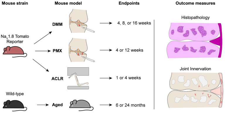
Methods: Sham surgery, destabilization of the medial meniscus (DMM), partial meniscectomy (PMX), or non-invasive anterior cruciate ligament rupture (ACLR) was performed in the right knee of 10-12-week old male C57BL/6 NaV1.8-tdTomato mice. Mice were euthanized (1) 4, 8 or 16 weeks after DMM or sham surgery; (2) 4 or 12 weeks after PMX or sham; (3) 1 or 4 weeks after ACLR injury or sham. Additionally, a cohort of naïve male wildtype mice was evaluated at age 6 and 24 months. Mid-joint cryosections were assessed qualitatively and quantitatively for NaV1.8+ or PGP9.5+ innervation. Cartilage damage, synovitis, and osteophytes were assessed.

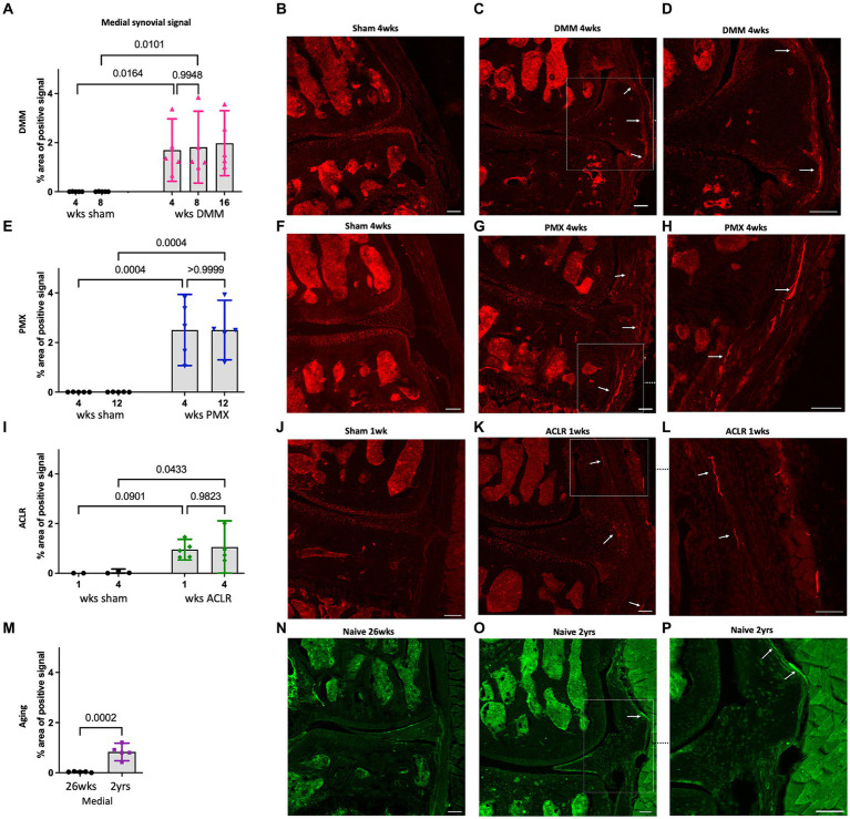
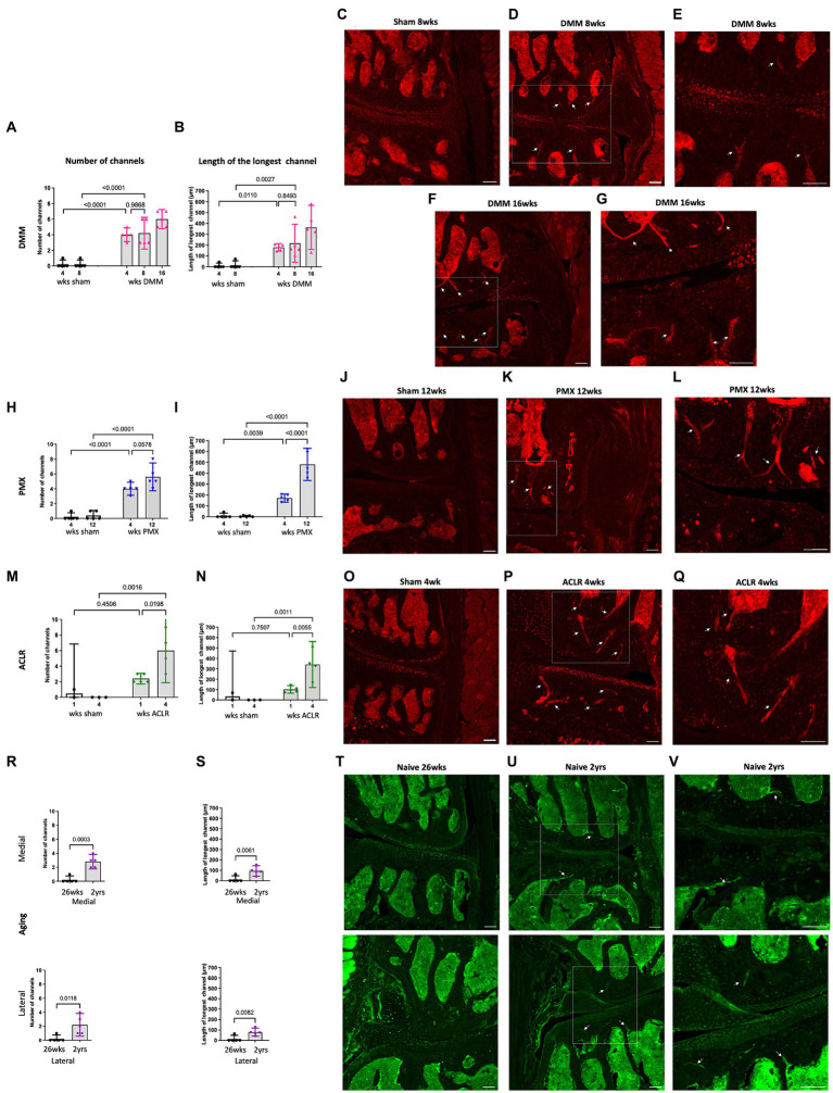
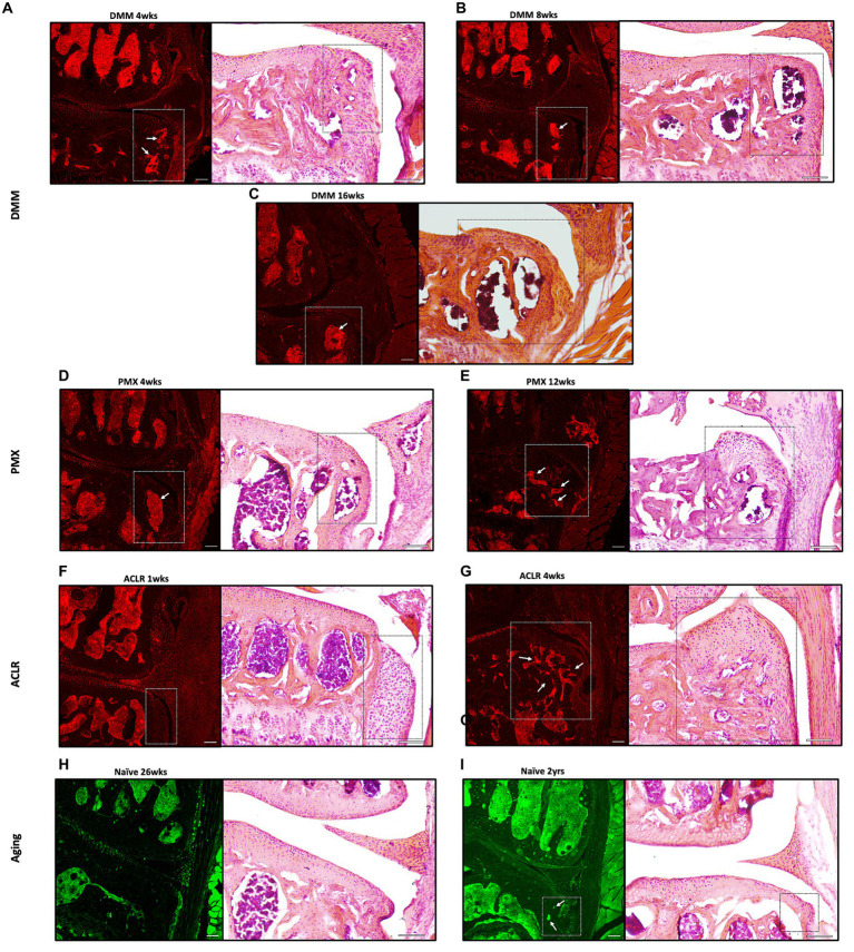
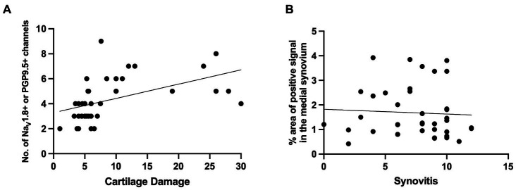
Results: Progressive OA developed in the medial compartment after DMM, PMX, and ACLR. Synovitis and associated neo-innervation of the synovium by nociceptors peaked in early-stage OA. In the subchondral bone, channels containing sprouting nociceptors appeared early, and progressed with worsening joint damage. Two-year old mice developed primary OA in the medial and the lateral compartment, accompanied by nociceptor sprouting in the synovium and the subchondral bone. All four models showed increased nerve signal in osteophytes.

Conclusion: These findings suggest that anatomical neuroplasticity of nociceptors is intrinsic to OA pathology. The detailed description of innervation of the OA joint and its relationship to joint damage might help in understanding OA pain.

Keywords: mouse models; neuroplasticity; nociceptive innervation; osteoarthritis; sprouting.


Conflict of interest statement

A-MM received consulting fees from Asahi Kasei Pharma Corporation, Orion, and 23andMe. The remaining authors declare that the research was conducted in the absence of any commercial or financial relationships that could be construed as a potential conflict of interest.

Figures

Figure 1
Figure 1
Schematic overview of male mouse strains, osteoarthritis models, timepoints, and outcome measures used in the study.
Figure 2
Figure 2
(A) Quantification of neuronal signal in the medial synovium 4, 8 and 16 weeks after sham and DMM surgeries (n = 5/group); (B–D) Representative confocal images of NaV1.8-tdTomato mouse knees showing the nerve fibers in the medial synovium (white arrows) at 4 weeks after sham and DMM surgery, with (D) showing a magnified image of (C); (E) Quantification of neuronal signal in the medial synovium 4 and 12 weeks after sham and PMX surgeries (n = 5/group); (F–H) 4 weeks after sham, 4 weeks after PMX, and zoomed in image, respectively; (I) Quantification of neuronal signal in the medial synovium 1 and 4 weeks after sham and ACLR injury (n = 4–5/group); (J–L) 1 week after sham, 1 week after ACLR injury, and zoomed in image, respectively; (M) Quantification of neuronal signal in the medial synovium of 26-week old and 2-year old naïve mice (n = 5/group); (N–P) PGP9.5 staining in a 26-week old mouse, a 2-year old mouse, and zoomed in image, respectively. Mean ± 95% CI. Scale bar = 100 μm.
Figure 3
Figure 3
Quantification of neuronal signal in the medial subchondral bone 4, 8 and 16 weeks after sham and DMM surgeries showing (A) the number of NaV1.8 + channels; (B) the length of the longest channel; (C–E) Representative confocal images of NaV1.8-tdTomato mouse knees showing fibers within medial subchondral bone channels (white arrows) at 8 weeks after sham and 8 and 16 weeks after DMM surgery; (E,G) magnified image of medial subchondral bone fibers at 8 and 16 weeks after DMM; (H,I) quantification of the number of positive channels and the length of the longest channel, respectively, at 4 and 12 weeks after sham and PMX surgeries; (J–L) 4 weeks after sham, 4 weeks after PMX surgery, and zoomed in image, respectively; (M,N) quantification of the number of positive channels and the length of the longest channel, respectively; (O–Q) 1 week after sham, 1 week after ACLR injury, and zoomed in image, respectively; (R,S) quantification of the number of positive channels and the length of the longest channel, respectively, in 26-week old and 2-year old naïve mice in the medial and lateral compartment; (T–V) PGP9.5 staining in the medial and lateral compartment of 26-week old mice, 2-year old naïve mice, and zoomed in image, respectively. Mean ± 95% CI. Scale bar = 100 μm.
Figure 4
Figure 4
Representative confocal and H&E stained images of NaV1.8-tdTomato and PGP9.5-stained WT knees showing neuronal signal within osteophytes (white and black insets respectively) at (A–C) 4,8 and 16 weeks after DMM; (D,E) 4 and 12 weeks after PMX; (F,G) 1 and 4 weeks after ACLR; (H,I) 26-week old and 2-year old naïve mice. White arrows indicate NaV1.8+ signal within the bone marrow of the osteophytes. Scale bar = 100 μm.
Figure 5
Figure 5
(A) Correlation between NaV1.8+ or PGP9.5+ medial subchondral bone channels and cartilage degeneration [r = 0.664, 95% confidence interval 0.434 to 0.813, P (two-tailed) <0.0001, Number of XY Pairs 39]. (B) Correlation between NaV1.8+ or PGP9.5+ signal in the medial synovium to synovitis scores in the medial synovium [r = −0.117, 95% confidence interval − 0.425 to 0.216, P (two-tailed) = 0.4787, Number of XY Pairs 39].

References

    1. Allen K. D., Thoma L. M., Golightly Y. M. (2022). Epidemiology of osteoarthritis. Osteoarthr. Cartil. 30, 184–195. doi: 10.1016/j.joca.2021.04.020, PMID: - DOI - PMC - PubMed
    1. Arshadi C., Günther U., Eddison M., Harrington K. I. S., Ferreira T. A. (2021). SNT: a unifying toolbox for quantification of neuronal anatomy. Nat. Methods 18, 374–377. doi: 10.1038/s41592-021-01105-7, PMID: - DOI - PubMed
    1. Aso K., Shahtaheri S. M., Hill R., Wilson D., McWilliams D. F., Nwosu L. N., et al. . (2020). Contribution of nerves within osteochondral channels to osteoarthritis knee pain in humans and rats. Osteoarthr. Cartil. 28, 1245–1254. doi: 10.1016/j.joca.2020.05.010, PMID: - DOI - PubMed
    1. Aso K., Shahtaheri S. M., Hill R., Wilson D., McWilliams D. F., Walsh D. A. (2019). Associations of symptomatic knee osteoarthritis with histopathologic features in subchondral bone. Arthritis Rheumatol. 71, 916–924. doi: 10.1002/art.40820, PMID: - DOI - PubMed
    1. Aso K., Walsh D. A., Wada H., Izumi M., Tomitori H., Fujii K., et al. . (2022). Time course and localization of nerve growth factor expression and sensory nerve growth during progression of knee osteoarthritis in rats. Osteoarthr. Cartil. 30, 1344–1355. doi: 10.1016/j.joca.2022.07.003, PMID: - DOI - PubMed

LinkOut - more resources